Domina Conceptos Médicos

Estudia para la escuela de medicina y tus examenes con Lecturio.

Tumores Hepáticos Benignos

Los LOS Neisseria tumores hepáticos benignos más comunes son los LOS Neisseria hemangiomas hepáticos, la hiperplasia nodular focal y los LOS Neisseria adenomas hepáticos. Estos tumores son en EN Erythema nodosum is an immune-mediated panniculitis (inflammation of the subcutaneous fat) caused by a type IV (delayed-type) hypersensitivity reaction. It commonly manifests in young women as tender, erythematous nodules on the shins. Erythema Nodosum su mayoría asintomáticos y/o se encuentran incidentalmente en EN Erythema nodosum is an immune-mediated panniculitis (inflammation of the subcutaneous fat) caused by a type IV (delayed-type) hypersensitivity reaction. It commonly manifests in young women as tender, erythematous nodules on the shins. Erythema Nodosum estudios de imagen abdominales. Aunque estos tumores son benignos, las lesiones de gran tamaño pueden causar síntomas como dolor Dolor Inflammation en EN Erythema nodosum is an immune-mediated panniculitis (inflammation of the subcutaneous fat) caused by a type IV (delayed-type) hypersensitivity reaction. It commonly manifests in young women as tender, erythematous nodules on the shins. Erythema Nodosum la parte superior del abdomen, o producir complicaciones como hemorragias. El potencial maligno es una preocupación para el adenoma hepático, dependiendo de los LOS Neisseria factores de riesgo. El diagnóstico se basa en EN Erythema nodosum is an immune-mediated panniculitis (inflammation of the subcutaneous fat) caused by a type IV (delayed-type) hypersensitivity reaction. It commonly manifests in young women as tender, erythematous nodules on the shins. Erythema Nodosum la imagenología, con hallazgos característicos que definen cada tumor Tumor Inflammation. La biopsia se reserva generalmente para los LOS Neisseria casos ambiguos. El tratamiento se basa en EN Erythema nodosum is an immune-mediated panniculitis (inflammation of the subcutaneous fat) caused by a type IV (delayed-type) hypersensitivity reaction. It commonly manifests in young women as tender, erythematous nodules on the shins. Erythema Nodosum la observación para la mayoría de los LOS Neisseria tumores pequeños, asintomáticos y que no crecen. Sin embargo, los LOS Neisseria factores de alto riesgo, la presencia de síntomas, el aumento del tamaño del tumor Tumor Inflammation y las complicaciones dictan la necesidad de una intervención quirúrgica.

Last updated: Dec 15, 2025

Editorial responsibility: Stanley Oiseth, Lindsay Jones, Evelin Maza

Descripción General

Introducción

  • Tumores hepáticos benignos más comunes:
    • Hemangioma Hemangioma A vascular anomaly due to proliferation of blood vessels that forms a tumor-like mass. The common types involve capillaries and veins. It can occur anywhere in the body but is most frequently noticed in the skin and subcutaneous tissue. Imaging of the Liver and Biliary Tract hepático
    • Hiperplasia nodular focal
    • Adenoma hepatocelular o adenoma hepático
  • Importancia clínica:
    • Estas lesiones pueden causar síntomas y complicaciones.
    • Algunas lesiones tienen riesgo de desarrollar un carcinoma hepatocelular.

Epidemiología

  • Los LOS Neisseria avances en EN Erythema nodosum is an immune-mediated panniculitis (inflammation of the subcutaneous fat) caused by a type IV (delayed-type) hypersensitivity reaction. It commonly manifests in young women as tender, erythematous nodules on the shins. Erythema Nodosum el campo de la imagenología han dado lugar a muchos hallazgos fortuitos.
  • Las lesiones hepáticas se observan en EN Erythema nodosum is an immune-mediated panniculitis (inflammation of the subcutaneous fat) caused by a type IV (delayed-type) hypersensitivity reaction. It commonly manifests in young women as tender, erythematous nodules on the shins. Erythema Nodosum los LOS Neisseria siguientes estudios de imagen:
    • Ultrasonido (US)
    • Tomografía computarizada (TC)
    • Resonancia magnética (RM)
  • La detección fortuita de hallazgos hepatobiliares en EN Erythema nodosum is an immune-mediated panniculitis (inflammation of the subcutaneous fat) caused by a type IV (delayed-type) hypersensitivity reaction. It commonly manifests in young women as tender, erythematous nodules on the shins. Erythema Nodosum la TC de tórax de tamizaje: prevalencia del 6%.
  • Hallazgos epidemiológicos de los LOS Neisseria tumores benignos:
    • Hemangioma Hemangioma A vascular anomaly due to proliferation of blood vessels that forms a tumor-like mass. The common types involve capillaries and veins. It can occur anywhere in the body but is most frequently noticed in the skin and subcutaneous tissue. Imaging of the Liver and Biliary Tract hepático o cavernoso:
      • El tumor Tumor Inflammation hepático benigno más común
      • Prevalencia: 0.4%–20% de la población
      • Mujeres > hombres
      • A menudo se encuentra a los LOS Neisseria 30–50 años de edad
    • Hiperplasia nodular focal:
      • 2do tumor Tumor Inflammation hepático benigno más frecuente
      • Prevalencia: 2%–3% de la población
      • Mujeres > hombres
      • A menudo se encuentra a los LOS Neisseria 35–50 años de edad
    • Adenoma hepatocelular:
      • Raros
      • Mujeres > hombres
      • Se diagnostica frecuentemente a los LOS Neisseria 35–40 años de edad

Comparación general de los LOS Neisseria tumores hepáticos

  • Es importante distinguir los LOS Neisseria tumores benignos de los LOS Neisseria malignos, ya que el tratamiento y el pronóstico se ven afectados.
  • A continuación se resume un esquema de las características que los LOS Neisseria diferencian.
Tabla: Comparación de los LOS Neisseria tumores hepáticos
Hemangioma Hemangioma A vascular anomaly due to proliferation of blood vessels that forms a tumor-like mass. The common types involve capillaries and veins. It can occur anywhere in the body but is most frequently noticed in the skin and subcutaneous tissue. Imaging of the Liver and Biliary Tract hepático Hiperplasia nodular focal Adenoma hepatocelular Carcinoma hepatocelular
Características y rasgos patológicos Espacios vasculares cavernosos Cicatriz estrellada central + tractos portales, ductos biliares, células de Kupffer Láminas de hepatocitos agrandados; sin tractos portales ni conductillos biliares Bien diferenciado (similar a los LOS Neisseria hepatocitos normales); mal diferenciado (marcada atipia citológica)
Género predominante afectado Mujeres Mujeres Mujeres Hombres
Historia clínica Anticonceptivos orales (ACO) pueden afectar al AL Amyloidosis crecimiento. No se ha HA Hemolytic anemia (HA) is the term given to a large group of anemias that are caused by the premature destruction/hemolysis of circulating red blood cells (RBCs). Hemolysis can occur within (intravascular hemolysis) or outside the blood vessels (extravascular hemolysis). Hemolytic Anemia demostrado el efecto de los LOS Neisseria ACO; poco o ningún efecto sobre el desarrollo o el crecimiento Los LOS Neisseria ACO y los LOS Neisseria esteroides anabólicos son factores de riesgo Antecedentes de cirrosis y factores de riesgo (e.g., hepatitis B Hepatitis B Hepatitis B virus (HBV) is a partially double-stranded DNA virus, which belongs to the Orthohepadnavirus genus and the Hepadnaviridae family. Most individuals with acute HBV infection are asymptomatic or have mild, self-limiting symptoms. Chronic infection can be asymptomatic or create hepatic inflammation, leading to liver cirrhosis and hepatocellular carcinoma (HCC). Hepatitis B Virus)
Potencial de malignidad Ninguno Ninguno N/A
RM (con contraste): fase arterial Realce nodular periférico Realce homogéneo/difuso temprano Realce bien delimitado; heterogéneo (debido a hemorragia, necrosis Necrosis The death of cells in an organ or tissue due to disease, injury or failure of the blood supply. Ischemic Cell Damage, esteatosis) Hiperrealce
RM (con contraste): fase venosa Llenado centrípeto progresivo Isointenso con realce de la cicatriz central Fase tardía variable Variable Variables represent information about something that can change. The design of the measurement scales, or of the methods for obtaining information, will determine the data gathered and the characteristics of that data. As a result, a variable can be qualitative or quantitative, and may be further classified into subgroups. Types of Variables
  • Inflamación: el realce persiste.
  • Mutación HNF1-ɑ: el realce arterial no persiste.
Lavado venoso portal
Puntos clave adicionales
  • Puede utilizar la gammagrafía con glóbulos rojos marcados con tecnecio-99m pertecnetato
  • Biopsia no recomendada (alto riesgo de hemorragia)
60%–70% captación positiva en EN Erythema nodosum is an immune-mediated panniculitis (inflammation of the subcutaneous fat) caused by a type IV (delayed-type) hypersensitivity reaction. It commonly manifests in young women as tender, erythematous nodules on the shins. Erythema Nodosum la gammagrafía con sulfuro coloidal No se identificó ningún patrón específico en EN Erythema nodosum is an immune-mediated panniculitis (inflammation of the subcutaneous fat) caused by a type IV (delayed-type) hypersensitivity reaction. It commonly manifests in young women as tender, erythematous nodules on the shins. Erythema Nodosum la RM para el adenoma hepatocelular con mutación de β-catenina. Realce del borde en EN Erythema nodosum is an immune-mediated panniculitis (inflammation of the subcutaneous fat) caused by a type IV (delayed-type) hypersensitivity reaction. It commonly manifests in young women as tender, erythematous nodules on the shins. Erythema Nodosum las imágenes poscontraste retardadas, que provoca una apariencia capsular: relativamente específico para el carcinoma hepatocelular

Hemangioma

Características

  • Examen macroscópico:
    • A menudo son solitarios, pero pueden presentarse como lesiones múltiples
    • Masa esponjosa de color rojo-marrón, frecuentemente con una cápsula
    • Tamaño:
      • La mayoría son < 5 cm
      • Si la lesión es ≥ 10 cm: hemangioma Hemangioma A vascular anomaly due to proliferation of blood vessels that forms a tumor-like mass. The common types involve capillaries and veins. It can occur anywhere in the body but is most frequently noticed in the skin and subcutaneous tissue. Imaging of the Liver and Biliary Tract gigante
    • Localización: frecuentemente en EN Erythema nodosum is an immune-mediated panniculitis (inflammation of the subcutaneous fat) caused by a type IV (delayed-type) hypersensitivity reaction. It commonly manifests in young women as tender, erythematous nodules on the shins. Erythema Nodosum el lóbulo derecho
  • Examen microscópico:
    • Consiste en EN Erythema nodosum is an immune-mediated panniculitis (inflammation of the subcutaneous fat) caused by a type IV (delayed-type) hypersensitivity reaction. It commonly manifests in young women as tender, erythematous nodules on the shins. Erythema Nodosum espacios vasculares cavernosos, con una capa de endotelio
    • Puede contener trombos
Gross and histological findings of hepatic hemangioma

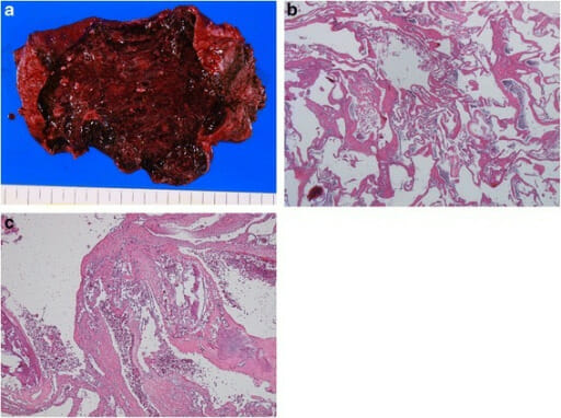
Hallazgos macroscópicos e histológicos del hemangioma hepático:
a: tumor elástico de color vino
b: tumor compuesto por espacios llenos de sangre revestidos por una sola capa de células endoteliales sin músculo liso (tinción H&E, objetivo 40x)
c: lesiones fibróticas, hialinizadas y calcificadas (tinción H&E, objetivo 40x)
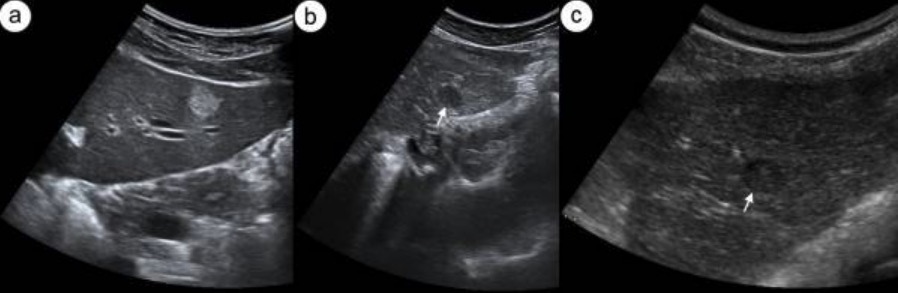
Imagen: “Fig2” por Yano T, Kobayashi T et al. Licencia: CC BY 4.0

Patogénesis

  • Proceso patogénico poco claro
  • Se cree que es el resultado de una malformación vascular congénita, con dilatación como el patrón de crecimiento
  • Factores de riesgo: Los LOS Neisseria estrógenos, como en EN Erythema nodosum is an immune-mediated panniculitis (inflammation of the subcutaneous fat) caused by a type IV (delayed-type) hypersensitivity reaction. It commonly manifests in young women as tender, erythematous nodules on the shins. Erythema Nodosum los LOS Neisseria ACO o el embarazo, pueden favorecer su crecimiento.

Presentación clínica

  • Típicamente asintomático
  • Frecuentemente se descubre incidentalmente en EN Erythema nodosum is an immune-mediated panniculitis (inflammation of the subcutaneous fat) caused by a type IV (delayed-type) hypersensitivity reaction. It commonly manifests in young women as tender, erythematous nodules on the shins. Erythema Nodosum los LOS Neisseria estudios de imagen
  • Puede causar dolor Dolor Inflammation/plenitud en EN Erythema nodosum is an immune-mediated panniculitis (inflammation of the subcutaneous fat) caused by a type IV (delayed-type) hypersensitivity reaction. It commonly manifests in young women as tender, erythematous nodules on the shins. Erythema Nodosum el cuadrante superior derecho
  • Si es lo suficientemente grande (> 10 cm), puede comprimir otros órganos y manifestarse como náuseas, saciedad temprana y distensión abdominal
  • La exploración física suele ser normal, pero puede mostrar una masa hepática palpable
  • Síndrome de Kasabach-Merritt:
    • Un hemangioma Hemangioma A vascular anomaly due to proliferation of blood vessels that forms a tumor-like mass. The common types involve capillaries and veins. It can occur anywhere in the body but is most frequently noticed in the skin and subcutaneous tissue. Imaging of the Liver and Biliary Tract gigante puede causar este raro trastorno de la coagulación.
    • Se asocia con trombocitopenia, coagulopatía de consumo y hemorragias
    • Tasa de mortalidad de hasta el 37%.

Diagnóstico

  • Ultrasonido:
    • Masa hiperecoica homogénea
    • El doppler Doppler Ultrasonography applying the doppler effect, with frequency-shifted ultrasound reflections produced by moving targets (usually red blood cells) in the bloodstream along the ultrasound axis in direct proportion to the velocity of movement of the targets, to determine both direction and velocity of blood flow. Ultrasound (Sonography) puede mostrar el flujo sanguíneo dentro del hemangioma Hemangioma A vascular anomaly due to proliferation of blood vessels that forms a tumor-like mass. The common types involve capillaries and veins. It can occur anywhere in the body but is most frequently noticed in the skin and subcutaneous tissue. Imaging of the Liver and Biliary Tract.
    • Con contraste:
      • Señal de ultrasonido mejorada por la sangre que fluye
      • Fase arterial: realce nodular periférico
      • Fase tardía: realce continuo, con llenado centrípeto
  • RM:
    • Masa homogénea bien delimitada
    • Lesión hipointensa en EN Erythema nodosum is an immune-mediated panniculitis (inflammation of the subcutaneous fat) caused by a type IV (delayed-type) hypersensitivity reaction. It commonly manifests in young women as tender, erythematous nodules on the shins. Erythema Nodosum T1; alta intensidad de señal en EN Erythema nodosum is an immune-mediated panniculitis (inflammation of the subcutaneous fat) caused by a type IV (delayed-type) hypersensitivity reaction. It commonly manifests in young women as tender, erythematous nodules on the shins. Erythema Nodosum imágenes ponderadas en EN Erythema nodosum is an immune-mediated panniculitis (inflammation of the subcutaneous fat) caused by a type IV (delayed-type) hypersensitivity reaction. It commonly manifests in young women as tender, erythematous nodules on the shins. Erythema Nodosum T2
    • Con contraste: realce nodular periférico, con progresión centrípeta
  • TC:
    • Sin contraste: masa hipodensa bien delimitada
    • Con contraste: realce periférico en EN Erythema nodosum is an immune-mediated panniculitis (inflammation of the subcutaneous fat) caused by a type IV (delayed-type) hypersensitivity reaction. It commonly manifests in young women as tender, erythematous nodules on the shins. Erythema Nodosum fase temprana y con llenado centrípeto visto en EN Erythema nodosum is an immune-mediated panniculitis (inflammation of the subcutaneous fat) caused by a type IV (delayed-type) hypersensitivity reaction. It commonly manifests in young women as tender, erythematous nodules on the shins. Erythema Nodosum fase tardía
  • No se recomienda la biopsia:
    • Riesgos de hemorragia o ruptura con el procedimiento invasivo
    • Bajo rendimiento diagnóstico

Tratamiento

  • Observación en EN Erythema nodosum is an immune-mediated panniculitis (inflammation of the subcutaneous fat) caused by a type IV (delayed-type) hypersensitivity reaction. It commonly manifests in young women as tender, erythematous nodules on the shins. Erythema Nodosum los LOS Neisseria casos asintomáticos:
    • ≤ 5 cm: no se requiere de otros estudios imagenológicos
    • > 5 cm:
      • Repetir estudios de imagen en EN Erythema nodosum is an immune-mediated panniculitis (inflammation of the subcutaneous fat) caused by a type IV (delayed-type) hypersensitivity reaction. It commonly manifests in young women as tender, erythematous nodules on the shins. Erythema Nodosum 6–12 meses.
      • Si la lesión es estable (tasa de crecimiento ≤ 3 mm/año), no se realizan más estudios
  • Considerar la cirugía (resección hepática o enucleación):
    • Síntomas persistentes y/o crecimiento rápido (> 3 mm/año)
    • Pacientes sintomáticos
  • La embolización arterial transcatéter puede utilizarse para reducir el tamaño de la lesión antes de la cirugía.

Hiperplasia Nodular Focal

Características

  • Examen macroscópico:
    • Lesión firme y solitaria, sin cápsula
    • Cicatriz estrellada central característica (múltiples ramas de una arteria que irradian hacia la periferia)
    • Tamaño: a menudo < 5 cm
    • Localización: normalmente subcapsular
    • La hiperplasia nodular focal tiene variantes atípicas.
  • Examen microscópico:
    • Hepatocitos agrupados, divididos por septos fibrosos, que irradian desde la cicatriz central
    • Los LOS Neisseria septos contienen arterias, venas portales, conductillos biliares y células de Kupffer.
    • Fuerte tinción citoplasmática de la glutamina sintetasa en EN Erythema nodosum is an immune-mediated panniculitis (inflammation of the subcutaneous fat) caused by a type IV (delayed-type) hypersensitivity reaction. It commonly manifests in young women as tender, erythematous nodules on the shins. Erythema Nodosum grupos de hepatocitos
Hiperplasia nodular focal clásica

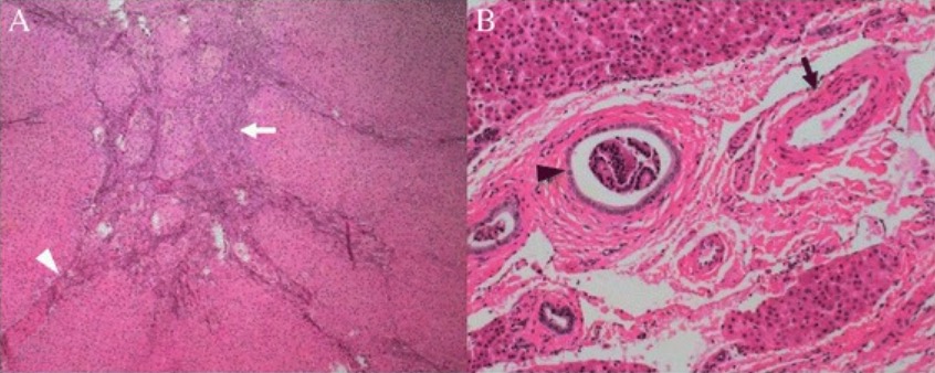
Características histológicas de la hiperplasia nodular focal clásica: El tumor estaba subdividido en nódulos por septos fibrosos (A, punta de flecha blanca) que se originaban de una cicatriz central (A, flecha blanca). Se muestran arterias malformadas (B, flecha negra) y la proliferación de conductos biliares (B, punta de flecha negra).

Imagen: “Histological features of the classic focal nodular hyperplasia” por Department of Radiology, PLA General Hospital, #28 Fuxing Road, Beijing, 100853, China. Licencia: CC BY 4.0

Patogénesis

  • Respuesta regenerativa de los LOS Neisseria hepatocitos ante la perfusión alterada de las arterias anómalas ( en EN Erythema nodosum is an immune-mediated panniculitis (inflammation of the subcutaneous fat) caused by a type IV (delayed-type) hypersensitivity reaction. It commonly manifests in young women as tender, erythematous nodules on the shins. Erythema Nodosum el centro del nódulo)
  • Sin potencial maligno
  • Mayor riesgo de hiperplasia nodular focal en EN Erythema nodosum is an immune-mediated panniculitis (inflammation of the subcutaneous fat) caused by a type IV (delayed-type) hypersensitivity reaction. It commonly manifests in young women as tender, erythematous nodules on the shins. Erythema Nodosum pacientes con telangiectasia Telangiectasia Permanent dilation of preexisting blood vessels creating small focal red lesions, most commonly in the skin or mucous membranes. It is characterized by the prominence of skin blood vessels, such as vascular spiders. Chronic Venous Insufficiency hemorrágica hereditaria

Presentación clínica

  • Asintomática
  • A menudo se encuentra incidentalmente en EN Erythema nodosum is an immune-mediated panniculitis (inflammation of the subcutaneous fat) caused by a type IV (delayed-type) hypersensitivity reaction. It commonly manifests in young women as tender, erythematous nodules on the shins. Erythema Nodosum estudios de imagen
  • En EN Erythema nodosum is an immune-mediated panniculitis (inflammation of the subcutaneous fat) caused by a type IV (delayed-type) hypersensitivity reaction. It commonly manifests in young women as tender, erythematous nodules on the shins. Erythema Nodosum los LOS Neisseria casos sintomáticos, el dolor Dolor Inflammation abdominal es la manifestación más común.
  • Examen físico: suele ser normal, pero puede mostrar una masa abdominal

Diagnóstico

  • US:
    • Sin contraste: masa isoecoica
    • Con contraste:
      • Hiperplasia nodular focal: realce sostenido en EN Erythema nodosum is an immune-mediated panniculitis (inflammation of the subcutaneous fat) caused by a type IV (delayed-type) hypersensitivity reaction. It commonly manifests in young women as tender, erythematous nodules on the shins. Erythema Nodosum la fase arterial y en EN Erythema nodosum is an immune-mediated panniculitis (inflammation of the subcutaneous fat) caused by a type IV (delayed-type) hypersensitivity reaction. It commonly manifests in young women as tender, erythematous nodules on the shins. Erythema Nodosum la fase venosa temprana
      • Las arterias centrales se ven con un patrón de rueda de carreta en EN Erythema nodosum is an immune-mediated panniculitis (inflammation of the subcutaneous fat) caused by a type IV (delayed-type) hypersensitivity reaction. It commonly manifests in young women as tender, erythematous nodules on the shins. Erythema Nodosum la fase arterial (centrífuga)
  • RM:
    • Máxima precisión diagnóstica
    • Precontraste: isointenso en EN Erythema nodosum is an immune-mediated panniculitis (inflammation of the subcutaneous fat) caused by a type IV (delayed-type) hypersensitivity reaction. It commonly manifests in young women as tender, erythematous nodules on the shins. Erythema Nodosum T1; ligeramente hiperintenso en EN Erythema nodosum is an immune-mediated panniculitis (inflammation of the subcutaneous fat) caused by a type IV (delayed-type) hypersensitivity reaction. It commonly manifests in young women as tender, erythematous nodules on the shins. Erythema Nodosum imágenes ponderadas en EN Erythema nodosum is an immune-mediated panniculitis (inflammation of the subcutaneous fat) caused by a type IV (delayed-type) hypersensitivity reaction. It commonly manifests in young women as tender, erythematous nodules on the shins. Erythema Nodosum T2
    • Con contraste:
      • Fase arterial: masa hipervascular de realce homogéneo rápido/temprano
      • Fase retardada: masa isointensa, con realce de la cicatriz central
  • TC:
    • Precontraste: isodenso
    • Con contraste:
      • Fase arterial: lesión hiperdensa homogénea
      • Fase venosa: isodensa, similar al AL Amyloidosis parénquima hepático; la cicatriz central se vuelve hiperdensa.
  • Medicina nuclear (gammagrafía de tecnecio-99m y azufre coloidal):
    • Se observa un aumento de la captación del azufre coloidal (actividad de las células de Kupffer) en EN Erythema nodosum is an immune-mediated panniculitis (inflammation of the subcutaneous fat) caused by a type IV (delayed-type) hypersensitivity reaction. It commonly manifests in young women as tender, erythematous nodules on the shins. Erythema Nodosum el 60%–70% de los LOS Neisseria pacientes.
    • Ayuda a diferenciar la hiperplasia nodular focal del adenoma (que no tiene actividad de las células de Kupffer)
  • Si los LOS Neisseria resultados de imagenología no son típicos de una hiperplasia nodular focal, puede ser necesaria una biopsia.
Hiperplasia Nodular Focal

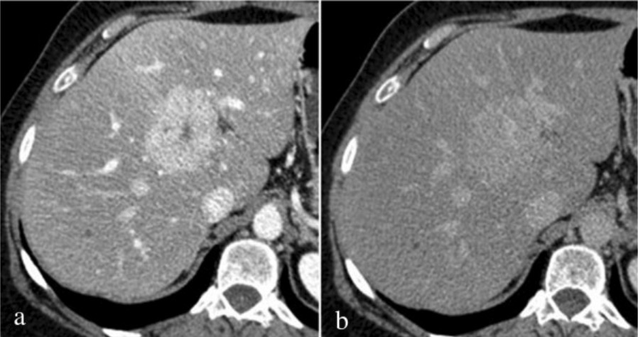
Hallazgos de la tomografía computarizada de la hiperplasia nodular focal: La fase mixta (fase arterial hepática/fase venosa portal durante el realce hepático) muestra un realce homogéneo intenso con una cicatriz central hipodensa (a); en la fase retardada (b), la lesión se observa sustancialmente isodensa al parénquima hepático con realce persistente de la cicatriz central.

Imagen: “Fig3” por Department of Surgical and Biomedical Sciences, Division of Radiology 2, Perugia University, S, Maria della Misericordia Hospital, S, Andrea delle Fratte, 06134 Perugia, Italy. Licencia: CC BY 4.0

Tratamiento

  • Para pacientes asintomáticos: no se recomienda la vigilancia rutinaria porque la lesión raramente crece
  • Para los LOS Neisseria pacientes sintomáticos: pueden ser sometidos a embolización transarterial, ablación por radiofrecuencia o resección quirúrgica

Adenoma Hepatocelular

Características

  • Examen macroscópico:
    • Suele tratarse de una lesión solitaria con un margen bien definido, pero puede ocurrir como lesiones múltiples
    • Tamaños variados, desde pequeños hasta varios centímetros
    • Carece de cápsula fibrosa (riesgo de rotura y hemorragia)
    • Localización: a menudo en EN Erythema nodosum is an immune-mediated panniculitis (inflammation of the subcutaneous fat) caused by a type IV (delayed-type) hypersensitivity reaction. It commonly manifests in young women as tender, erythematous nodules on the shins. Erythema Nodosum el lóbulo derecho del hígado
  • Examen microscópico:
    • Láminas de hepatocitos agrandados con núcleos pequeños, glucógeno y lípidos
    • Presencia de grandes arterias
    • Generalmente, no hay tractos portales o conductillos biliares (lo que diferencia el adenoma hepatocelular de la hiperplasia nodular focal); el subtipo inflamatorio es una excepción
Adenoma hepático

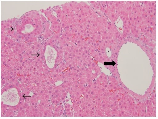
Adenoma hepático: neoplasia bien diferenciada compuesta por hepatocitos de apariencia normal dispuestos en láminas y cordones delgados con un patrón de crecimiento pseudoacinar en parches (flechas finas), focos inflamatorios dispersos y bandas de fibrosis con arterias grandes no apareadas (flecha gruesa).

Imagen: “Hepatic adenoma” por M. I. Montenovo. Licencia: CC BY 4.0

Patogénesis

  • Tumor Tumor Inflammation epitelial glandular benigno, con los LOS Neisseria siguientes factores de riesgo:
    • Uso de ACO, esteroides anabólicos
    • Síndromes genéticos como las enfermedades de almacenamiento de glucógeno y la poliposis adenomatosa familiar
    • Obesidad, síndrome metabólico
  • Subtipos basados en EN Erythema nodosum is an immune-mediated panniculitis (inflammation of the subcutaneous fat) caused by a type IV (delayed-type) hypersensitivity reaction. It commonly manifests in young women as tender, erythematous nodules on the shins. Erythema Nodosum el comportamiento molecular del adenoma hepatocelular:
    • Adenomas con mutación del factor nuclear hepático 1 alfa (HNF1-ɑ, por sus siglas en EN Erythema nodosum is an immune-mediated panniculitis (inflammation of the subcutaneous fat) caused by a type IV (delayed-type) hypersensitivity reaction. It commonly manifests in young women as tender, erythematous nodules on the shins. Erythema Nodosum inglés):
      • 35%–40%
      • Tienen mutaciones de pérdida de función del gen HNF1-ɑ
      • Hallazgos: esteatosis o grasa prominente en EN Erythema nodosum is an immune-mediated panniculitis (inflammation of the subcutaneous fat) caused by a type IV (delayed-type) hypersensitivity reaction. It commonly manifests in young women as tender, erythematous nodules on the shins. Erythema Nodosum los LOS Neisseria hepatocitos
      • Afecta predominantemente a las mujeres
      • Bajo riesgo de transformación maligna
      • Asociada a la diabetes Diabetes Diabetes mellitus (DM) is a metabolic disease characterized by hyperglycemia and dysfunction of the regulation of glucose metabolism by insulin. Type 1 DM is diagnosed mostly in children and young adults as the result of autoimmune destruction of β cells in the pancreas and the resulting lack of insulin. Type 2 DM has a significant association with obesity and is characterized by insulin resistance. Diabetes Mellitus del adulto de inicio juvenil ( MODY MODY Diabetes Mellitus 3, por sus siglas en EN Erythema nodosum is an immune-mediated panniculitis (inflammation of the subcutaneous fat) caused by a type IV (delayed-type) hypersensitivity reaction. It commonly manifests in young women as tender, erythematous nodules on the shins. Erythema Nodosum inglés)
    • Adenoma hepatocelular con β-catenina activada:
      • 10%–20%
      • Asociado a mutaciones del gen CTNNB1 (β-catenina) o de otros componentes de la vía Wnt
      • Frecuente en EN Erythema nodosum is an immune-mediated panniculitis (inflammation of the subcutaneous fat) caused by a type IV (delayed-type) hypersensitivity reaction. It commonly manifests in young women as tender, erythematous nodules on the shins. Erythema Nodosum los LOS Neisseria hombres
      • Vinculado al AL Amyloidosis uso de esteroides anabólicos
      • Hallazgos: características citológicas atípicas
      • Alto riesgo de desarrollar un carcinoma hepatocelular
    • Adenomas inflamatorios:
      • 40%–50%
      • Hallazgos: infiltrados inflamatorios, dilatación sinusoidal
      • Más frecuente en EN Erythema nodosum is an immune-mediated panniculitis (inflammation of the subcutaneous fat) caused by a type IV (delayed-type) hypersensitivity reaction. It commonly manifests in young women as tender, erythematous nodules on the shins. Erythema Nodosum mujeres con obesidad o diabetes Diabetes Diabetes mellitus (DM) is a metabolic disease characterized by hyperglycemia and dysfunction of the regulation of glucose metabolism by insulin. Type 1 DM is diagnosed mostly in children and young adults as the result of autoimmune destruction of β cells in the pancreas and the resulting lack of insulin. Type 2 DM has a significant association with obesity and is characterized by insulin resistance. Diabetes Mellitus
    • Sin clasificar: hasta el 10%.

Presentación clínica

  • Puede ser asintomático, pero el 25% de los LOS Neisseria pacientes presentan dolor Dolor Inflammation o una masa en EN Erythema nodosum is an immune-mediated panniculitis (inflammation of the subcutaneous fat) caused by a type IV (delayed-type) hypersensitivity reaction. It commonly manifests in young women as tender, erythematous nodules on the shins. Erythema Nodosum el cuadrante superior derecho.
  • Hemorragia: mayor riesgo cuando el tamaño es > 5 cm
    • La hemorragia en EN Erythema nodosum is an immune-mediated panniculitis (inflammation of the subcutaneous fat) caused by a type IV (delayed-type) hypersensitivity reaction. It commonly manifests in young women as tender, erythematous nodules on the shins. Erythema Nodosum la lesión puede manifestarse como dolor Dolor Inflammation abdominal.
    • En EN Erythema nodosum is an immune-mediated panniculitis (inflammation of the subcutaneous fat) caused by a type IV (delayed-type) hypersensitivity reaction. It commonly manifests in young women as tender, erythematous nodules on the shins. Erythema Nodosum los LOS Neisseria casos graves, la ruptura hacia el peritoneo se presenta con dolor Dolor Inflammation intenso e hipotensión.

Diagnóstico

  • US:
    • Sin contraste: masa heterogénea inespecífica
    • Con contraste: hiperrealce de la periferia al AL Amyloidosis centro (centrípeta)
  • TC:
    • Sin contraste: lesión isodensa
    • Con contraste:
      • Fase arterial: puede haber realce periférico
      • Fase venosa portal: con flujo centrípeto
    • Si tiene áreas de hemorragia, necrosis Necrosis The death of cells in an organ or tissue due to disease, injury or failure of the blood supply. Ischemic Cell Damage y calcificación, el adenoma parece heterogéneo.
  • RM con contraste:
    • Superior a otras modalidades para el diagnóstico del adenoma hepatocelular
    • Fase arterial: lesión bien delimitada, con realce (heterogéneo por hemorragia, necrosis Necrosis The death of cells in an organ or tissue due to disease, injury or failure of the blood supply. Ischemic Cell Damage, esteatosis)
    • El patrón en EN Erythema nodosum is an immune-mediated panniculitis (inflammation of the subcutaneous fat) caused by a type IV (delayed-type) hypersensitivity reaction. It commonly manifests in young women as tender, erythematous nodules on the shins. Erythema Nodosum las fases posteriores se correlaciona con los LOS Neisseria subtipos moleculares en EN Erythema nodosum is an immune-mediated panniculitis (inflammation of the subcutaneous fat) caused by a type IV (delayed-type) hypersensitivity reaction. It commonly manifests in young women as tender, erythematous nodules on the shins. Erythema Nodosum la mayoría de los LOS Neisseria casos (tipo HNF1-ɑ e inflamatorio):
      • Adenoma inflamatorio: El realce arterial persiste en EN Erythema nodosum is an immune-mediated panniculitis (inflammation of the subcutaneous fat) caused by a type IV (delayed-type) hypersensitivity reaction. It commonly manifests in young women as tender, erythematous nodules on the shins. Erythema Nodosum la fase venosa portal y en EN Erythema nodosum is an immune-mediated panniculitis (inflammation of the subcutaneous fat) caused by a type IV (delayed-type) hypersensitivity reaction. It commonly manifests in young women as tender, erythematous nodules on the shins. Erythema Nodosum la fase retardada.
      • Adenoma hepatocelular con mutación HNF1-ɑ: El realce arterial no persiste.
      • Adenoma hepatocelular con β-catenina activada: no hay características específicas en EN Erythema nodosum is an immune-mediated panniculitis (inflammation of the subcutaneous fat) caused by a type IV (delayed-type) hypersensitivity reaction. It commonly manifests in young women as tender, erythematous nodules on the shins. Erythema Nodosum la RM
  • Biopsia con aguja gruesa: para aclarar si la imagenología es ambigua
Enfermedad hepática benigna RMN con adenoma con hemorragia

Resonancia magnética de un paciente con un adenoma con sangrado: flecha blanca, hematoma; flecha negra, adenoma hepático.
A: La secuencia T2 con saturación grasa muestra un nódulo subcapsular hepático con hiposeñal.
B: La secuencia T1 previa al contraste muestra una lesión con hiperseñal, indicando la presencia de productos de la degradación de la hemoglobina.
C: La secuencia T1 post-contraste en la fase arterial resalta la lesión hepática.

Imagen: “MRI” por Enio Campos AMICO, José Roberto ALVES et al. Licencia: CC BY 4.0

Tratamiento

  • Suspender los LOS Neisseria esteroides anabólicos o los LOS Neisseria ACO.
  • Pérdida de peso
  • Observación y vigilancia:
    • Mujeres con tumor Tumor Inflammation < 5 cm: RM con contraste a los LOS Neisseria 6 meses y luego anualmente si no hay crecimiento
    • Mujeres asintomáticas, tumor Tumor Inflammation > 5 cm con uso de ACO:
      • Puede probar la interrupción del ACO
      • Si la RM muestra que la lesión se mantiene > 5 cm después de 6 meses, se procede a la resección quirúrgica.
  • Resección quirúrgica:
    • Tumor Tumor Inflammation > 5 cm: resección quirúrgica debido a un alto riesgo de rotura, hemorragia o transformación maligna
    • Hombres con adenoma hepatocelular (independientemente del tamaño)
    • Mutación probada de β-catenina
  • Embolización transarterial:
    • Tumor Tumor Inflammation complicado con hemorragia e inestabilidad hemodinámica
    • Si después del procedimiento se observa una lesión residual en EN Erythema nodosum is an immune-mediated panniculitis (inflammation of the subcutaneous fat) caused by a type IV (delayed-type) hypersensitivity reaction. It commonly manifests in young women as tender, erythematous nodules on the shins. Erythema Nodosum la imagenología de seguimiento, se debe realizar una resección quirúrgica.

Diagnóstico Diferencial

  • Carcinoma hepatocelular: un tipo de cáncer que se desarrolla en EN Erythema nodosum is an immune-mediated panniculitis (inflammation of the subcutaneous fat) caused by a type IV (delayed-type) hypersensitivity reaction. It commonly manifests in young women as tender, erythematous nodules on the shins. Erythema Nodosum pacientes con cirrosis hepática. Suele presentarse con dolor Dolor Inflammation abdominal y se encuentra en EN Erythema nodosum is an immune-mediated panniculitis (inflammation of the subcutaneous fat) caused by a type IV (delayed-type) hypersensitivity reaction. It commonly manifests in young women as tender, erythematous nodules on the shins. Erythema Nodosum estudios de imagen hepáticos. El tratamiento es con cirugía, radioterapia y quimioterapia. Existen características imagenológicas específicas del hemangioma Hemangioma A vascular anomaly due to proliferation of blood vessels that forms a tumor-like mass. The common types involve capillaries and veins. It can occur anywhere in the body but is most frequently noticed in the skin and subcutaneous tissue. Imaging of the Liver and Biliary Tract hepático (realce periférico) y de la hiperplasia nodular focal (cicatriz estrellada central) que pueden ayudar a diferenciar estos tumores del carcinoma hepatocelular. Una mayor diferenciación puede requerir una biopsia de la lesión.
  • Metástasis hepáticas: se presentan como masas en EN Erythema nodosum is an immune-mediated panniculitis (inflammation of the subcutaneous fat) caused by a type IV (delayed-type) hypersensitivity reaction. It commonly manifests in young women as tender, erythematous nodules on the shins. Erythema Nodosum el parénquima hepático. Estas lesiones se ven como múltiples lesiones anulares del hígado en EN Erythema nodosum is an immune-mediated panniculitis (inflammation of the subcutaneous fat) caused by a type IV (delayed-type) hypersensitivity reaction. It commonly manifests in young women as tender, erythematous nodules on the shins. Erythema Nodosum la TC con contraste, lo que ayuda a distinguirlas de las lesiones benignas. Los LOS Neisseria pacientes suelen tener una historia de malignidad extrahepática que hace HACE Altitude Sickness que la metástasis sea una causa más probable de las lesiones hepáticas.
  • Absceso hepático: se presenta como una masa en EN Erythema nodosum is an immune-mediated panniculitis (inflammation of the subcutaneous fat) caused by a type IV (delayed-type) hypersensitivity reaction. It commonly manifests in young women as tender, erythematous nodules on the shins. Erythema Nodosum el parénquima hepático debido a una infección en EN Erythema nodosum is an immune-mediated panniculitis (inflammation of the subcutaneous fat) caused by a type IV (delayed-type) hypersensitivity reaction. It commonly manifests in young women as tender, erythematous nodules on the shins. Erythema Nodosum el hígado. Los LOS Neisseria pacientes suelen presentar diarrea ( en EN Erythema nodosum is an immune-mediated panniculitis (inflammation of the subcutaneous fat) caused by a type IV (delayed-type) hypersensitivity reaction. It commonly manifests in young women as tender, erythematous nodules on the shins. Erythema Nodosum los LOS Neisseria casos de absceso hepático amebiano), dolor Dolor Inflammation abdominal y/o fiebre. Los LOS Neisseria síntomas sistémicos ayudan a diferenciar los LOS Neisseria abscesos de otros tumores.
  • Quistes hepáticos: lesiones benignas llenas de líquido en EN Erythema nodosum is an immune-mediated panniculitis (inflammation of the subcutaneous fat) caused by a type IV (delayed-type) hypersensitivity reaction. It commonly manifests in young women as tender, erythematous nodules on the shins. Erythema Nodosum el hígado, que a menudo se presentan como masas hepáticas incidentales. En EN Erythema nodosum is an immune-mediated panniculitis (inflammation of the subcutaneous fat) caused by a type IV (delayed-type) hypersensitivity reaction. It commonly manifests in young women as tender, erythematous nodules on the shins. Erythema Nodosum el ultrasonido, el quiste es una lesión anecoica y redonda con realce acústico posterior. En EN Erythema nodosum is an immune-mediated panniculitis (inflammation of the subcutaneous fat) caused by a type IV (delayed-type) hypersensitivity reaction. It commonly manifests in young women as tender, erythematous nodules on the shins. Erythema Nodosum la TC, el quiste se ve VE Ventilation: Mechanics of Breathing como una lesión bien delimitada sin realce con el contraste, lo que ayuda a diferenciar los LOS Neisseria quistes hepáticos de los LOS Neisseria tumores hepáticos benignos.

Referencias

  1. Chopra, S. (2019). Focal nodular hyperplasia. Runyon, B., Robson, K. (Eds). UpToDate. Retrieved 22 Nov 2020 from https://www.uptodate.com/contents/focal-nodular-hyperplasia
  2. Colombo, M. (2020). EASL Clinical Practice Guidelines on the Management of Benign Liver Tumors. Retrieved 22 Nov 2020 from https://aasldpubs.onlinelibrary.wiley.com/doi/full/10.1002/cld.933
  3. Curry, M., Chopra, S. (2019). Hepatic hemangioma. Lindor, K., Robson, K. (Eds). UpToDate. Retrieved 22 Nov 2020 from https://www.uptodate.com/contents/hepatic-hemangioma
  4. Friedman L.S. (2021). Benign liver neoplasms. Papadakis M.A., & McPhee S.J., & Rabow M.W.(Eds.), Current Medical Diagnosis & Treatment 2021. McGraw-Hill.
  5. Gill, R., Kakar, S. (2020). In Kumar, V., Abbas, A. K., Aster, J.C., (Eds.), Robbins & Cotran Pathologic Basis of Disease. (10th ed., pp. 865-867). Elsevier, Inc.
  6. Leon, M., Chavez, L., Surani, S. (2020). Hepatic hemangioma: what internists need to know. World J Gastroenterol. 26(1): 11–20. doi: 10.3748/wjg.v26.i1.11
  7. Llovet J.M. (2018). Tumors of the liver and biliary tree. Jameson J, & Fauci A.S., & Kasper D.L., & Hauser S.L., & Longo D.L., & Loscalzo J (Eds.), Harrison’s Principles of Internal Medicine, 20e. McGraw-Hill.
  8. Raveendran, S., Lu, Z. (2018). A short review on early HCC: MRI findings and pathological diagnosis. Radiology of Infectious Diseases, Volume 5, Issue 2, pp. 91-97. https://doi.org/10.1016/j.jrid.2017.08.008
  9. Shreenath, A., Kahloon, A. (2020). Hepatic Adenoma. StatPearls. https://www.ncbi.nlm.nih.gov/books/NBK513264/

¡Crea tu cuenta gratis o inicia una sesión para seguir leyendo!

Regístrate ahora y obtén acceso gratuito a Lecturio con páginas de concepto, videos médicos y cuestionarios para tu educación médica.

User Reviews

Que tengas una sesión de estudio alegre y navideña 🎁 Ahorra 50% en todos los planes >>

Details