Malassezia Malassezia Malassezia is a lipophilic yeast commonly found on the skin surfaces of many animals, including humans. In the presence of certain environments or triggers, this fungus can cause pathologic diseases ranging from superficial skin conditions (tinea versicolor and dermatitis) to invasive disease (e.g., Malassezia folliculitis, catheter-associated fungemia, meningitis, and urinary tract infections). Malassezia Fungi es una levadura lipofílica que se encuentra habitualmente en EN Erythema nodosum is an immune-mediated panniculitis (inflammation of the subcutaneous fat) caused by a type IV (delayed-type) hypersensitivity reaction. It commonly manifests in young women as tender, erythematous nodules on the shins. Erythema Nodosum la superficie de la piel de muchos animales, e inclusive humanos. En EN Erythema nodosum is an immune-mediated panniculitis (inflammation of the subcutaneous fat) caused by a type IV (delayed-type) hypersensitivity reaction. It commonly manifests in young women as tender, erythematous nodules on the shins. Erythema Nodosum presencia de determinadas condiciones ambientales o desencadenantes, este hongo puede causar enfermedades patológicas que van desde condiciones cutáneas superficiales (tiña versicolor y dermatitis Dermatitis Any inflammation of the skin. Atopic Dermatitis (Eczema)) hasta enfermedades invasivas (e.g., foliculitis por Malassezia Malassezia Malassezia is a lipophilic yeast commonly found on the skin surfaces of many animals, including humans. In the presence of certain environments or triggers, this fungus can cause pathologic diseases ranging from superficial skin conditions (tinea versicolor and dermatitis) to invasive disease (e.g., Malassezia folliculitis, catheter-associated fungemia, meningitis, and urinary tract infections). Malassezia Fungi, fungemia Fungemia The presence of fungi circulating in the blood. Opportunistic fungal sepsis is seen most often in immunosuppressed patients with severe neutropenia or in postoperative patients with intravenous catheters and usually follows prolonged antibiotic therapy. Chronic Granulomatous Disease asociada a catéter, meningitis Meningitis Meningitis is inflammation of the meninges, the protective membranes of the brain, and spinal cord. The causes of meningitis are varied, with the most common being bacterial or viral infection. The classic presentation of meningitis is a triad of fever, altered mental status, and nuchal rigidity. Meningitis e infecciones del tracto urinario). Los LOS Neisseria pacientes con tiña versicolor desarrollan una erupción hipopigmentada o hiperpigmentada asintomática o ligeramente pruriginosa en EN Erythema nodosum is an immune-mediated panniculitis (inflammation of the subcutaneous fat) caused by a type IV (delayed-type) hypersensitivity reaction. It commonly manifests in young women as tender, erythematous nodules on the shins. Erythema Nodosum el tórax, la espalda, el abdomen o la cara. La dermatitis Dermatitis Any inflammation of the skin. Atopic Dermatitis (Eczema) seborreica se presenta con una erupción eritematosa y pruriginosa con escamas grasientas y costras amarillas, que afecta con mayor frecuencia a zonas de la cara, la parte superior del tronco o a las regiones intertriginosas. La presentación de la foliculitis por Malassezia Malassezia Malassezia is a lipophilic yeast commonly found on the skin surfaces of many animals, including humans. In the presence of certain environments or triggers, this fungus can cause pathologic diseases ranging from superficial skin conditions (tinea versicolor and dermatitis) to invasive disease (e.g., Malassezia folliculitis, catheter-associated fungemia, meningitis, and urinary tract infections). Malassezia Fungi es similar a la de la foliculitis bacteriana, con pápulas o pústulas pruriginosas y monomorfas en EN Erythema nodosum is an immune-mediated panniculitis (inflammation of the subcutaneous fat) caused by a type IV (delayed-type) hypersensitivity reaction. It commonly manifests in young women as tender, erythematous nodules on the shins. Erythema Nodosum un patrón folicular. El diagnóstico de las condiciones cutáneas superficiales se realiza principalmente mediante el examen físico, pero también puede confirmarse mediante la microscopía de raspados cutáneos. El tratamiento implica el uso de agentes antifúngicos tópicos y orales.
Last updated: Dec 15, 2025
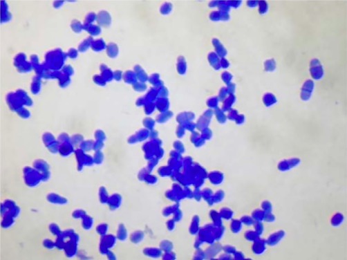
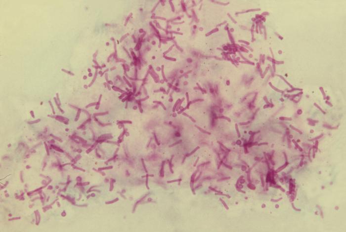
Tinción con azul de metileno de la levadura Malassezia
Imagen: “2916” por Allergy Unit, Department of Dermatology, University Hospital of Zurich, Gloriastrasse 31, 8091 Zurich, Switzerland. Licencia: CC BY 4.0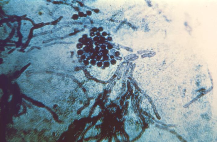
Microfotografía de una muestra de raspado de piel en un paciente con tiña versicolor:
Obsérvese la presencia de células fúngicas esféricas tipo levadura y de hifas cortas de la especie Malassezia.
Aspecto microscópico de las células e hifas del hongo Malassezia furfur a partir del raspado de la piel en la tiña versicolor
Imagen: “3938” por CDC. Licencia: Dominio PúblicoHay numerosas especies reconocidas:
Tiña versicolor:
Foliculitis por Malassezia Malassezia Malassezia is a lipophilic yeast commonly found on the skin surfaces of many animals, including humans. In the presence of certain environments or triggers, this fungus can cause pathologic diseases ranging from superficial skin conditions (tinea versicolor and dermatitis) to invasive disease (e.g., Malassezia folliculitis, catheter-associated fungemia, meningitis, and urinary tract infections). Malassezia Fungi:
Malassezia Malassezia Malassezia is a lipophilic yeast commonly found on the skin surfaces of many animals, including humans. In the presence of certain environments or triggers, this fungus can cause pathologic diseases ranging from superficial skin conditions (tinea versicolor and dermatitis) to invasive disease (e.g., Malassezia folliculitis, catheter-associated fungemia, meningitis, and urinary tract infections). Malassezia Fungi forma parte de la flora cutánea normal de los LOS Neisseria humanos y los LOS Neisseria animales.
Entre los LOS Neisseria factores de riesgo que predisponen a la enfermedad se encuentran:
Tiña versicolor:
Dermatitis Dermatitis Any inflammation of the skin. Atopic Dermatitis (Eczema) seborreica:
Foliculitis por Malassezia Malassezia Malassezia is a lipophilic yeast commonly found on the skin surfaces of many animals, including humans. In the presence of certain environments or triggers, this fungus can cause pathologic diseases ranging from superficial skin conditions (tinea versicolor and dermatitis) to invasive disease (e.g., Malassezia folliculitis, catheter-associated fungemia, meningitis, and urinary tract infections). Malassezia Fungi:
Síntomas:
Examen físico:
Erupción eritematosa en el tórax, el cuello y el abdomen de un paciente, consistente con tiña versicolor
Imagen: “22849” por CDC/Dr. Lucille K. Georg. Licencia: Dominio Público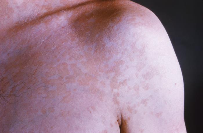
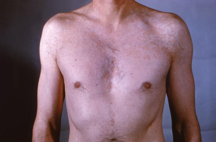
Erupción eritematosa, en forma de placa, en el hombro izquierdo de un paciente debida a una tiña versicolor
Imagen: “22847” por CDC/Dr. Lucille K. Georg. Licencia: Dominio Público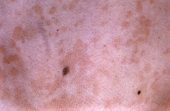
Vista de cerca de la superficie de la piel de un paciente que muestra lesiones hiperpigmentadas de tiña versicolor
Imagen: “22848” por CDC/Dr. Lucille K. Georg. Licencia: Dominio Público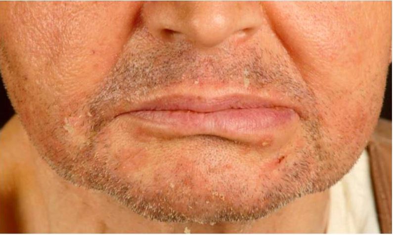
Dermatitis seborreica en un receptor de trasplante renal:
Obsérvense las pápulas eritematosas escamosas en el mentón y en los pliegues nasolabiales.
Esta afección puede parecer similar al AL Amyloidosis acné vulgar o a la foliculitis bacteriana:
El diagnóstico suele hacerse clínicamente con base en EN Erythema nodosum is an immune-mediated panniculitis (inflammation of the subcutaneous fat) caused by a type IV (delayed-type) hypersensitivity reaction. It commonly manifests in young women as tender, erythematous nodules on the shins. Erythema Nodosum los LOS Neisseria antecedentes y el examen físico. Evaluaciones adicionales pueden incluir:
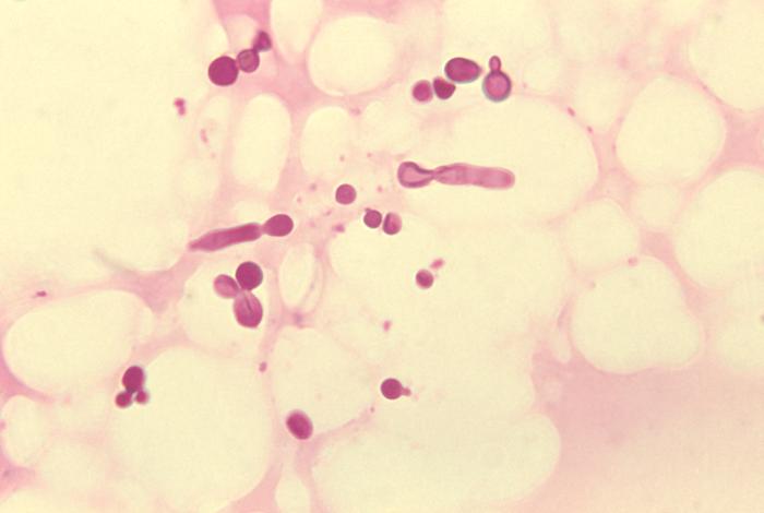
Malassezia furfur organismos fúngicos:
Microfotografía de una muestra de tejido de escamas de la piel que revela la presencia de numerosos organismos fúngicos de M. furfur. Obsérvese el aspecto de espaguetis y albóndigas debido a la presencia tanto de levaduras como de hifas.
Las recurrencias de la tiña versicolor y la dermatitis Dermatitis Any inflammation of the skin. Atopic Dermatitis (Eczema) seborreica pueden prevenirse mediante profilaxis antifúngica tópica.