Las Escherichia coli Escherichia coli The gram-negative bacterium Escherichia coli is a key component of the human gut microbiota. Most strains of E. coli are avirulent, but occasionally they escape the GI tract, infecting the urinary tract and other sites. Less common strains of E. coli are able to cause disease within the GI tract, most commonly presenting as abdominal pain and diarrhea. Escherichia coli diarreogénicas son una cepa de bacterias patógenas que puede causar infecciones intestinales. La transmisión se produce principalmente por el consumo de alimentos o agua contaminados, el contacto con personas o animales infectados y el baño en EN Erythema nodosum is an immune-mediated panniculitis (inflammation of the subcutaneous fat) caused by a type IV (delayed-type) hypersensitivity reaction. It commonly manifests in young women as tender, erythematous nodules on the shins. Erythema Nodosum aguas no tratadas. La patogénesis varía en EN Erythema nodosum is an immune-mediated panniculitis (inflammation of the subcutaneous fat) caused by a type IV (delayed-type) hypersensitivity reaction. It commonly manifests in young women as tender, erythematous nodules on the shins. Erythema Nodosum función de la cepa, pero puede incluir la producción de toxinas, la invasión de la superficie de la mucosa y la adhesión con alteración de la estructura de los LOS Neisseria enterocitos. La enfermedad no invasiva suele presentarse con diarrea acuosa, mientras que las infecciones invasivas provocan diarrea sanguinolenta. El diagnóstico puede establecerse con la reacción en EN Erythema nodosum is an immune-mediated panniculitis (inflammation of the subcutaneous fat) caused by a type IV (delayed-type) hypersensitivity reaction. It commonly manifests in young women as tender, erythematous nodules on the shins. Erythema Nodosum cadena de la polimerasa. El tratamiento suele consistir en EN Erythema nodosum is an immune-mediated panniculitis (inflammation of the subcutaneous fat) caused by a type IV (delayed-type) hypersensitivity reaction. It commonly manifests in young women as tender, erythematous nodules on the shins. Erythema Nodosum terapia de soporte (líquidos y electrolitos). Los LOS Neisseria antibióticos se reservan para las infecciones graves o persistentes y están contraindicados con E. coli enterohemorrágica por el riesgo de síndrome urémico hemolítico.
Last updated: Dec 15, 2025
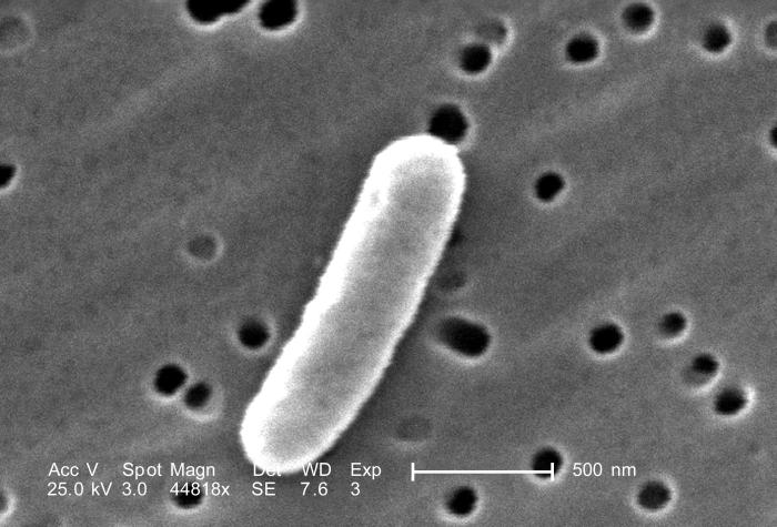
Imagen de microscopio electrónico de barrido de Escherichia coli enterotoxigénica
Imagen: “Under an extremely high magnification of 44, 818X, twice that of PHIL 10574 and 10575, this scanning electron microscopic (SEM) image revealed some of the morphologic details displayed by a single Gram-negative, rod-shaped, Escherichia coli bacterium.” por Janice Haney Carr. Licencia: Dominio PúblicoLas cepas diarreagénicas de E. coli pueden clasificarse en 5 “patotipos” clave, cada uno de los LOS Neisseria cuales tiene factores de virulencia y mecanismos patológicos únicos:
| Patógeno | ¿Invasiva? | ¿Toxina? | Tipo de diarrea |
|---|---|---|---|
| E. coli enterotoxigénica | No |
|
|
| E. coli enteropatogénica | No | No |
|
| E. coli enteroagregativa | No |
|
|
| E. coli enteroinvasiva | Sí |
|
|
| E. coli enterohemorrágica | Sí | Toxina Shiga |
|
La E. coli enterotoxigénica es un patógeno no invasivo. Utiliza las adhesinas en EN Erythema nodosum is an immune-mediated panniculitis (inflammation of the subcutaneous fat) caused by a type IV (delayed-type) hypersensitivity reaction. It commonly manifests in young women as tender, erythematous nodules on the shins. Erythema Nodosum sus fimbrias para unirse a los LOS Neisseria enterocitos del intestino delgado y produce las siguientes enterotoxinas:
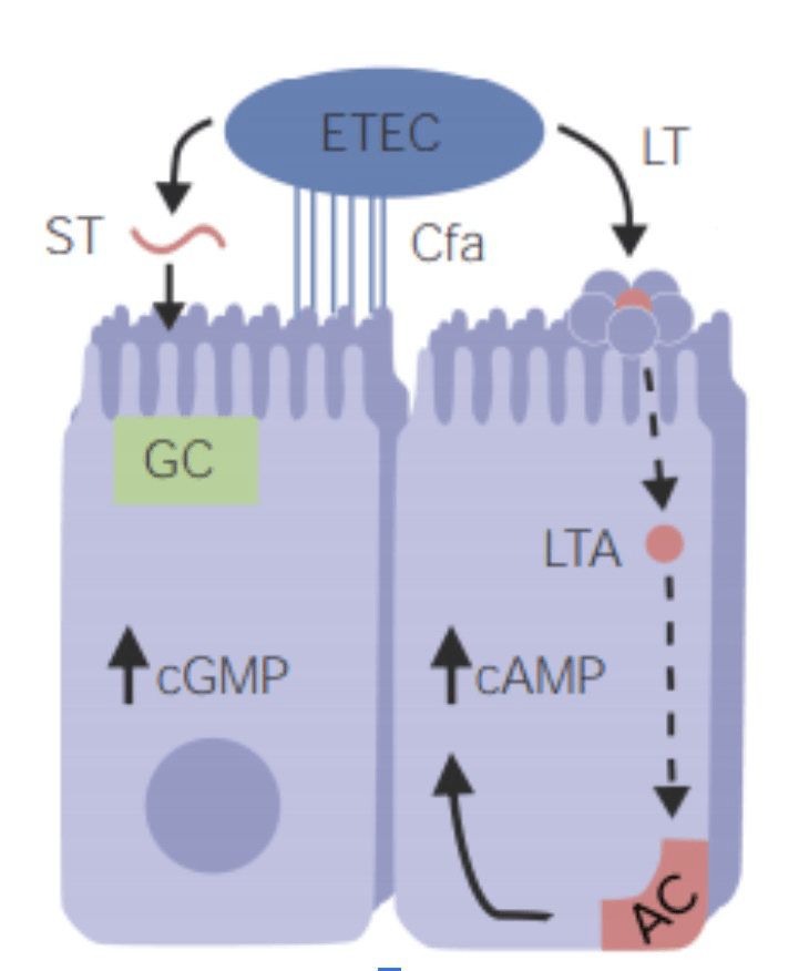
Patogénesis de la Escherichia coli enterotoxigénica (ETEC, por sus siglas en inglés):
La E. coli enterotoxigénica se adhiere a los enterocitos a través del antígeno del factor de colonización (CFA, por sus siglas en inglés); adhesina fimbrial. La enterotoxina termoestable (ST) provoca la acumulación de guanosina monofosfato cíclico (cGMP, por sus siglas en inglés) en las células y la secreción de líquido y electrolitos en la luz intestinal. La enterotoxina termolábil (LT) actúa como la toxina del cólera, que aumenta el monofosfato de adenosina cíclico (cAMP, por sus siglas en inglés) mediante la activación de la adenil ciclasa (AC). El efecto general es la hipersecreción de agua y cloruro y la inhibición de la reabsorción de sodio. Las enterotoxinas no invasivas permanecen en la luz intestinal y no invaden las células epiteliales.br>GC: guanilil ciclasa
LTA: Una subunidad de la enterotoxina termolábil
Esta enfermedad es autolimitada, por lo que no suele ser necesario un estudio diagnóstico. Sin embargo, la E. coli enterotoxigénica puede diagnosticarse mediante la identificación de los LOS Neisseria genes Genes A category of nucleic acid sequences that function as units of heredity and which code for the basic instructions for the development, reproduction, and maintenance of organisms. DNA Types and Structure enterotoxina termolábil o enterotoxina termoestable en EN Erythema nodosum is an immune-mediated panniculitis (inflammation of the subcutaneous fat) caused by a type IV (delayed-type) hypersensitivity reaction. It commonly manifests in young women as tender, erythematous nodules on the shins. Erythema Nodosum la reacción en EN Erythema nodosum is an immune-mediated panniculitis (inflammation of the subcutaneous fat) caused by a type IV (delayed-type) hypersensitivity reaction. It commonly manifests in young women as tender, erythematous nodules on the shins. Erythema Nodosum cadena de la polimerasa para los LOS Neisseria individuos con enfermedad grave.
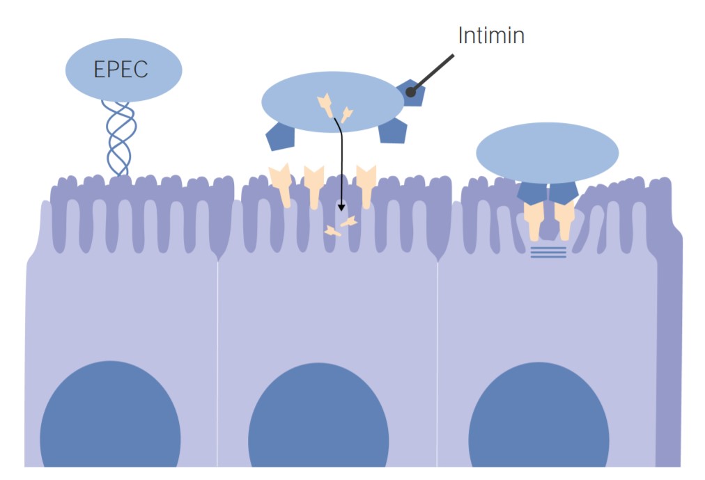
Patogénesis de Escherichia coli enteropatógena:
La E. coli enteropatogénica utiliza la adhesina intimina para adherirse a las células intestinales. La unión provoca la deformación de las células (degeneración del borde en cepillo y pérdida de microvellosidades). Se cree que el efecto característico de la fijación y el borrado es la causa principal de la diarrea.
La E. coli enteropatogénica puede diagnosticarse mediante la identificación de genes Genes A category of nucleic acid sequences that function as units of heredity and which code for the basic instructions for the development, reproduction, and maintenance of organisms. DNA Types and Structure específicos utilizando la reacción en EN Erythema nodosum is an immune-mediated panniculitis (inflammation of the subcutaneous fat) caused by a type IV (delayed-type) hypersensitivity reaction. It commonly manifests in young women as tender, erythematous nodules on the shins. Erythema Nodosum cadena de la polimerasa.
La E. coli enteroagregativa se asocia con mayor frecuencia a la diarrea persistente en EN Erythema nodosum is an immune-mediated panniculitis (inflammation of the subcutaneous fat) caused by a type IV (delayed-type) hypersensitivity reaction. It commonly manifests in young women as tender, erythematous nodules on the shins. Erythema Nodosum:
El diagnóstico de la E. coli enteroagregativa puede realizarse mediante la identificación de genes Genes A category of nucleic acid sequences that function as units of heredity and which code for the basic instructions for the development, reproduction, and maintenance of organisms. DNA Types and Structure específicos utilizando la reacción en EN Erythema nodosum is an immune-mediated panniculitis (inflammation of the subcutaneous fat) caused by a type IV (delayed-type) hypersensitivity reaction. It commonly manifests in young women as tender, erythematous nodules on the shins. Erythema Nodosum cadena de la polimerasa.
La E. coli enteroinvasiva se presenta de forma muy similar a la shigelosis y puede ser grave.
El diagnóstico de la E. coli enteroinvasiva puede realizarse mediante la identificación de genes Genes A category of nucleic acid sequences that function as units of heredity and which code for the basic instructions for the development, reproduction, and maintenance of organisms. DNA Types and Structure específicos utilizando la reacción en EN Erythema nodosum is an immune-mediated panniculitis (inflammation of the subcutaneous fat) caused by a type IV (delayed-type) hypersensitivity reaction. It commonly manifests in young women as tender, erythematous nodules on the shins. Erythema Nodosum cadena de la polimerasa.
La E. coli enterohemorrágica da lugar a manifestaciones clínicas a través de la producción de toxina shiga:
El diagnóstico de E. coli enterohemorrágica se realiza mediante la identificación: