Actinomyces es un bacilo anaerobio, gram-positivo, ramificado y filamentoso. Actinomyces israelii es la especie más comúnmente implicada en enfermedades humanas. El organismo suele encontrarse como parte de la flora normal en la cavidad oral, el tracto gastrointestinal y el tracto reproductivo. La enfermedad se produce cuando el organismo se desplaza incluso por un traumatismo o procedimiento menor, lo que permite que el organismo se mueva más allá de la barrera mucosa. El alcanzar áreas de bajo oxígeno conduce a la multiplicación del organismo. Actinomyces se asocia con infección cervicofacial, que forma tractos sinusales que drenan. Actinomyces también puede afectar las áreas torácica, abdominal y pélvica. El drenaje o el tejido infectado pueden tener los característicos gránulos amarillos de azufre asociados con Actinomyces. El tratamiento es con penicilina a largo plazo y cirugía, si es necesario.
Last updated: Dec 15, 2025

Bacterias gram-positivas:
La mayoría de las bacterias se pueden clasificar de acuerdo con un procedimiento de laboratorio llamado tinción de Gram.
Las bacterias con paredes celulares que tienen una capa gruesa de peptidoglicano retienen la tinción cristal violeta utilizada en la tinción de Gram, pero no se ven afectadas por la contratinción de safranina. Estas bacterias aparecen como azul púrpura en la tinción, lo que indica que son gram-positivas. Las bacterias pueden clasificarse además según su morfología (filamentos ramificados, bacilos y cocos en grupos o cadenas) y su capacidad para crecer en presencia de oxígeno (aeróbicos versus anaeróbicos). Los cocos también pueden identificarse con mayor profundidad. Los estafilococos pueden reducirse en función de la presencia de la enzima coagulasa y de su sensibilidad al antibiótico novobiocina. Los estreptococos se cultivan en agar sangre y se clasifican según la forma de hemólisis que emplean (α, β o γ). Los estreptococos se reducen aún más en función de su respuesta a la prueba de pirrolidonil-β-naftilamida, su sensibilidad a antimicrobianos específicos (optoquina y bacitracina) y su capacidad para crecer en medios de cloruro de sodio (NaCl).
Cambios histopatológicos de un absceso cerebral debido a la bacteria A. naeslundii (en tinción de plata). Se observan bacilos ramificados.
Imagen: “Actinomyces naeslundii 01” por CDC/Dr. Lucille Georg. Licencia: Dominio PúblicoReservorio:
Transmisión:

Patogenia de Actinomyces
Cuando hay una ruptura de la barrera mucosa, los Actinomyces de la orofaringe (que se dirige hacia áreas respiratorias), el tracto gastrointestinal y el tracto reproductivo causan una infección y se diseminan a los tejidos contiguos mientras crean tractos sinusales.
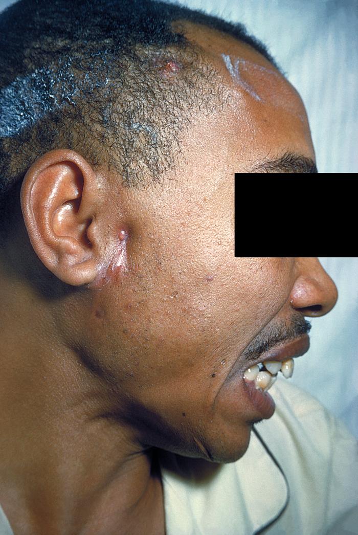
Un paciente con actinomicosis en el lado derecho de la cara (anterior al pabellón auricular)
Imagen: “Actinomycosis PHIL 2856 lores” por CDC/Dr. Thomas F. Sellers. Licencia: Dominio Público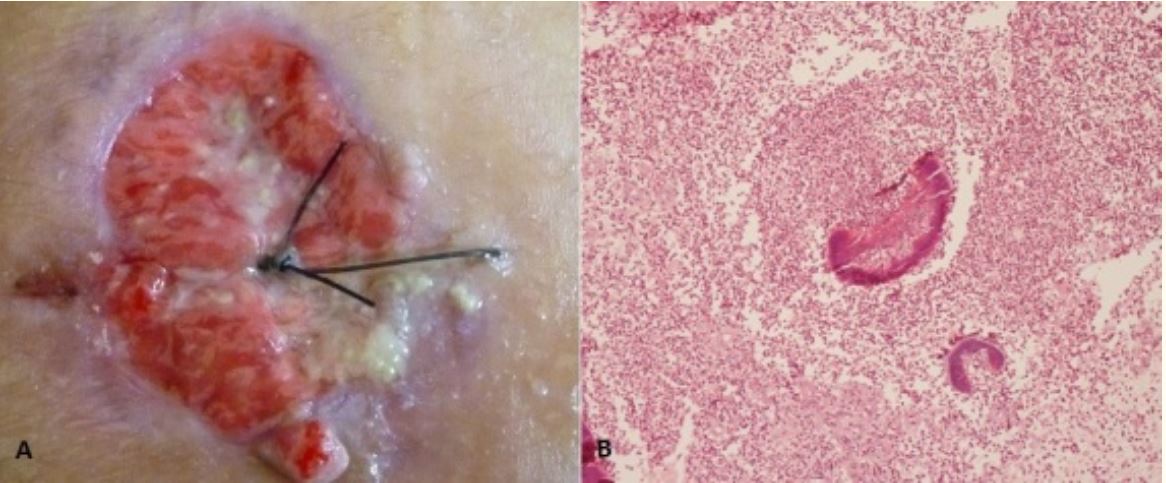
Actinomicosis de la pared torácica: A: Lesión ulcerada y protuberante en la región paraesternal derecha, con pus mezclado con gránulos de azufre. B: las células inflamatorias rodean los gránulos basófilos de azufre (compuestos por filamentos dispuestos en un patrón radiante).
Imagen: “Unusual actinomycosis of the chest wall” por Bouaddi M, Hassam B. Licencia: CC BY 2.0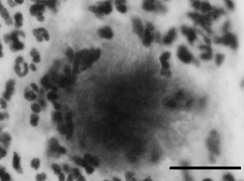
Actinomicosis pélvica por un DIU.
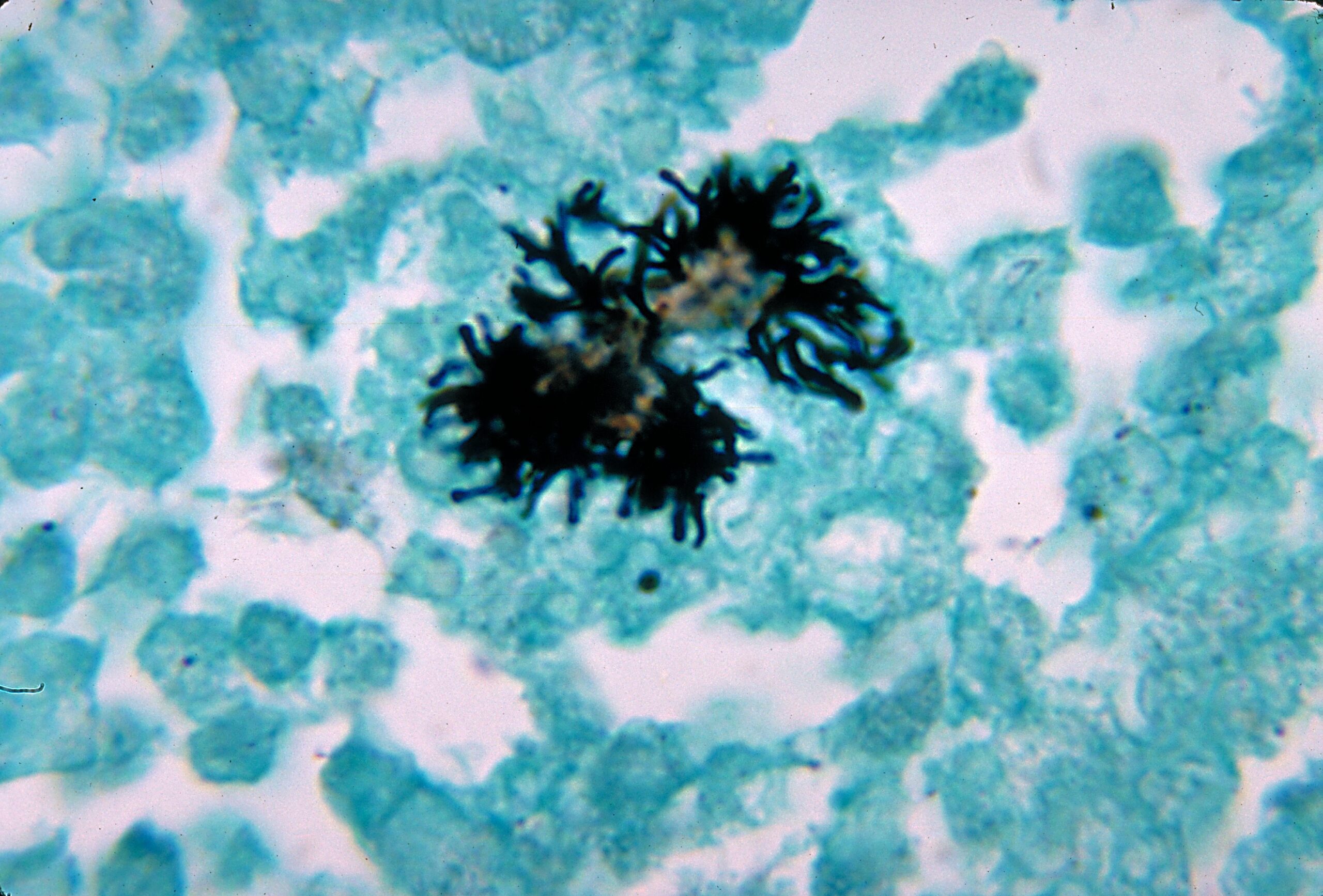
Un agregado bacteriano típico de un frotis cervical de Papanicolaou que muestra una colonia similar a una bola de algodón con filamentos miceliales que sobresalen, lo que sugiere una infección por especies de Actinomyces. La barra indica 20 μm.
Osteonecrosis de la mandíbula asociada a Actinomyces
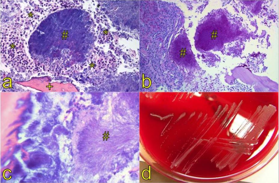
(a) Tinción con hematoxilina y eosina de un agregado compuesto de filamentos, los llamados gránulos de azufre (#). Estos gránulos aparecen macroscópicamente como gránulos amarillos rodeados de granulocitos neutrofílicos (*) y una trabécula ósea necrótica (+, aumento x 100).
(b) Los gránulos (#) se tiñen ácido periódico de Schiff positivo (aumento x 200).
(c) Un gran aumento muestra la estructura filamentosa (#, morfología de rayos solares) de los organismos (aumento x 400).
(d) Patrón de crecimiento típico de Actinomyces spp. en cultivo microbiológico.
Dos bacilos filamentosos, gram-positivos y ramificados clínicamente relevantes que deben distinguirse:
| Factores diferenciadores | Actinomyces Actinomyces Actinomyces is an anaerobic, gram-positive, branching, filamentous rod. Actinomyces israelii is the most common species involved in human disease. The organism is commonly found as part of the normal flora in the oral cavity, gastrointestinal tract, and reproductive tract. Actinomyces/Actinomycosis | Nocardia Nocardia Nocardia is a branching, filamentous, gram-positive bacilli. It is partially acid fast due to the presence of mycolic acids in the cell wall. Nocardia is a ubiquitous soil organism that most commonly affects immunocompromised patients. Nocardia is transmitted via inhalation of aerosolized bacteria or less commonly, via direct contact with wounds. Nocardia/Nocardiosis |
|---|---|---|
| Requerimiento de oxígeno | Anaeróbico | Aeróbico |
| Tinción de ácido-alcohol resistencia | No ácido-alcohol resistente | Ácido-alcohol resistente (parcialmente) |
| Reservorio/hábitat | Flora oral, gastrointestinal y del tracto reproductor normal | Se encuentra en EN Erythema nodosum is an immune-mediated panniculitis (inflammation of the subcutaneous fat) caused by a type IV (delayed-type) hypersensitivity reaction. It commonly manifests in young women as tender, erythematous nodules on the shins. Erythema Nodosum tierra, agua, materia orgánica en EN Erythema nodosum is an immune-mediated panniculitis (inflammation of the subcutaneous fat) caused by a type IV (delayed-type) hypersensitivity reaction. It commonly manifests in young women as tender, erythematous nodules on the shins. Erythema Nodosum descomposición |
| Infecciones causadas |
|
|
| Tratamiento | Penicilina | Trimetoprim-sulfametoxazol |
Mnemotecnia:
“SNAP” (Sulfonamidas-Nocardia, Actinomyces–Penicilina)