Shigella Shigella Shigella is a genus of gram-negative, non-lactose-fermenting facultative intracellular bacilli. Infection spreads most commonly via person-to-person contact or through contaminated food and water. Humans are the only known reservoir. Shigella es un género de bacilos intracelulares facultativos gramnegativos que no fermentan la lactosa. La infección se propaga más comúnmente a través del contacto de persona a persona o a través de alimentos y agua contaminados. Los LOS Neisseria humanos son el único reservorio conocido. Debido a que es resistente al AL Amyloidosis ácido, Shigella Shigella Shigella is a genus of gram-negative, non-lactose-fermenting facultative intracellular bacilli. Infection spreads most commonly via person-to-person contact or through contaminated food and water. Humans are the only known reservoir. Shigella spp. sobrevive al AL Amyloidosis tránsito por el estómago; por lo tanto, solo se necesita una pequeña cantidad del inóculo para causar la enfermedad. La shigelosis (disentería por Shigella Shigella Shigella is a genus of gram-negative, non-lactose-fermenting facultative intracellular bacilli. Infection spreads most commonly via person-to-person contact or through contaminated food and water. Humans are the only known reservoir. Shigella) produce fiebre, dolor Dolor Inflammation abdominal y diarrea sanguinolenta, que son efectos de las toxinas y la invasión de células epiteliales del organismo. En EN Erythema nodosum is an immune-mediated panniculitis (inflammation of the subcutaneous fat) caused by a type IV (delayed-type) hypersensitivity reaction. It commonly manifests in young women as tender, erythematous nodules on the shins. Erythema Nodosum la mayoría de los LOS Neisseria casos, los LOS Neisseria síntomas se resuelven en EN Erythema nodosum is an immune-mediated panniculitis (inflammation of the subcutaneous fat) caused by a type IV (delayed-type) hypersensitivity reaction. It commonly manifests in young women as tender, erythematous nodules on the shins. Erythema Nodosum unos pocos días. Sin embargo, pueden surgir complicaciones de deshidratación, síndrome urémico hemolítico, megacólon tóxico o artritis reactiva. El tratamiento consiste principalmente en EN Erythema nodosum is an immune-mediated panniculitis (inflammation of the subcutaneous fat) caused by a type IV (delayed-type) hypersensitivity reaction. It commonly manifests in young women as tender, erythematous nodules on the shins. Erythema Nodosum reposición de líquidos y electrolitos y antibióticos.
Last updated: Dec 28, 2025
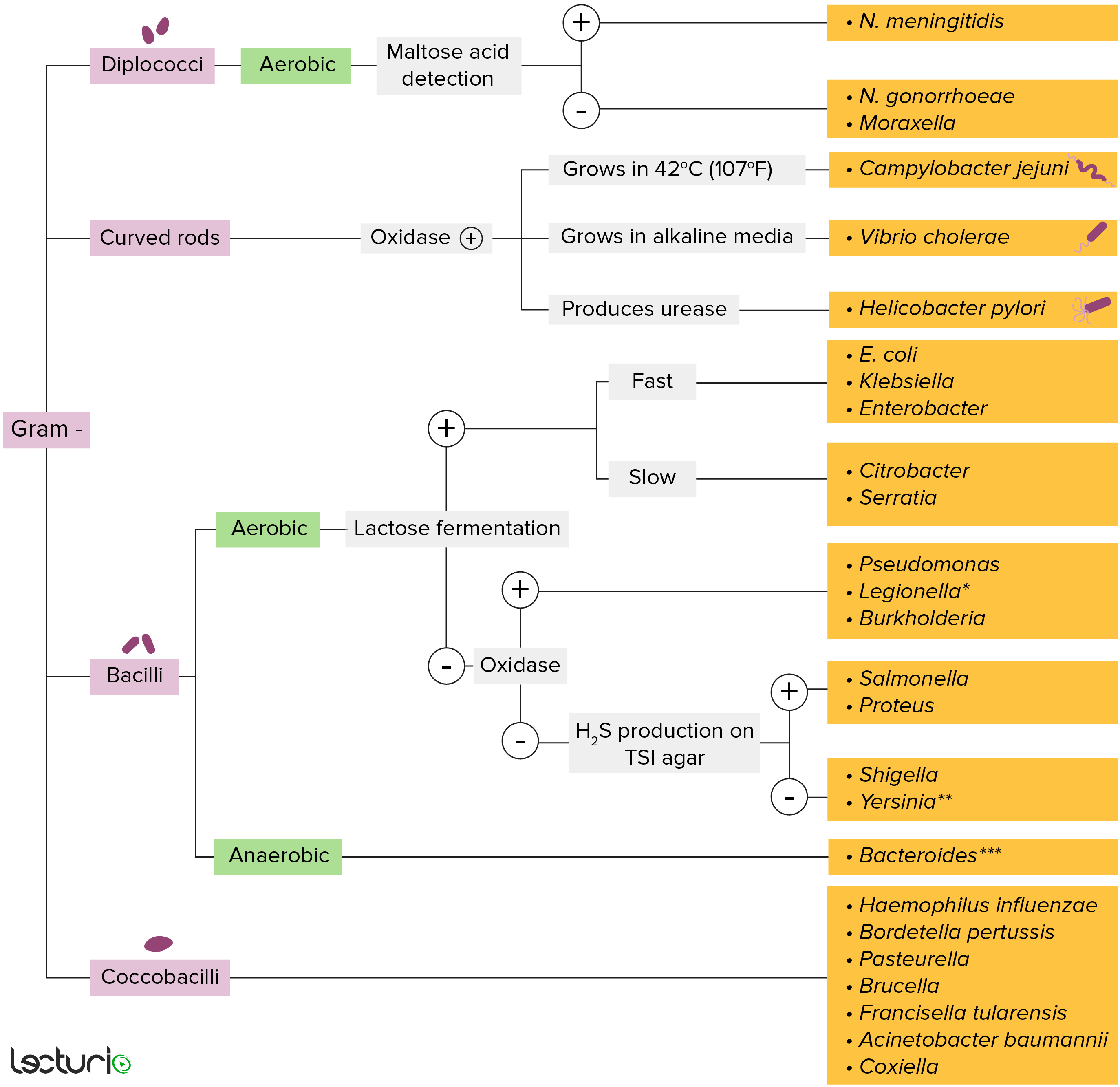
Bacterias gram-negativas:
La mayoría de las bacterias se pueden clasificar de acuerdo con un procedimiento de laboratorio llamado tinción de Gram.
Las bacterias con paredes celulares que tienen una capa delgada de peptidoglicano no retienen la tinción de cristal violeta utilizada en la tinción de Gram. Sin embargo, estas bacterias retienen la contratinción de safranina y, por lo tanto, se tiñen de color rojo rosado, lo que las convierte en gram negativas. Estas bacterias pueden clasificarse además según su morfología (diplococos, bastoncillos curvos, bacilos y cocobacilos) y su capacidad para crecer en presencia de oxígeno (aeróbicos frente a anaeróbicos). Las bacterias se pueden identificar de manera más estrecha cultivándolas en medios específicos (agar triple azúcar hierro) donde se pueden identificar sus enzimas (ureasa, oxidasa) y se puede probar su capacidad para fermentar lactosa.
* Se tiñe mal en la tinción de Gram
** Baston pleomórfico/cocobacilo
*** Requiere medios de transporte especiales
Serogrupos definidos por antígenos O específicos:
Para ayudar a recordar los LOS Neisseria modos de transmisión de Shigella Shigella Shigella is a genus of gram-negative, non-lactose-fermenting facultative intracellular bacilli. Infection spreads most commonly via person-to-person contact or through contaminated food and water. Humans are the only known reservoir. Shigella, recuerde las “4 Fs” ( en EN Erythema nodosum is an immune-mediated panniculitis (inflammation of the subcutaneous fat) caused by a type IV (delayed-type) hypersensitivity reaction. It commonly manifests in young women as tender, erythematous nodules on the shins. Erythema Nodosum ingles):

Invasión y propagación de célula a célula por Shigella:
1. El patógeno invade y es engullido por la célula M (transcitosis).
2. El patógeno alcanza los macrófagos subepiteliales y las células dendríticas y luego induce la apoptosis de los macrófagos. Shigella sp. se libera junto con la interleucina-1 (IL-1) y otras citocinas. Luego, el patógeno es absorbido por la célula epitelial adyacente en un compartimento unido a la membrana (entrada de la célula epitelial).
3. Las interleucinas liberadas también reclutan leucocitos polimorfonucleares (PMN), que desestabilizan las uniones celulares. Esta es otra vía de entrada para el patógeno (transmigración de PMN).
4. La Shigella sp. pasa a través de las uniones estrechas interrumpidas y luego el patógeno ingresa a la célula epitelial. Una vez dentro, los nucleadores de actina de la célula huésped son secuestrados. La motilidad basada en actina (ABM, por sus siglas en inglés) impulsa al patógeno a alcanzar la membrana plasmática, donde la célula infectada entra en contacto con otra célula.
5. La protrusión celular (con Shigella) y la elongación hacia la célula adyacente facilitan la multiplicación y la diseminación intercelular. La protuberancia se resuelve como una vacuola de doble membrana. El patógeno lisa la membrana, escapa y entra en la célula adyacente. La célula adyacente se infecta y este proceso se repite en otras células.
6. A medida que muere cada célula epitelial invadida, se pierden líquidos.
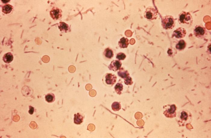
Shigellosis (“disentería por Shigella”): muestra de heces de un paciente con shigellosis (diarrea sanguinolenta en las heces, fiebre y calambres abdominales). Shigella vista como bastones en el frotis
Imagen: “Shigella stool” por Centers for Disease Control and Prevention Public Health Image Library. Licencia: Dominio Público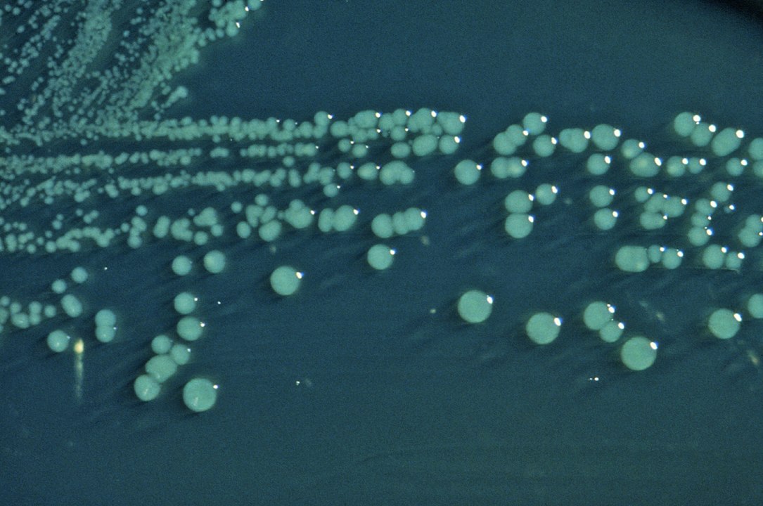
Shigella en agar entérico Hektoen (HE): Las colonias de S. boydii cultivadas en agar HE tienen un aspecto abultado, verde y húmedo.
Imagen: “Shigella boydii 01” por CDC. Licencia: Dominio PúblicoShigella Shigella Shigella is a genus of gram-negative, non-lactose-fermenting facultative intracellular bacilli. Infection spreads most commonly via person-to-person contact or through contaminated food and water. Humans are the only known reservoir. Shigella y Salmonella Salmonella Salmonellae are gram-negative bacilli of the family Enterobacteriaceae. Salmonellae are flagellated, non-lactose-fermenting, and hydrogen sulfide-producing microbes. Salmonella enterica, the most common disease-causing species in humans, is further classified based on serotype as typhoidal (S. typhi and paratyphi) and nontyphoidal (S. enteritidis and typhimurium). Salmonella invaden el tracto gastrointestinal y causan diarrea.
| Shigela | Salmonela | |
|---|---|---|
| Tinción de Gram/estructura | Bacilos gramnegativos | Bacilos gramnegativos |
| Fermentación de lactosa | No fermentadores de lactosa | No fermentadores de lactosa |
| Oxidasa | Negativa | Negativa |
| Producción de H2S | No | Si |
| Motilidad | No | Sí (con flagelos) |
| Factores virulentos | Endotoxina, toxina Shiga | Endotoxina, antígeno capsular Vi |
| Reservorio | Humanos | Humanos (S. typhi), animales |
| Dosis para producir la enfermedad | Inóculo pequeño (estable en EN Erythema nodosum is an immune-mediated panniculitis (inflammation of the subcutaneous fat) caused by a type IV (delayed-type) hypersensitivity reaction. It commonly manifests in young women as tender, erythematous nodules on the shins. Erythema Nodosum ácido) | Gran dosis (inactivado por ácidos) |
| Propagación de infecciones | De célula a célula (sin diseminación hematógena) | Puede diseminarse hematógenamente |