Rickettsiae son una colección diversa de bacterias gram-negativas intracelulares obligadas que tienen un tropismo por las células endoteliales vasculares. Los LOS Neisseria vectores de transmisión varían según la especie, pero incluyen garrapatas, pulgas, ácaros y piojos. Los LOS Neisseria patógenos clínicamente más relevantes son R. rickettsii, que causa la fiebre maculosa de las Montañas Rocosas; R. prowazekii, que causa el tifus epidémico (transmitido por piojos); R. typhi, que causa el tifus endémico; y R. akari, que causa la rickettsiosis vesiculosa. Todas estas enfermedades son una forma de vasculitis Vasculitis Inflammation of any one of the blood vessels, including the arteries; veins; and rest of the vasculature system in the body. Systemic Lupus Erythematosus inflamatoria y comúnmente se presentan con fiebre, cefalea y erupción cutánea. El tratamiento es con doxiciclina.
Last updated: Dec 15, 2025
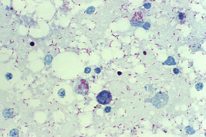
R. rickettsii: microfotografía de un frotis de saco vitelino, que muestra varias bacterias intracelulares teñidas de rojo por la tinción de Giemsa
Imagen: “10955” por CDC/Billie Ruth Bird. Licencia: Dominio Público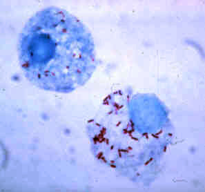
R. rickettsii teñida con Giemsa en las células de una garrapata
Imagen: “Rickettsia rickettsii” por CDC. Licencia: Dominio Público| R. rickettsii | R. prowazekii | R. typhi | R. akari | |
|---|---|---|---|---|
| Vector | Garrapatas duras (familia Ixodidae Ixodidae A family of hardbacked ticks, in the subclass Acari. Genera include Dermacentor and Ixodes among others. Rickettsia): Dermacentor Dermacentor A widely distributed genus of ticks, in the family ixodidae, including a number that infest humans and other mammals. Several are vectors of diseases such as tularemia; rocky mountain spotted fever; colorado tick fever; and anaplasmosis. Rickettsia (garrapata de perro), Amblyoma (garrapata de madera) | Piojos humanos (
Pediculus humanus corporis
Pediculus Humanus Corporis
Epidemic Typhus):
|
Picaduras de pulgas de ratas y gatos | Ácaros de ratones |
| Enfermedad | Fiebre maculosa de las Montañas Rocosas (la enfermedad por rickettsia Rickettsia Rickettsiae are a diverse collection of obligate intracellular, gram-negative bacteria that have a tropism for vascular endothelial cells. The vectors for transmission vary by species but include ticks, fleas, mites, and lice. Rickettsia más grave) | Tifus epidémico (transmitido por piojos) | Tifus endémico | Rickettsiosis vesiculosa (la enfermedad rickettsial menos grave) |
| Variaciones geográficas |
|
|
|
|
Epidemiología:
Fisiopatología:
Presentación clínica:
Pronóstico:
Identificación:
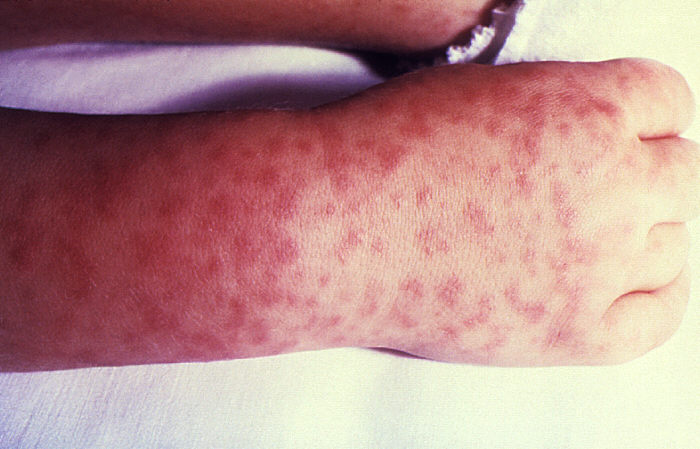
Erupción característica de la fiebre maculosa de las Montañas Rocosas: mano y muñeca de un niño afectado
Imagen: “Rocky Mountain spotted fever PHIL 1962 lores” por CDC. Licencia: Dominio PúblicoEl tifus epidémico (transmitido por piojos) es ahora una enfermedad rara.
Patogénesis:
Presentación clínica:
Enfermedad de Brill-Zinsser:
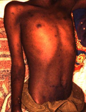
Erupción en un paciente con tifus epidémico
Imagen: “Epidemic typhus Burundi” por D. Raoult, V. Roux, JB Ndihokubwayo, G. Bise, D. Baudon, G. Martet y R. Birtles. Licencia: Dominio Público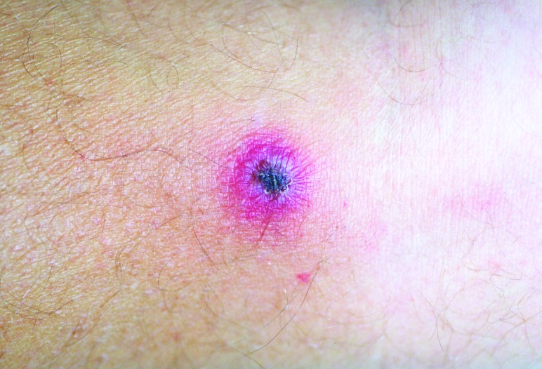
Escara indolora, negra y costrosa de la rickettsiosis vesiculosa, que se desarrolla como el último estadio de la erupción típica (máculas → pápulas → vesículas → costras/escaras que cicatrizan sin dejar cicatrices).
Imagen: “Rickettsialpox lesion” por Krusell A, Comer JA, Sexton DJ. Licencia: Dominio Público