Domina Conceptos Médicos

Estudia para la escuela de medicina y tus examenes con Lecturio.

Micología

Los LOS Neisseria hongos pertenecen al AL Amyloidosis dominio eucariota y, al AL Amyloidosis igual que las plantas, tienen paredes celulares y vacuolas, exhiben flujo citoplasmático y son inmóviles. Sin embargo, casi todos los LOS Neisseria hongos tienen paredes celulares compuestas de quitina y no de celulosa. Los LOS Neisseria hongos no realizan la fotosíntesis, sino que obtienen sus sustratos para el metabolismo como saprofitos (obtienen su alimento de la materia muerta). La micosis es una infección causada por hongos.

Last updated: Dec 15, 2025

Editorial responsibility: Stanley Oiseth, Lindsay Jones, Evelin Maza

Formas de Crecimiento y Reproducción

Formas de crecimiento

  • Las formas multicelulares incluyen mohos y hongos:
    • Compuesto principalmente de hifas en EN Erythema nodosum is an immune-mediated panniculitis (inflammation of the subcutaneous fat) caused by a type IV (delayed-type) hypersensitivity reaction. It commonly manifests in young women as tender, erythematous nodules on the shins. Erythema Nodosum forma de hilo:
      • Tabicado: Las paredes celulares subdividen las hifas.
      • Coenocítico: Las hifas carecen de paredes celulares.
    • Los LOS Neisseria “hongos” familiares son órganos reproductivos (talos) de hongos.
    • Sistemas de redes tubulares en EN Erythema nodosum is an immune-mediated panniculitis (inflammation of the subcutaneous fat) caused by a type IV (delayed-type) hypersensitivity reaction. It commonly manifests in young women as tender, erythematous nodules on the shins. Erythema Nodosum las que las hifas forman redes ramificadas (micelio)
  • Las formas unicelulares incluyen levaduras:
    • Las levaduras no tienen hifas ni micelio.
    • Algunas especies forman estructuras pseudomulticelulares (levaduras en EN Erythema nodosum is an immune-mediated panniculitis (inflammation of the subcutaneous fat) caused by a type IV (delayed-type) hypersensitivity reaction. It commonly manifests in young women as tender, erythematous nodules on the shins. Erythema Nodosum brotes).
  • Hongos dimórficos:
    • Pueden existir como formas unicelulares o multicelulares
    • Los LOS Neisseria factores ambientales (temperatura, humedad, presencia de nutrientes) pueden desencadenar cambios.

Estructura

  • Las células fúngicas tienen características similares a las células vegetales y animales:
    • Características que son comunes con las células animales:
      • Presencia de un núcleo unido a la membrana
      • Organelos como las mitocondrias
      • Falta de cloroplastos
    • Características que son comunes con las células vegetales:
      • Presencia de una pared celular
      • Contienen vacuolas
  • Los LOS Neisseria hongos tienen propiedades únicas que los LOS Neisseria distinguen de los LOS Neisseria animales y las plantas:
    • Las paredes celulares contienen quitina, a diferencia de las paredes celulares vegetales que contienen celulosa.
    • Las membranas celulares contienen ergosterol Ergosterol A steroid occurring in fungi. Irradiation with ultraviolet rays results in formation of ergocalciferol (vitamin d2). Azoles, a diferencia de las membranas celulares animales que contienen colesterol.

Reproducción

La reproducción es sexual o asexual.

  • Tipos de reproducción asexual:
    • Fisión binaria
    • Desglose de hifas, dispersión y rebrote
    • Brotes (levaduras)
    • Formación de conidios (que contienen esporas mitóticas asexuales)
  • Reproducción sexual:
    • Fusión de 2 células haploides:
      • Autofecundación: 2 células del mismo organismo
      • Fertilización cruzada: células de diferentes organismos
    • El cigoto diploide emergente sufre meiosis Meiosis The creation of eukaryotic gametes involves a DNA replication phase followed by 2 cellular division stages: meiosis I and meiosis II. Meiosis I separates homologous chromosomes into separate cells (1n, 2c), while meiosis II separates sister chromatids into gametes (1n, 1c). Meiosis para formar esporas haploides.
    • Las esporas germinan para crear más micelios haploides.
Brotación en hongos

Brotes en hongos:
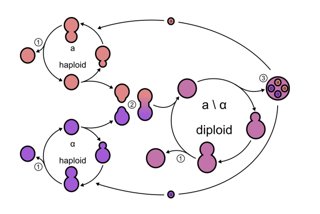
Las levaduras en brotes se dividen asimétricamente. Hay estados haploides y diploides (2 tipos de apareamiento, a y α). Cada tipo de apareamiento secreta su propio tipo de feromona.
1: Brote: la división celular mitótica puede ocurrir en los estados haploide y diploide, lo que da como resultado células hijas genéticamente idénticas.
2: Apareamiento: Cada tipo de apareamiento secreta su propio tipo de feromona, iniciando el proceso de apareamiento. Esto da como resultado una célula diploide.
3: Esporulación: las células diploides pueden sufrir meiosis, lo que resulta en la formación de esporas. Estas esporas pueden germinar en células haploides.

Imagen: “Yeast lifecycle” por Masur. Licencia: Dominio Público

Productos Tóxicos de Hongos

Tabla: Productos tóxicos de hongos
Toxina Hongo Efecto
Aflatoxina
  • Aspergillus Aspergillus A genus of mitosporic fungi containing about 100 species and eleven different teleomorphs in the family trichocomaceae. Echinocandins flavus
  • A. parasiticus
Altamente cancerígeno y, a menudo, la causa de intoxicación alimentaria (rastros en EN Erythema nodosum is an immune-mediated panniculitis (inflammation of the subcutaneous fat) caused by a type IV (delayed-type) hypersensitivity reaction. It commonly manifests in young women as tender, erythematous nodules on the shins. Erythema Nodosum nueces, granos, especias)
Amanitina Amanita phalloides Amanita Phalloides Toxicology of Plants (hongo del sombrero de la muerte) Inhibición de la ácido ribonucleico (ARN) polimerasa II, letal incluso en EN Erythema nodosum is an immune-mediated panniculitis (inflammation of the subcutaneous fat) caused by a type IV (delayed-type) hypersensitivity reaction. It commonly manifests in young women as tender, erythematous nodules on the shins. Erythema Nodosum pequeñas dosis
Muscarina A. muscaria (seta venenosa u hongo del agárico de mosca) Afecta la regulación parasimpática del sistema nervioso
Ergotamina Hongo cornezuelo (Claviceps purpurea) Afecta el sistema nervioso autónomo, causa alucinaciones y afecta las contracciones uterinas
Ciclosporina A
  • Cylindrocarpon
  • Tolipocladio
Inmunosupresor (uso clínico: después del trasplante de órganos)

Efecto Patógeno de los Hongos

  • Micosis: enfermedades infecciosas causadas por hongos:
    • No suelen plantear problemas en EN Erythema nodosum is an immune-mediated panniculitis (inflammation of the subcutaneous fat) caused by a type IV (delayed-type) hypersensitivity reaction. It commonly manifests in young women as tender, erythematous nodules on the shins. Erythema Nodosum individuos sanos
    • Puede causar infecciones oportunistas en EN Erythema nodosum is an immune-mediated panniculitis (inflammation of the subcutaneous fat) caused by a type IV (delayed-type) hypersensitivity reaction. It commonly manifests in young women as tender, erythematous nodules on the shins. Erythema Nodosum personas inmunocomprometidas
  • Clases:
    • Micosis superficiales:
      • Orofaringe: comúnmente visto en EN Erythema nodosum is an immune-mediated panniculitis (inflammation of the subcutaneous fat) caused by a type IV (delayed-type) hypersensitivity reaction. It commonly manifests in young women as tender, erythematous nodules on the shins. Erythema Nodosum lactantes, ancianos o personas inmunodeprimidas
      • Área anogenital: las infecciones vaginales por hongos se observan comúnmente incluso en EN Erythema nodosum is an immune-mediated panniculitis (inflammation of the subcutaneous fat) caused by a type IV (delayed-type) hypersensitivity reaction. It commonly manifests in young women as tender, erythematous nodules on the shins. Erythema Nodosum personas sanas.
      • Piel: hongos queratinófilos (también conocidos como dermatofitos) que afectan la piel, el cabello y las uñas de los LOS Neisseria humanos
    • Micosis sistémicas:
      • Se desarrollan cuando las esporas de hongos se inhalan o se introducen en EN Erythema nodosum is an immune-mediated panniculitis (inflammation of the subcutaneous fat) caused by a type IV (delayed-type) hypersensitivity reaction. It commonly manifests in young women as tender, erythematous nodules on the shins. Erythema Nodosum el torrente sanguíneo
      • Puede manifestarse en EN Erythema nodosum is an immune-mediated panniculitis (inflammation of the subcutaneous fat) caused by a type IV (delayed-type) hypersensitivity reaction. It commonly manifests in young women as tender, erythematous nodules on the shins. Erythema Nodosum diferentes órganos internos, lo que resulta en EN Erythema nodosum is an immune-mediated panniculitis (inflammation of the subcutaneous fat) caused by a type IV (delayed-type) hypersensitivity reaction. It commonly manifests in young women as tender, erythematous nodules on the shins. Erythema Nodosum infecciones letales graves
      • Los LOS Neisseria pacientes con virus Virus Viruses are infectious, obligate intracellular parasites composed of a nucleic acid core surrounded by a protein capsid. Viruses can be either naked (non-enveloped) or enveloped. The classification of viruses is complex and based on many factors, including type and structure of the nucleoid and capsid, the presence of an envelope, the replication cycle, and the host range. Virology de inmunodeficiencia humana (VIH) positivo o inmunosuprimidos son susceptibles a micosis sistémicas.
  • Ejemplos:
    • Muguet: infección de la mucosa bucal; especies de Candida Candida Candida is a genus of dimorphic, opportunistic fungi. Candida albicans is part of the normal human flora and is the most common cause of candidiasis. The clinical presentation varies and can include localized mucocutaneous infections (e.g., oropharyngeal, esophageal, intertriginous, and vulvovaginal candidiasis) and invasive disease (e.g., candidemia, intraabdominal abscess, pericarditis, and meningitis). Candida/Candidiasis
    • Onicomicosis: infección de la uña; Trichophyton rubrum Trichophyton rubrum Dermatophytes/Tinea Infections
    • Dermatofitosis: infección de la piel ( tinea pedis Tinea pedis Dermatological pruritic lesion in the feet, caused by Trichophyton rubrum, T. mentagrophytes, or Epidermophyton floccosum. Dermatophytes/Tinea Infections, corporis o cruris)
      • Microsporum Microsporum A mitosporic oxygenales fungal genus causing various diseases of the skin and hair. The species microsporum canis produces tinea capitis and tinea corporis, which usually are acquired from domestic cats and dogs. Teleomorphs includes arthroderma (nannizzia). Dermatophytes/Tinea Infections canis, M. audouinii, M. gypseum
      • Epidermophyton Epidermophyton A fungal genus which grows in the epidermis and is the cause of tinea. Dermatophytes/Tinea Infections floccosum
      • Trichophyton Trichophyton A mitosporic fungal genus and an anamorphic form of arthroderma. Various species attack the skin, nails, and hair. Dermatophytes/Tinea Infections tonsurans, T. mentagrophytes, T. verrucosum, T. schoenlenii
    • Coccidioidomicosis: infección fúngica pulmonar o diseminada
tiña del pie

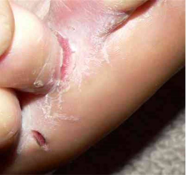
Tinea pedis también se conoce como pie de atleta.

Imagen: “Tinea pedis interdigitalis” por Falloonb. Licencia: Dominio Público

Síntesis de Antimicrobianos

Algunos hongos son capaces de producir sustancias que son eficaces como antimicrobianos:

  • Penicilina de Penicillium notatum Penicillium notatum Mycology
  • Cefalosporina de Acremonium Acremonium A mitosporic fungal genus with many reported ascomycetous teleomorphs. Cephalosporin antibiotics are derived from this genus. Mycology
  • Estreptomicina de Streptomyces griseus Streptomyces griseus An actinomycete from which the antibiotics streptomycin, grisein, and candicidin are obtained. Mycology
  • Griseofulvina de P. griseofulvum P. griseofulvum Mycology
  • 50 de aproximadamente 2 000 sustancias caracterizadas como antibióticos se utilizan como quimioterapéuticos.

Relevancia Clínica

Infecciones fúngicas oportunistas

Muchos hongos son oportunistas y son especialmente patógenos en EN Erythema nodosum is an immune-mediated panniculitis (inflammation of the subcutaneous fat) caused by a type IV (delayed-type) hypersensitivity reaction. It commonly manifests in young women as tender, erythematous nodules on the shins. Erythema Nodosum pacientes inmunocomprometidos. Las infecciones fúngicas sistémicas oportunistas (micosis) incluyen candidiasis Candidiasis Candida is a genus of dimorphic, opportunistic fungi. Candida albicans is part of the normal human flora and is the most common cause of candidiasis. The clinical presentation varies and can include localized mucocutaneous infections (e.g., oropharyngeal, esophageal, intertriginous, and vulvovaginal candidiasis) and invasive disease (e.g., candidemia, intraabdominal abscess, pericarditis, and meningitis). Candida/Candidiasis, aspergilosis, mucormicosis y fusariosis, y típicamente se manifiestan con neumonía o fungemia Fungemia The presence of fungi circulating in the blood. Opportunistic fungal sepsis is seen most often in immunosuppressed patients with severe neutropenia or in postoperative patients with intravenous catheters and usually follows prolonged antibiotic therapy. Chronic Granulomatous Disease rápidamente progresiva.

Infecciones fúngicas primarias

Causadas por la inhalación de esporas de hongos, lo que resulta en EN Erythema nodosum is an immune-mediated panniculitis (inflammation of the subcutaneous fat) caused by a type IV (delayed-type) hypersensitivity reaction. It commonly manifests in young women as tender, erythematous nodules on the shins. Erythema Nodosum neumonía. Diferentes infecciones tienen una distribución geográfica específica:

  • Coccidioidomicosis: suroeste de Estados Unidos, Washington, norte de México y América Central y del Sur
  • Histoplasmosis Histoplasmosis Histoplasmosis is an infection caused by Histoplasma capsulatum, a dimorphic fungus. Transmission is through inhalation, and exposure to soils containing bird or bat droppings increases the risk of infection. Most infections are asymptomatic; however, immunocompromised individuals generally develop acute pulmonary infection, chronic infection, or even disseminated disease. Histoplasma/Histoplasmosis: este y medio oeste de Estados Unidos y partes de América Central y del Sur, África, Asia ASIA Spinal Cord Injuries y Australia
  • Blastomicosis: confinada a América del Norte y África
  • Paracoccidioidomicosis: Sudamérica

Referencias

  1. Centers for Disease Control and Prevention. (2019). Types of Fungal Diseases. http://www.cdc.gov/fungal/diseases/index.html
  2. Blackwell, M. (2011). The fungi: 1, 2, 3 … 5.1 million species? American Journal of Botany. 98(3), 426–438. https://pubmed.ncbi.nlm.nih.gov/21613136/
  3. Centers for Disease Control and Prevention. (2024). Vaginal Candidiasis. https://www.cdc.gov/candidiasis/about/index.html
  4. Delaloye, J., Thierry, C. (2014). Invasive candidiasis as a cause of sepsis in the critically ill patient. Virulence. 5(1), 161–169. https://pubmed.ncbi.nlm.nih.gov/24157707/
  5. Centers for Disease Control and Prevention. (2024). Ringworm. https://www.cdc.gov/ringworm/about/
  6. Centers for Disease Control and Prevention. (2024). Sporotrichosis. https://www.cdc.gov/sporotrichosis/about/?CDC_AAref_Val=https://www.cdc.gov/fungal/diseases/sporotrichosis/index.html
  7. Centers for Disease Control and Prevention. (2015). Aspergillosis. https://www.cdc.gov/aspergillosis/
  8. Centers for Disease Control and Prevention. (2024). Blastomycosis. https://www.cdc.gov/blastomycosis/index.html
  9. van de Peppel, R.J., et al. (2018). The burden of invasive aspergillosis in patients with haematological malignancy: A meta-analysis and systematic review. The Journal of Infection. 76(6), 550–562. https://pubmed.ncbi.nlm.nih.gov/29727605/
  10. Fernández-Cruz, A., et al. (2018). Bronchoalveolar lavage fluid cytology in culture-documented invasive pulmonary aspergillosis in patients with hematologic diseases: Analysis of 67 episodes. Journal of Clinical Microbiology. 56(10), e00962–e00918. https://pubmed.ncbi.nlm.nih.gov/30021823/
  11. Mohedano Del Pozo, R.B., et al. (2018). Diagnosis of invasive fungal disease in hospitalized patients with chronic obstructive pulmonary disease. Revista Iberoamericana de Micología. 35(3), 117–122. https://pubmed.ncbi.nlm.nih.gov/30078525/
  12. Alanio, A., Bretagne, S. (2017). Challenges in microbiological diagnosis of invasive Aspergillus infections. F1000Res. 6(F1000) Faculty Rev-157. https://pubmed.ncbi.nlm.nih.gov/28299183/
  13. Zilberberg, M.D., et al. (2018). Epidemiology and outcomes of hospitalizations with invasive aspergillosis in the United States, 2009-2013. Clinical Infectious Diseases. 67(5), 727–735. https://pubmed.ncbi.nlm.nih.gov/29718296/
  14. Kanj, A., et al. (2018). The spectrum of pulmonary aspergillosis. Respiratory Medicine. 141, 121–131. https://pubmed.ncbi.nlm.nih.gov/30053957/
  15. Davda, S., et al. (2018). The development of pulmonary aspergillosis and its histologic, clinical, and radiologic manifestations. Clinical Radiology. 73(11), 913–921. https://pubmed.ncbi.nlm.nih.gov/30075854/
  16. Hoenigl, M., et al. (2018). Global guidelines and initiatives from the European Confederation of Medical Mycology to improve patient care and research worldwide: New leadership is about working together. Mycoses. 61(11), 885–894. https://pubmed.ncbi.nlm.nih.gov/30086186/
  17. Jenks, J.D., Hoenigl, M. (2018). Treatment of aspergillosis. Journal of Fungi (Basel). 4(3), 98. https://pubmed.ncbi.nlm.nih.gov/30126229/
  18. Fang, J., et al. (2021). Efficacy of antifungal drugs in the treatment of oral candidiasis: A Bayesian network meta-analysis. The Journal of Prosthetic Dentistry. 125(2), 257–265. https://pubmed.ncbi.nlm.nih.gov/32165010/
  19. Valente, P., et al. (2020). Management of laryngeal candidiasis: An evidence-based approach for the otolaryngologist. European Archives of Otorhinolaryngology. 277(5), 1267–1272. https://pubmed.ncbi.nlm.nih.gov/32107617/
  20. Hurvitz, A.Z., et al. (2020). Palatal erythema with histological psoriasiform pattern: An enigmatic oral finding shared by a range of conditions. Head and Neck Pathology 14(4), 1111–1116. https://pubmed.ncbi.nlm.nih.gov/31989432/
  21. Willems, H.M.E., et al. (2020). Vulvovaginal candidiasis: A current understanding and burning questions. Journal of Fungi (Basel). 6(1), 27. https://pubmed.ncbi.nlm.nih.gov/32106438/
  22. Dubey, A.K., Singla, R.K. (2019). Current trends in anti-Candida drug development. Current Topics in Medicinal Chemistry. 19(28), 2525–2526. https://pubmed.ncbi.nlm.nih.gov/31876267/
  23. Hashemi, S.E., et al. (2019). Species distribution and susceptibility profiles of Candida species isolated from vulvovaginal candidiasis, emergence of C. lusitaniae. Current Medical Mycology. 5(4), 26–34. https://pubmed.ncbi.nlm.nih.gov/32104741/
  24. Bertolini, M., Dongari-Bagtzoglou, A. (2019). The relationship of Candida albicans with the oral bacterial microbiome in health and disease. Advances in Experimental Medicine and Biology. 1197, 69–78. https://pubmed.ncbi.nlm.nih.gov/31732935/
  25. Raesi Vanani, A., et al. (2019). Antifungal effect of the effect of Securigera securidaca L. vaginal gel on Candida species. Current Medical Mycology. 5(3), 31–35. https://pubmed.ncbi.nlm.nih.gov/31850394/

¡Crea tu cuenta gratis o inicia una sesión para seguir leyendo!

Regístrate ahora y obtén acceso gratuito a Lecturio con páginas de concepto, videos médicos y cuestionarios para tu educación médica.

User Reviews

Que tengas una sesión de estudio alegre y navideña 🎁 Ahorra 50% en todos los planes >>

Details