Domina Conceptos Médicos

Estudia para la escuela de medicina y tus examenes con Lecturio.

Lesiones Abdominales Contusas

Las lesiones abdominales se clasifican según su mecanismo de lesión como contusas o penetrantes. En EN Erythema nodosum is an immune-mediated panniculitis (inflammation of the subcutaneous fat) caused by a type IV (delayed-type) hypersensitivity reaction. It commonly manifests in young women as tender, erythematous nodules on the shins. Erythema Nodosum los LOS Neisseria traumatismos abdominales contusos, pueden lesionarse el intestino, el bazo, el hígado, los LOS Neisseria riñones y los LOS Neisseria órganos pélvicos. La extensión y el tipo específico de lesión abdominal traumática pueden identificarse mediante los LOS Neisseria antecedentes clínicos y el examen físico adecuado y confirmarse mediante la imagenología apropiada. El tratamiento depende de la estabilidad del paciente y del tipo específico de lesión.

Last updated: Dec 15, 2025

Editorial responsibility: Stanley Oiseth, Lindsay Jones, Evelin Maza

Descripción General

Definición

Las lesiones abdominales contusas se definen como daños en EN Erythema nodosum is an immune-mediated panniculitis (inflammation of the subcutaneous fat) caused by a type IV (delayed-type) hypersensitivity reaction. It commonly manifests in young women as tender, erythematous nodules on the shins. Erythema Nodosum el abdomen y/o en EN Erythema nodosum is an immune-mediated panniculitis (inflammation of the subcutaneous fat) caused by a type IV (delayed-type) hypersensitivity reaction. It commonly manifests in young women as tender, erythematous nodules on the shins. Erythema Nodosum los LOS Neisseria órganos abdominales secundarios al AL Amyloidosis impacto con un objeto o superficie contundente (no penetrante).
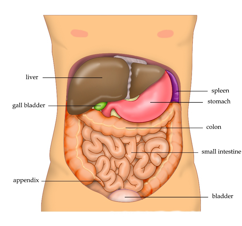
Epidemiología y etiología

  • Las lesiones abdominales contusas constituyen el 80% de todas las lesiones abdominales observadas en EN Erythema nodosum is an immune-mediated panniculitis (inflammation of the subcutaneous fat) caused by a type IV (delayed-type) hypersensitivity reaction. It commonly manifests in young women as tender, erythematous nodules on the shins. Erythema Nodosum las salas de emergencia de Estados Unidos.
  • Causas:
    • La causa más común es una colisión de vehículo motorizado
    • Los LOS Neisseria golpes en EN Erythema nodosum is an immune-mediated panniculitis (inflammation of the subcutaneous fat) caused by a type IV (delayed-type) hypersensitivity reaction. It commonly manifests in young women as tender, erythematous nodules on the shins. Erythema Nodosum el abdomen y las caídas también son causas comunes.
  • Órganos que se lesionan con mayor frecuencia: bazo e hígado

Anatomía

  • Cavidad peritoneal:
    • Subdividida en EN Erythema nodosum is an immune-mediated panniculitis (inflammation of the subcutaneous fat) caused by a type IV (delayed-type) hypersensitivity reaction. It commonly manifests in young women as tender, erythematous nodules on the shins. Erythema Nodosum segmentos intratorácicos y abdominales
    • El segmento intratorácico incluye:
      • Diafragma
      • Hígado
      • Bazo
      • Estómago
      • Colon Colon The large intestines constitute the last portion of the digestive system. The large intestine consists of the cecum, appendix, colon (with ascending, transverse, descending, and sigmoid segments), rectum, and anal canal. The primary function of the colon is to remove water and compact the stool prior to expulsion from the body via the rectum and anal canal. Colon, Cecum, and Appendix: Anatomy transverso
    • El segmento abdominal incluye:
      • La mayor parte del intestino delgado
      • Partes del colon Colon The large intestines constitute the last portion of the digestive system. The large intestine consists of the cecum, appendix, colon (with ascending, transverse, descending, and sigmoid segments), rectum, and anal canal. The primary function of the colon is to remove water and compact the stool prior to expulsion from the body via the rectum and anal canal. Colon, Cecum, and Appendix: Anatomy
  • Retroperitoneo:
    • De difícil acceso en EN Erythema nodosum is an immune-mediated panniculitis (inflammation of the subcutaneous fat) caused by a type IV (delayed-type) hypersensitivity reaction. It commonly manifests in young women as tender, erythematous nodules on the shins. Erythema Nodosum el examen físico y el lavado peritoneal
    • Contiene:
      • Aorta Aorta The main trunk of the systemic arteries. Mediastinum and Great Vessels: Anatomy
      • Vena cava
      • Páncreas
      • Riñón
      • Uréteres
      • Porciones del duodeno y del colon Colon The large intestines constitute the last portion of the digestive system. The large intestine consists of the cecum, appendix, colon (with ascending, transverse, descending, and sigmoid segments), rectum, and anal canal. The primary function of the colon is to remove water and compact the stool prior to expulsion from the body via the rectum and anal canal. Colon, Cecum, and Appendix: Anatomy
  • Compartimento pélvico:
    • Separado de los LOS Neisseria órganos intraperitoneales por los LOS Neisseria pliegues peritoneales
    • Contiene los LOS Neisseria órganos pélvicos:
      • Vejiga
      • Útero/ovarios
      • Próstata

Fisiopatología

El traumatismo abdominal cerrado puede ocurrir debido a varios procesos patológicos:

  • Desaceleración:
    • Las fuerzas de cizallamiento hacen que los LOS Neisseria órganos y la vasculatura se desgarren de sus puntos de unión al AL Amyloidosis peritoneo.
    • Los LOS Neisseria órganos con fijaciones fijas (p. ej., mesenterio, bazo, hígado, riñones, grandes vasos) son particularmente vulnerables.
  • Aplastamiento:
    • Las fuerzas anteriores atrapan los LOS Neisseria órganos entre la pared abdominal anterior y la caja torácica posterior.
    • Los LOS Neisseria órganos sólidos son sensibles al AL Amyloidosis mecanismo de lesión por aplastamiento.
  • Compresión externa:
    • Un golpe directo o una compresión externa contra una estructura fija y rígida
    • Las vísceras huecas son más vulnerables debido al AL Amyloidosis repentino ↑ en EN Erythema nodosum is an immune-mediated panniculitis (inflammation of the subcutaneous fat) caused by a type IV (delayed-type) hypersensitivity reaction. It commonly manifests in young women as tender, erythematous nodules on the shins. Erythema Nodosum la presión intraabdominal

Diagnóstico

Debido a la gran variedad y gravedad de las lesiones asociadas a los LOS Neisseria traumatismos abdominales cerrados, es necesario realizar una rápida pero cuidadosa toma de antecedentes y examen físico para dirigir la investigación con imagenología.

Antecedentes

  • Mecanismo del traumatismo:
    • Puede indicar la gravedad de la lesión
    • Colisión de vehículo motorizado:
      • Posición sentada en EN Erythema nodosum is an immune-mediated panniculitis (inflammation of the subcutaneous fat) caused by a type IV (delayed-type) hypersensitivity reaction. It commonly manifests in young women as tender, erythematous nodules on the shins. Erythema Nodosum el vehículo
      • Otras víctimas fallecidas en EN Erythema nodosum is an immune-mediated panniculitis (inflammation of the subcutaneous fat) caused by a type IV (delayed-type) hypersensitivity reaction. It commonly manifests in young women as tender, erythematous nodules on the shins. Erythema Nodosum el vehículo
      • Cantidad de intrusión en EN Erythema nodosum is an immune-mediated panniculitis (inflammation of the subcutaneous fat) caused by a type IV (delayed-type) hypersensitivity reaction. It commonly manifests in young women as tender, erythematous nodules on the shins. Erythema Nodosum el vehículo (> 15 cm (6 pulgadas) predice lesiones graves)
      • Cantidad de intrusión en EN Erythema nodosum is an immune-mediated panniculitis (inflammation of the subcutaneous fat) caused by a type IV (delayed-type) hypersensitivity reaction. It commonly manifests in young women as tender, erythematous nodules on the shins. Erythema Nodosum el compartimento del ocupante (≥ 12 pulgadas en EN Erythema nodosum is an immune-mediated panniculitis (inflammation of the subcutaneous fat) caused by a type IV (delayed-type) hypersensitivity reaction. It commonly manifests in young women as tender, erythematous nodules on the shins. Erythema Nodosum el sitio del ocupante o ≥ 18 pulgadas en EN Erythema nodosum is an immune-mediated panniculitis (inflammation of the subcutaneous fat) caused by a type IV (delayed-type) hypersensitivity reaction. It commonly manifests in young women as tender, erythematous nodules on the shins. Erythema Nodosum cualquier sitio aumenta el riesgo de lesiones graves)
  • Antecedentes de traumatismos previos
  • Consumo de drogas (ilícitas y de prescripción) o de alcohol por parte del paciente
  • Lo último que ha HA Hemolytic anemia (HA) is the term given to a large group of anemias that are caused by the premature destruction/hemolysis of circulating red blood cells (RBCs). Hemolysis can occur within (intravascular hemolysis) or outside the blood vessels (extravascular hemolysis). Hemolytic Anemia comido o bebido el paciente: importante para considerar la intubación/anestesia
  • Antecedentes quirúrgicos previos

Examen físico

  • Evaluación de la vía aérea, la respiración y la circulación (ABC, por sus siglas en EN Erythema nodosum is an immune-mediated panniculitis (inflammation of the subcutaneous fat) caused by a type IV (delayed-type) hypersensitivity reaction. It commonly manifests in young women as tender, erythematous nodules on the shins. Erythema Nodosum inglés):
    • Vía aérea:
      • Buscar objetos extraños que obstruyan la vía aérea ( los LOS Neisseria dientes caídos son cuerpos extraños comunes en EN Erythema nodosum is an immune-mediated panniculitis (inflammation of the subcutaneous fat) caused by a type IV (delayed-type) hypersensitivity reaction. It commonly manifests in young women as tender, erythematous nodules on the shins. Erythema Nodosum los LOS Neisseria traumatismos de alto impacto).
      • Evaluar si existen lesiones en EN Erythema nodosum is an immune-mediated panniculitis (inflammation of the subcutaneous fat) caused by a type IV (delayed-type) hypersensitivity reaction. It commonly manifests in young women as tender, erythematous nodules on the shins. Erythema Nodosum la tráquea (una lesión traqueal significa que la intubación será compleja).
      • Escuchar si existen sonidos respiratorios inusuales (el estridor sugiere un estrechamiento por un cuerpo extraño o edema Edema Edema is a condition in which excess serous fluid accumulates in the body cavity or interstitial space of connective tissues. Edema is a symptom observed in several medical conditions. It can be categorized into 2 types, namely, peripheral (in the extremities) and internal (in an organ or body cavity). Edema).
    • Respiración:
      • Observar el movimiento de la pared torácica para evaluar si la respiración es uniforme y espontánea (un movimiento torácico irregular sugiere un tórax inestable).
      • Escuchar los LOS Neisseria ruidos respiratorios (si están amortiguados o son irregulares pueden sugerir un neumotórax o hemotórax).
    • Circulación:
      • Palpar los LOS Neisseria pulsos en EN Erythema nodosum is an immune-mediated panniculitis (inflammation of the subcutaneous fat) caused by a type IV (delayed-type) hypersensitivity reaction. It commonly manifests in young women as tender, erythematous nodules on the shins. Erythema Nodosum las 4 extremidades (la taquicardia sugiere inestabilidad hemodinámica o neumotórax).
      • Evaluar el llenado capilar en EN Erythema nodosum is an immune-mediated panniculitis (inflammation of the subcutaneous fat) caused by a type IV (delayed-type) hypersensitivity reaction. It commonly manifests in young women as tender, erythematous nodules on the shins. Erythema Nodosum las extremidades.
  • Evaluación secundaria:
    • Examinar al AL Amyloidosis paciente desde la cabeza hasta los LOS Neisseria pies.
    • La decisión de la imagenología se basa en EN Erythema nodosum is an immune-mediated panniculitis (inflammation of the subcutaneous fat) caused by a type IV (delayed-type) hypersensitivity reaction. It commonly manifests in young women as tender, erythematous nodules on the shins. Erythema Nodosum los LOS Neisseria hallazgos del examen físico.
    • Una lesión externa debe hacer que se investigue la lesión interna correspondiente.
    • Si el paciente está alerta y sin dolor Dolor Inflammation que lo distraiga, los LOS Neisseria síntomas más específicos de un traumatismo abdominal cerrado son:
    • Signos de alarma de una lesión intraabdominal grave:
      • Signo del cinturón de seguridad (esquimosis en EN Erythema nodosum is an immune-mediated panniculitis (inflammation of the subcutaneous fat) caused by a type IV (delayed-type) hypersensitivity reaction. It commonly manifests in young women as tender, erythematous nodules on the shins. Erythema Nodosum el tórax o la pared abdominal; corresponde a la correa diagonal u horizontal de un cinturón de seguridad)
      • Hipotensión
      • Sensibilidad de rebote
      • Distensión abdominal
      • Rigidez abdominal
      • Fractura de fémur concomitante

Imagenología

  • FAST (evaluación enfocada con ultrasonido para traumatismos): se utiliza en EN Erythema nodosum is an immune-mediated panniculitis (inflammation of the subcutaneous fat) caused by a type IV (delayed-type) hypersensitivity reaction. It commonly manifests in young women as tender, erythematous nodules on the shins. Erythema Nodosum todos los LOS Neisseria pacientes (estables o inestables) para buscar sangre intraperitoneal Intraperitoneal Peritoneum: Anatomy y derrame pericárdico:
    • Pacientes hemodinámicamente inestables:
      • FAST positivo → laparotomía de emergencia
      • FAST negativo → búsqueda de fuentes de hemorragia extraabdominal (fractura de fémur)
      • FAST ambiguo → lavado peritoneal diagnóstico o estabilizar al AL Amyloidosis paciente y realizar una tomografía computarizada (TC)
    • Pacientes hemodinámicamente estables:
      • FAST positivo → laparotomía de emergencia
      • FAST negativo, bajo riesgo de lesión intraabdominal → observación
      • FAST negativo o equívoco con alto riesgo de lesión intraabdominal → TC
  • Radiografía (dirigida por los LOS Neisseria hallazgos del examen físico):
    • Radiografía de tórax puede mostrar:
      • Aire libre intraperitoneal Intraperitoneal Peritoneum: Anatomy
      • Hernia Hernia Protrusion of tissue, structure, or part of an organ through the bone, muscular tissue, or the membrane by which it is normally contained. Hernia may involve tissues such as the abdominal wall or the respiratory diaphragm. Hernias may be internal, external, congenital, or acquired. Abdominal Hernias de contenido abdominal
    • Radiografía de pelvis Pelvis The pelvis consists of the bony pelvic girdle, the muscular and ligamentous pelvic floor, and the pelvic cavity, which contains viscera, vessels, and multiple nerves and muscles. The pelvic girdle, composed of 2 “hip” bones and the sacrum, is a ring-like bony structure of the axial skeleton that links the vertebral column with the lower extremities. Pelvis: Anatomy: las fracturas de los LOS Neisseria huesos pélvicos pueden ser fuentes de hemorragias o lesiones vesicales.
  • TC de abdomen con contraste:
    • Imagenología de elección para pacientes estables
    • Proporciona información sobre:
      • Estructuras retroperitoneales
      • Diafragma
      • Órganos abdominales sólidos

Otros estudios diagnósticos

  • Hematocrito:
    • < 30% sugiere una lesión intra-abdominal
    • Un hematocrito normal no descarta una lesión grave.
  • Análisis de orina: la presencia de sangre sugiere una lesión renal/tracto urinario grave.
  • Pruebas de función hepática
  • Lavado peritoneal diagnóstico:
    • Procedimiento invasivo utilizado para evaluar la presencia de sangre en EN Erythema nodosum is an immune-mediated panniculitis (inflammation of the subcutaneous fat) caused by a type IV (delayed-type) hypersensitivity reaction. It commonly manifests in young women as tender, erythematous nodules on the shins. Erythema Nodosum la cavidad abdominal: se introduce un catéter en EN Erythema nodosum is an immune-mediated panniculitis (inflammation of the subcutaneous fat) caused by a type IV (delayed-type) hypersensitivity reaction. It commonly manifests in young women as tender, erythematous nodules on the shins. Erythema Nodosum la cavidad peritoneal, se aspira y se evalúa el fluido.
    • No se utiliza a menudo: alternativa cuando no se dispone de TC y FAST

Características de las lesiones abdominales por órgano

Tabla: Características de las lesiones abdominales por órgano
Órgano lesionado Características
Duodeno
  • Signo del “cinturón de seguridad”
  • Posible presentación tardía
  • Asociado a la fractura de Chance (lesión por hiperflexión que afecta a la columna vertebral en EN Erythema nodosum is an immune-mediated panniculitis (inflammation of the subcutaneous fat) caused by a type IV (delayed-type) hypersensitivity reaction. It commonly manifests in young women as tender, erythematous nodules on the shins. Erythema Nodosum L1 → se produce cuando se lleva solamente el cinturón de seguridad en EN Erythema nodosum is an immune-mediated panniculitis (inflammation of the subcutaneous fat) caused by a type IV (delayed-type) hypersensitivity reaction. It commonly manifests in young women as tender, erythematous nodules on the shins. Erythema Nodosum la cintura)
Bazo
  • Hipotensión, taquicardia
  • Asociado a fracturas de costillas inferiores izquierdas
  • Dolor Dolor Inflammation en EN Erythema nodosum is an immune-mediated panniculitis (inflammation of the subcutaneous fat) caused by a type IV (delayed-type) hypersensitivity reaction. It commonly manifests in young women as tender, erythematous nodules on the shins. Erythema Nodosum la pared torácica izquierda
Hígado
  • Hipotensión, taquicardia
  • Asociado a fracturas de costillas derechas
  • Dolor Dolor Inflammation en EN Erythema nodosum is an immune-mediated panniculitis (inflammation of the subcutaneous fat) caused by a type IV (delayed-type) hypersensitivity reaction. It commonly manifests in young women as tender, erythematous nodules on the shins. Erythema Nodosum el cuadrante superior derecho
Pelvis Pelvis The pelvis consists of the bony pelvic girdle, the muscular and ligamentous pelvic floor, and the pelvic cavity, which contains viscera, vessels, and multiple nerves and muscles. The pelvic girdle, composed of 2 “hip” bones and the sacrum, is a ring-like bony structure of the axial skeleton that links the vertebral column with the lower extremities. Pelvis: Anatomy
Riñón
Traumatismo abdominal cerrado

Signo del cinturón de seguridad:
Las lesiones cutáneas o los hematomas en el patrón de un cinturón de seguridad sugieren que existieron fuerzas importantes en un accidente de vehículo motorizado y pueden implicar una lesión subyacente grave.

Imagen: “Figure 2” por Abbas et al.  Licencia: CC BY 2.0.

Related videos

Tratamiento

Una investigación cuidadosa mostrará evidencia de los LOS Neisseria órganos lesionados, dirigiendo así el tratamiento posterior.

Tabla: Tratamiento de las lesiones abdominales por órgano
Órgano lesionado Tratamiento
Duodeno
  • Pacientes estables:
    • Tratamiento no quirúrgico
    • Período de observación de 9 horas
    • Exámenes abdominales seriados
  • Pacientes de alto riesgo con TC negativa:
    • Período de observación de 9 horas
    • Exámenes abdominales seriados
  • Pacientes de alto riesgo con TC positiva:
    • Ingreso en EN Erythema nodosum is an immune-mediated panniculitis (inflammation of the subcutaneous fat) caused by a type IV (delayed-type) hypersensitivity reaction. It commonly manifests in young women as tender, erythematous nodules on the shins. Erythema Nodosum el hospital
    • Puede requerir laparotomía
Bazo
  • Pacientes estables:
    • Tratamiento no quirúrgico (abordaje conservador)
    • Observar al AL Amyloidosis paciente.
    • Evitar la actividad vigorosa durante 6–8 semanas.
  • Pacientes con múltiples lesiones graves: extirpación inmediata del bazo (esplenectomía)
  • Vacunación: todos los LOS Neisseria pacientes con lesiones extensas del bazo necesitan vacunarse contra microorganismos encapsulados.
    • Haemophilus influenzae Haemophilus Influenzae A species of Haemophilus found on the mucous membranes of humans and a variety of animals. The species is further divided into biotypes I through viii. Haemophilus
    • Streptococcus Streptococcus Streptococcus is one of the two medically important genera of gram-positive cocci, the other being Staphylococcus. Streptococci are identified as different species on blood agar on the basis of their hemolytic pattern and sensitivity to optochin and bacitracin. There are many pathogenic species of streptococci, including S. pyogenes, S. agalactiae, S. pneumoniae, and the viridans streptococci. Streptococcus pneumoniae
    • Neisseria meningitidis Neisseria meningitidis A species of gram-negative, aerobic bacteria. It is a commensal and pathogen only of humans, and can be carried asymptomatically in the nasopharynx. When found in cerebrospinal fluid it is the causative agent of cerebrospinal meningitis. It is also found in venereal discharges and blood. There are at least 13 serogroups based on antigenic differences in the capsular polysaccharides; the ones causing most meningitis infections being a, b, c, y, and w-135. Each serogroup can be further classified by serotype, serosubtype, and immunotype. Neisseria
Hígado
  • Pacientes estables: tratamiento conservador
  • Pacientes inestables o con evidencia de empeoramiento del daño hepático:
    • Embolización transcatéter
    • Empaquetamiento y cirugía limitada
Pelvis Pelvis The pelvis consists of the bony pelvic girdle, the muscular and ligamentous pelvic floor, and the pelvic cavity, which contains viscera, vessels, and multiple nerves and muscles. The pelvic girdle, composed of 2 “hip” bones and the sacrum, is a ring-like bony structure of the axial skeleton that links the vertebral column with the lower extremities. Pelvis: Anatomy
  • Determinar la presencia de hemoperitoneo empleando FAST, TC, +/- lavado peritoneal diagnóstico.
  • La fijación externa minimiza la hemorragia.
  • Angiografía con embolización para controlar la hemorragia arterial
  • Uroperitoneo (orina en EN Erythema nodosum is an immune-mediated panniculitis (inflammation of the subcutaneous fat) caused by a type IV (delayed-type) hypersensitivity reaction. It commonly manifests in young women as tender, erythematous nodules on the shins. Erythema Nodosum la cavidad peritoneal) → requiere reparación quirúrgica de la vejiga
Riñón
  • Pacientes estables: tratamiento conservador
  • En EN Erythema nodosum is an immune-mediated panniculitis (inflammation of the subcutaneous fat) caused by a type IV (delayed-type) hypersensitivity reaction. It commonly manifests in young women as tender, erythematous nodules on the shins. Erythema Nodosum las lesiones graves:
    • Pueden necesitar stents si existe una obstrucción con un coágulo.
    • La nefrectomía es el último recurso.

Relevancia Clínica

  • Lesiones abdominales penetrantes: suelen consistir en EN Erythema nodosum is an immune-mediated panniculitis (inflammation of the subcutaneous fat) caused by a type IV (delayed-type) hypersensitivity reaction. It commonly manifests in young women as tender, erythematous nodules on the shins. Erythema Nodosum la transgresión de la cavidad abdominal por una herida de bala o una puñalada. Las estructuras más comúnmente lesionadas son el intestino delgado, seguido de colon Colon The large intestines constitute the last portion of the digestive system. The large intestine consists of the cecum, appendix, colon (with ascending, transverse, descending, and sigmoid segments), rectum, and anal canal. The primary function of the colon is to remove water and compact the stool prior to expulsion from the body via the rectum and anal canal. Colon, Cecum, and Appendix: Anatomy, hígado y estructuras vasculares. El tratamiento es una laparotomía exploratoria.
  • Lesión esplénica: en EN Erythema nodosum is an immune-mediated panniculitis (inflammation of the subcutaneous fat) caused by a type IV (delayed-type) hypersensitivity reaction. It commonly manifests in young women as tender, erythematous nodules on the shins. Erythema Nodosum las lesiones contusas, el hígado y el bazo son los LOS Neisseria órganos que se lesionan con mayor frecuencia. Generalmente, la lesión esplénica se asocia a las fracturas de las costillas inferiores izquierdas. Las características de la lesión esplénica incluyen hipotensión, taquicardia, dolor Dolor Inflammation abdominal, dolor Dolor Inflammation en EN Erythema nodosum is an immune-mediated panniculitis (inflammation of the subcutaneous fat) caused by a type IV (delayed-type) hypersensitivity reaction. It commonly manifests in young women as tender, erythematous nodules on the shins. Erythema Nodosum la pared torácica izquierda y dolor Dolor Inflammation en EN Erythema nodosum is an immune-mediated panniculitis (inflammation of the subcutaneous fat) caused by a type IV (delayed-type) hypersensitivity reaction. It commonly manifests in young women as tender, erythematous nodules on the shins. Erythema Nodosum el hombro izquierdo ( dolor Dolor Inflammation referido debido a la irritación del nervio frénico por la hemorragia esplénica).
  • Lesión pélvica: las lesiones pélvicas y las fracturas pélvicas se encuentran entre las peores complicaciones de las lesiones abdominales contusas. Las características clínicas incluyen hipotensión, dolor Dolor Inflammation con el movimiento, hematuria Hematuria Presence of blood in the urine. Renal Cell Carcinoma macroscópica y equimosis peripélvica. El tacto rectal es importante para identificar lesiones en EN Erythema nodosum is an immune-mediated panniculitis (inflammation of the subcutaneous fat) caused by a type IV (delayed-type) hypersensitivity reaction. It commonly manifests in young women as tender, erythematous nodules on the shins. Erythema Nodosum el recto y localizar la próstata. El tratamiento generalmente se limita a los LOS Neisseria cuidados de soporte, pero a veces puede ser necesaria la estabilización quirúrgica.
  • Evaluación ABC: la evaluación ABC es un abordaje para el tratamiento de los LOS Neisseria pacientes críticos. La vía aérea, la respiración y la circulación es un 1er paso esencial que se debe dar al AL Amyloidosis encontrarse con un paciente. Los LOS Neisseria pasos se adaptan fácilmente a muchas situaciones, como pacientes que no responden, paros cardíacos, pacientes críticos o pacientes con traumatismos. En EN Erythema nodosum is an immune-mediated panniculitis (inflammation of the subcutaneous fat) caused by a type IV (delayed-type) hypersensitivity reaction. It commonly manifests in young women as tender, erythematous nodules on the shins. Erythema Nodosum el caso de los LOS Neisseria pacientes con traumatismos, el ABC incluye la evaluación primaria, la valoración inicial y el tratamiento de las lesiones.

Referencias

  1. Nishijima, D. K., Simel, D. L., Wisner, D. H., & Holmes, J. F. (2012). Does this adult patient have a blunt intra-abdominal injury?. JAMA307(14), 1517–1527. https://doi.org/10.1001/jama.2012.422
  2. Isenhour, J. L., & Marx, J. (2007). Advances in abdominal trauma. Emergency medicine clinics of North America25(3), 713–ix. https://doi.org/10.1016/j.emc.2007.06.002
  3. Newgard, C. D., Lewis, R. J., & Jolly, B. T. (2002). Use of out-of-hospital variables to predict severity of injury in pediatric patients involved in motor vehicle crashes. Annals of emergency medicine39(5), 481–491. https://doi.org/10.1067/mem.2002.123549

¡Crea tu cuenta gratis o inicia una sesión para seguir leyendo!

Regístrate ahora y obtén acceso gratuito a Lecturio con páginas de concepto, videos médicos y cuestionarios para tu educación médica.

User Reviews

Que tengas una sesión de estudio alegre y navideña 🎁 Ahorra 50% en todos los planes >>

Details