El abdomen agudo, que en EN Erythema nodosum is an immune-mediated panniculitis (inflammation of the subcutaneous fat) caused by a type IV (delayed-type) hypersensitivity reaction. It commonly manifests in young women as tender, erythematous nodules on the shins. Erythema Nodosum muchos casos es una emergencia quirúrgica, se define como la aparición súbita de dolor Dolor Inflammation abdominal que puede ser causado por inflamación, infección, perforación, isquemia u obstrucción. La localización del dolor Dolor Inflammation, sus características y los LOS Neisseria síntomas asociados (e.g., ictericia) son herramientas importantes que ayudan a dirigir el diagnóstico. Los LOS Neisseria pacientes suelen tener una sensibilidad severa con rigidez asociada y dolor Dolor Inflammation de rebote. Los LOS Neisseria exámenes de laboratorios mostrarán leucocitosis, acidosis Acidosis A pathologic condition of acid accumulation or depletion of base in the body. The two main types are respiratory acidosis and metabolic acidosis, due to metabolic acid build up. Respiratory Acidosis y, en EN Erythema nodosum is an immune-mediated panniculitis (inflammation of the subcutaneous fat) caused by a type IV (delayed-type) hypersensitivity reaction. It commonly manifests in young women as tender, erythematous nodules on the shins. Erythema Nodosum algunos casos, pruebas de función hepática anormales. Los LOS Neisseria estudios de imagen ayudan a reducir el diagnóstico diferencial; la primera línea de estudio de imagen siempre es una radiografía de tórax de pie para evaluar por neumoperitoneo. El tratamiento y el pronóstico del abdomen agudo dependen en EN Erythema nodosum is an immune-mediated panniculitis (inflammation of the subcutaneous fat) caused by a type IV (delayed-type) hypersensitivity reaction. It commonly manifests in young women as tender, erythematous nodules on the shins. Erythema Nodosum gran medida de la causa subyacente, pero la gran mayoría de casos constituyen una emergencia quirúrgica con morbilidad y mortalidad asociadas.
Last updated: Dec 15, 2025
El abdomen agudo es la constelación de signos y síntomas asociados a un dolor Dolor Inflammation abdominal severo y peritonitis Peritonitis Inflammation of the peritoneum lining the abdominal cavity as the result of infectious, autoimmune, or chemical processes. Primary peritonitis is due to infection of the peritoneal cavity via hematogenous or lymphatic spread and without intra-abdominal source. Secondary peritonitis arises from the abdominal cavity itself through rupture or abscess of intra-abdominal organs. Penetrating Abdominal Injury que con frecuencia requiere una intervención quirúrgica de emergencia.
Causas no quirúrgicas de abdomen agudo:
Causas quirúrgicas del abdomen agudo:
Diagnósticos diferenciales de un dolor abdominal agudo
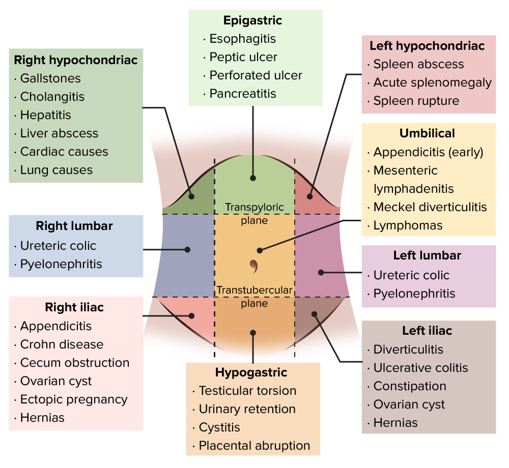
Imagen por Lecturio.Para efectos descriptivos, el abdomen puede dividirse en EN Erythema nodosum is an immune-mediated panniculitis (inflammation of the subcutaneous fat) caused by a type IV (delayed-type) hypersensitivity reaction. It commonly manifests in young women as tender, erythematous nodules on the shins. Erythema Nodosum 4 cuadrantes o 9 regiones.
Cuadrantes:
Dividido en EN Erythema nodosum is an immune-mediated panniculitis (inflammation of the subcutaneous fat) caused by a type IV (delayed-type) hypersensitivity reaction. It commonly manifests in young women as tender, erythematous nodules on the shins. Erythema Nodosum 4 cuadrantes por 2 líneas perpendiculares que se cruzan en EN Erythema nodosum is an immune-mediated panniculitis (inflammation of the subcutaneous fat) caused by a type IV (delayed-type) hypersensitivity reaction. It commonly manifests in young women as tender, erythematous nodules on the shins. Erythema Nodosum el ombligo:
Cuadrante superior derecho:
|
Cuadrante superior izquierdo:
|
Cuadrante inferior derecho:
|
Cuadrante inferior izquierdo:
|
Regiones:
El abdomen puede dividirse en EN Erythema nodosum is an immune-mediated panniculitis (inflammation of the subcutaneous fat) caused by a type IV (delayed-type) hypersensitivity reaction. It commonly manifests in young women as tender, erythematous nodules on the shins. Erythema Nodosum regiones por 4 líneas:
| Hipocondrio derecho | Región epigástrica | Hipocondrio izquierdo |
| Región lumbar derecha | Región umbilical | Región lumbar izquierda |
| Fosa ilíaca derecha | Región hipogástrica | Fosa ilíaca izquierda |
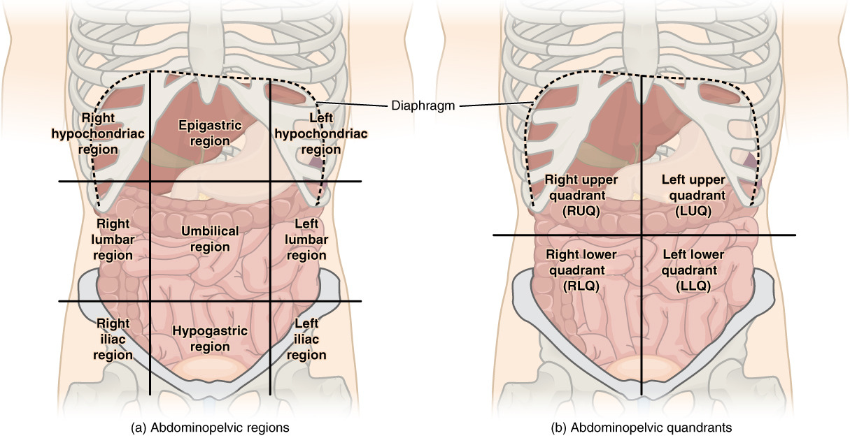
Cuadrantes abdominales:
Hay (a) 9 regiones abdominales y (b) 4 cuadrantes abdominales en la cavidad peritoneal.
Relación de los LOS Neisseria órganos intraabdominales con la cavidad peritoneal:
La característica distintiva del abdomen agudo es la aparición aguda de dolor Dolor Inflammation abdominal intenso que puede o no estar asociado a otros síntomas. Se debe realizar una historia clínica detallada y un examen físico para determinar el curso de acción correcto.
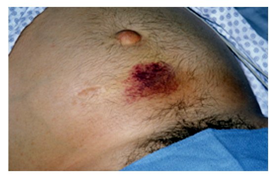
Hematoma periumbilical consistente con el signo de Cullen (pancreatitis hemorrágica)
Imagen: “Acude pancreatitis with Cullen’s sign”, por Herbert L. Fred, MD. Licencia: CC BY 2.0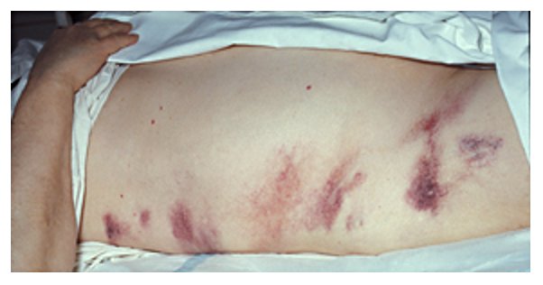
Hematomas alrededor de los flancos consistentes con el signo de Grey-Turner (pancreatitis hemorrágica)
Imagen: “Hemorrhagic pancreatitis – Grey Turner’s sign” por Herbert L. Fred, MD y Hendrik A. van Dijk. Licencia: CC BY 2.0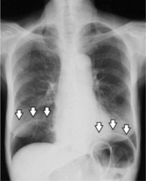
Radiografía de tórax de pie que muestra un neumoperitoneo (flechas)
Imagen: “A chest radiograph demonstrating pneumoperitoneum” por Manabu Kaneko et al. Licencia: CC BY 4.0El abdomen agudo es una emergencia quirúrgica en EN Erythema nodosum is an immune-mediated panniculitis (inflammation of the subcutaneous fat) caused by a type IV (delayed-type) hypersensitivity reaction. It commonly manifests in young women as tender, erythematous nodules on the shins. Erythema Nodosum la gran mayoría de los LOS Neisseria casos. La evaluación inicial debe determinar los LOS Neisseria casos que no requieren tratamiento quirúrgico.
Reanimación:
Evaluación:
El tratamiento quirúrgico suele ser necesario a menos que se haya establecido una causa no quirúrgica. Si se justifica la intervención quirúrgica, hay dos abordajes: