


O olho humano é um órgão sensorial que tem a visão como a sua função primária. O olho tem uma forma esférica e é estruturado em 3 camadas: uma camada fibrosa externa de suporte, uma camada vascular média e uma camada neuronal interna. O olho também pode ser subdividido em 3 compartimentos: as câmaras anterior, posterior e vítrea. Ao redor do globo ocular estão os músculos extraoculares, o aparelho lacrimal, vários nervos e vasos e a estrutura óssea da órbita. A luz viaja através dos compartimentos do olho para focar na retina Retina The ten-layered nervous tissue membrane of the eye. It is continuous with the optic nerve and receives images of external objects and transmits visual impulses to the brain. Its outer surface is in contact with the choroid and the inner surface with the vitreous body. The outermost layer is pigmented, whereas the inner nine layers are transparent. Eye: Anatomy, que é o local onde os fotorrecetores convertem o estímulo num impulso neuronal, que é transportado pelo nervo ótico até o cérebro.
Last updated: Dec 15, 2025
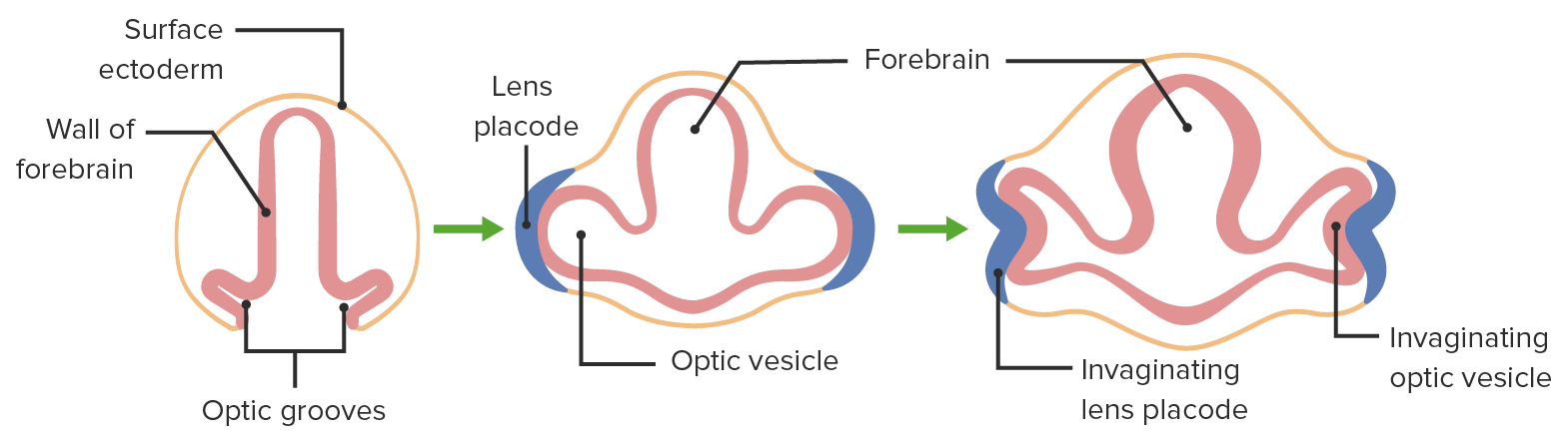
Desenvolvimento embrionário do olho
Imagem por Lecturio.O olho adulto é um órgão complexo contido na cavidade orbitária (composta por 7 ossos). Cada olho tem várias camadas e câmaras e é rodeado por 6 músculos extraoculares.

Anatomia do olho
Imagem por Lecturio.O olho é composto por 3 camadas (fibrosa, vascular, neuronal) e uma cobertura de tecido conjuntivo transparente (conjuntiva).
Conjuntiva :
Camada fibrosa:
Camada vascular:
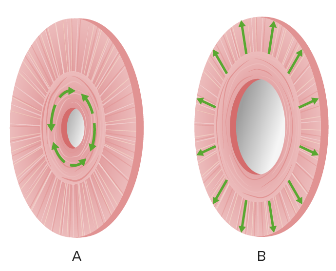
Os músculos do esfíncter da íris são responsáveis pela constrição (A) e dilatação (B) da pupila.
Imagem por Lecturio.Camada neuronal:
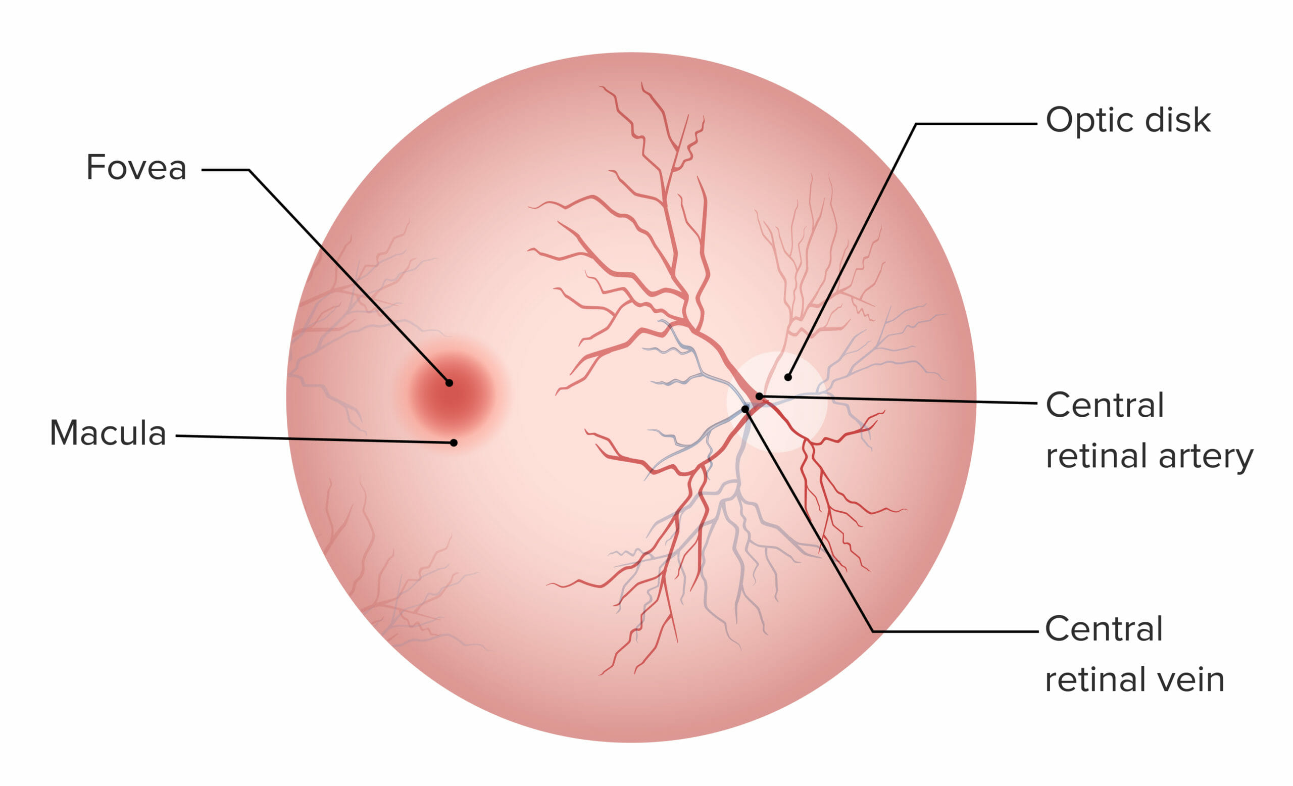
Retina:
A fóvea e a mácula não possuem vasos sanguíneos, sendo responsáveis pela visão de alta acuidade. O disco ótico é o ponto de entrada da vasculatura do olho e não contém fotorrecetores.
Características da retina Retina The ten-layered nervous tissue membrane of the eye. It is continuous with the optic nerve and receives images of external objects and transmits visual impulses to the brain. Its outer surface is in contact with the choroid and the inner surface with the vitreous body. The outermost layer is pigmented, whereas the inner nine layers are transparent. Eye: Anatomy e as suas camadas:
| Camada da retina Retina The ten-layered nervous tissue membrane of the eye. It is continuous with the optic nerve and receives images of external objects and transmits visual impulses to the brain. Its outer surface is in contact with the choroid and the inner surface with the vitreous body. The outermost layer is pigmented, whereas the inner nine layers are transparent. Eye: Anatomy (externa para interna) | Características |
|---|---|
| Epitélio pigmentado |
|
| Camada de bastonetes e cones |
|
| Membrana limitante externa | Suporta as células fotorrecetoras |
| Membrana nuclear externa | Núcleos de células fotorrecetoras (neurónio de 1ª ordem) |
| Camada plexiforme externa | 1ª sinapse, entre cones e bastonetes e células bipolares |
| Membrana limitante média | Membrana de suporte |
| Camada nuclear interna | Corpos celulares e núcleos de células bipolares (neurónio de 2ª ordem), transmitem informações para células ganglionares |
| Camada plexiforme interna | Segunda sinapse, entre células bipolares e células ganglionares |
| Camada de células ganglionares |
|
| Fibras do nervo ótico | Axónios das células ganglionares |
| Membrana limitante interna | Camada mais MAIS Androgen Insensitivity Syndrome interna, mais MAIS Androgen Insensitivity Syndrome próxima do humor Humor Defense Mechanisms vítreo |
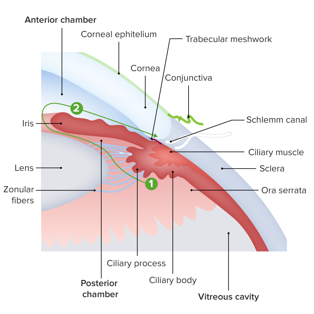
Diagrama da estrutura da região límbica:
O humor aquoso é produzido pelos processos ciliares (1), circula pela pupila da íris para a câmara anterior (2) e, finalmente, pelo canal de Schlemm para o seio venoso escleral.
Todo o suprimento arterial do olho é fornecido por ramos da artéria oftálmica e drenado por um sistema de veias que se unem para formar a veia central da retina Retina The ten-layered nervous tissue membrane of the eye. It is continuous with the optic nerve and receives images of external objects and transmits visual impulses to the brain. Its outer surface is in contact with the choroid and the inner surface with the vitreous body. The outermost layer is pigmented, whereas the inner nine layers are transparent. Eye: Anatomy.
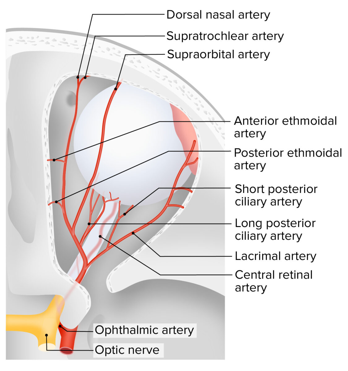
Suprimento arterial do olho
Imagem por Lecturio.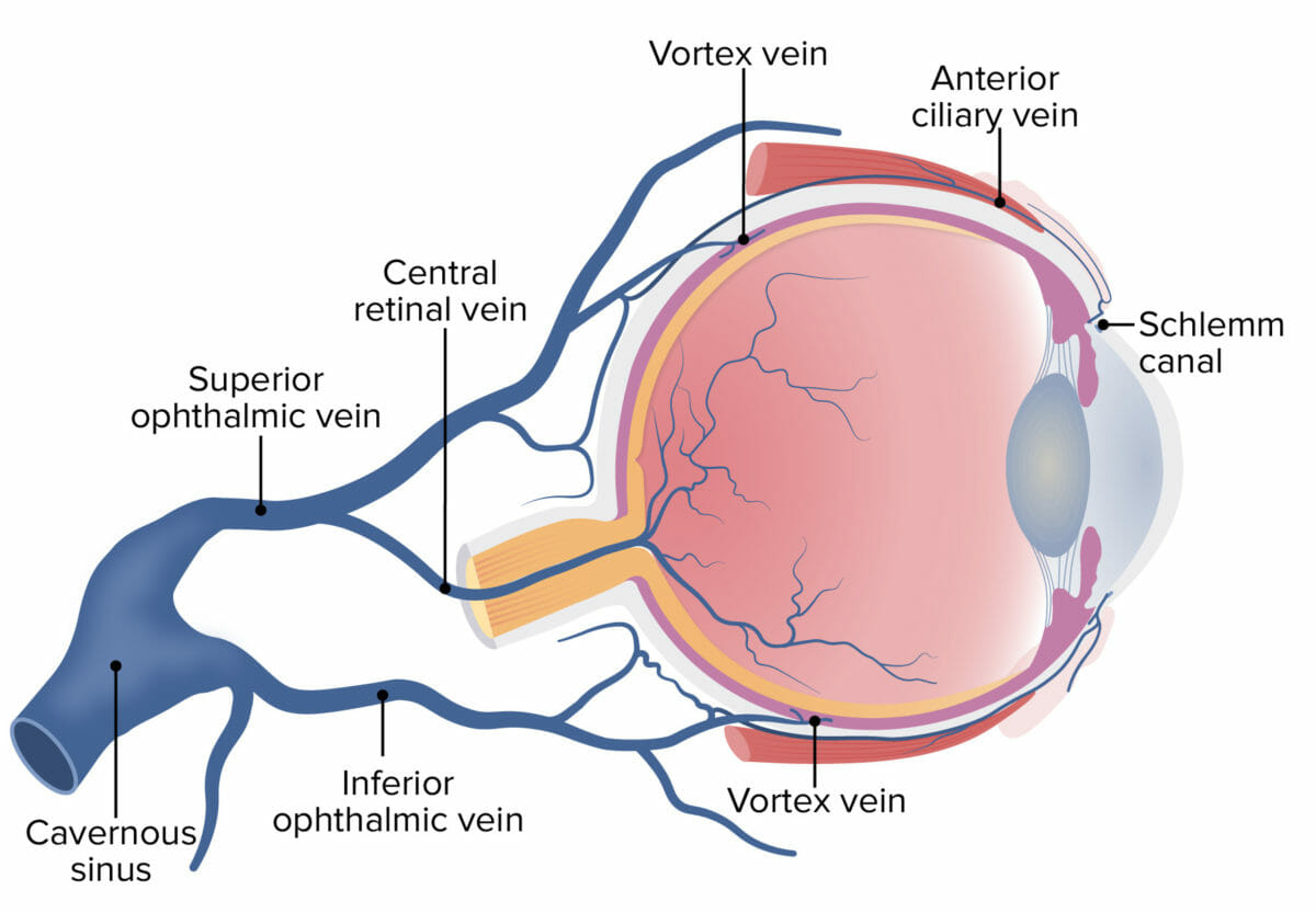
Drenagem venosa do olho
Imagem por Lecturio.