Domina Conceptos Médicos

Estudia para la escuela de medicina y tus examenes con Lecturio.

Rotura del Bazo

La rotura esplénica es una emergencia médica que conlleva un riesgo importante de shock Shock Shock is a life-threatening condition associated with impaired circulation that results in tissue hypoxia. The different types of shock are based on the underlying cause: distributive (↑ cardiac output (CO), ↓ systemic vascular resistance (SVR)), cardiogenic (↓ CO, ↑ SVR), hypovolemic (↓ CO, ↑ SVR), obstructive (↓ CO), and mixed. Types of Shock hipovolémico y muerte. La lesión del bazo representa casi la mitad de todas las lesiones de los LOS Neisseria órganos intraabdominales. La razón más común para una ruptura del bazo es un traumatismo abdominal cerrado, específicamente, los LOS Neisseria accidentes automovilísticos. Sin embargo, en EN Erythema nodosum is an immune-mediated panniculitis (inflammation of the subcutaneous fat) caused by a type IV (delayed-type) hypersensitivity reaction. It commonly manifests in young women as tender, erythematous nodules on the shins. Erythema Nodosum el caso de las personas con esplenomegalia, incluso un traumatismo mínimo puede provocar una lesión o rotura esplénica. Los LOS Neisseria pacientes suelen presentar dolor Dolor Inflammation en EN Erythema nodosum is an immune-mediated panniculitis (inflammation of the subcutaneous fat) caused by a type IV (delayed-type) hypersensitivity reaction. It commonly manifests in young women as tender, erythematous nodules on the shins. Erythema Nodosum el cuadrante superior izquierdo del abdomen; sin embargo, el dolor Dolor Inflammation puede irradiarse al AL Amyloidosis hombro izquierdo. Los LOS Neisseria pacientes corren el riesgo de sufrir inestabilidad hemodinámica debido a la pérdida de sangre. El diagnóstico se hace HACE Altitude Sickness generalmente con imagenología de TC; y el tratamiento, que va VA Ventilation: Mechanics of Breathing desde la observación hasta la esplenectomía, depende de la estabilidad hemodinámica del paciente.

Last updated: Dec 15, 2025

Editorial responsibility: Stanley Oiseth, Lindsay Jones, Evelin Maza

Descripción General

Definición

La rotura esplénica suele estar asociada a un traumatismo (e.g., un accidente de tráfico) que provoca una laceración del órgano.

Epidemiología

  • El bazo es el órgano intraabdominal que sufre lesiones y roturas con más frecuencia:
    • 50%–75% de los LOS Neisseria casos provocados por un traumatismo cerrado (causa más común)
    • La tasa de lesiones tras un traumatismo abdominal cerrado puede llegar al AL Amyloidosis 7,5%.
  • Más común en EN Erythema nodosum is an immune-mediated panniculitis (inflammation of the subcutaneous fat) caused by a type IV (delayed-type) hypersensitivity reaction. It commonly manifests in young women as tender, erythematous nodules on the shins. Erythema Nodosum los LOS Neisseria hombres
  • Pico de incidencia en EN Erythema nodosum is an immune-mediated panniculitis (inflammation of the subcutaneous fat) caused by a type IV (delayed-type) hypersensitivity reaction. It commonly manifests in young women as tender, erythematous nodules on the shins. Erythema Nodosum adultos jóvenes (18–34 años)

Etiología

  • Traumáticas:
    • Accidente de tráfico (causa más común)
    • Golpe abdominal directo
    • Caída
  • No traumáticas:
    • Malignidad
    • Infecciones
    • Iatrogenia (e.g., manipulación quirúrgica del órgano)

Fisiopatología

La rotura esplénica puede tener graves consecuencias debido a la función fisiológica del bazo.

  • Órgano linfático secundario altamente vascular y central para el sistema inmunitario
  • Filtra la sangre y elimina los LOS Neisseria eritrocitos viejos o dañados
  • Situado posterolateralmente en EN Erythema nodosum is an immune-mediated panniculitis (inflammation of the subcutaneous fat) caused by a type IV (delayed-type) hypersensitivity reaction. It commonly manifests in young women as tender, erythematous nodules on the shins. Erythema Nodosum el hipocondrio izquierdo, debajo del hemidiafragma izquierdo y lateral a la curvatura mayor del estómago
  • Esplenomegalia: aumento del tamaño del bazo
    • Numerosas causas:
      • Congestión de la vena porta
      • Infiltración esplénica
      • Infección
    • Conduce a ↑ del riesgo de rotura esplénica
Anatomy of the spleen all views

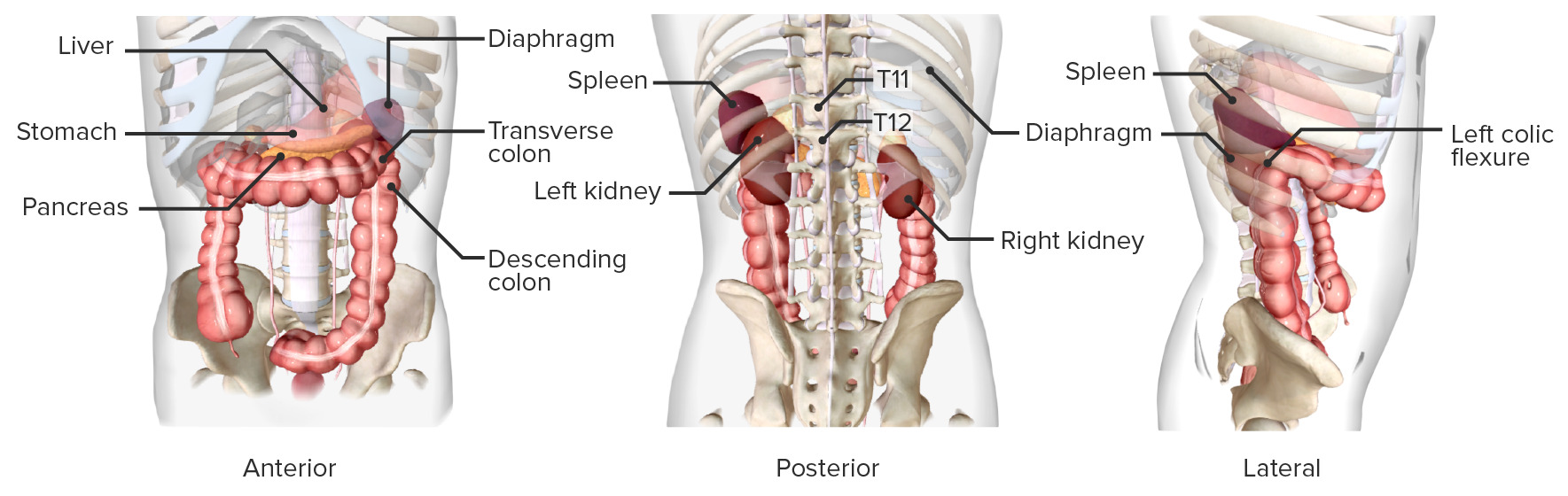
Localización del bazo

Imagen por BioDigital, editado por Lecturio

Presentación Clínica

Antecedentes

  • Traumatismos en EN Erythema nodosum is an immune-mediated panniculitis (inflammation of the subcutaneous fat) caused by a type IV (delayed-type) hypersensitivity reaction. It commonly manifests in young women as tender, erythematous nodules on the shins. Erythema Nodosum hipocondrio izquierdo
  • Esplenomegalia
  • Antecedentes de enfermedades asociadas a la esplenomegalia:
    • Enfermedad de células falciformes
    • Malaria Malaria Malaria is an infectious parasitic disease affecting humans and other animals. Most commonly transmitted via the bite of a female Anopheles mosquito infected with microorganisms of the Plasmodium genus. Patients present with fever, chills, myalgia, headache, and diaphoresis. Plasmodium/Malaria
    • Linfoma
    • VEB

Examen físico

  • Signos vitales:
    • Taquicardia e hipotensión: Pueden aparecer signos de shock Shock Shock is a life-threatening condition associated with impaired circulation that results in tissue hypoxia. The different types of shock are based on the underlying cause: distributive (↑ cardiac output (CO), ↓ systemic vascular resistance (SVR)), cardiogenic (↓ CO, ↑ SVR), hypovolemic (↓ CO, ↑ SVR), obstructive (↓ CO), and mixed. Types of Shock por la pérdida de grandes volúmenes de sangre.
  • Examen abdominal:
    • Laceración visible o signo de traumatismo cerrado en EN Erythema nodosum is an immune-mediated panniculitis (inflammation of the subcutaneous fat) caused by a type IV (delayed-type) hypersensitivity reaction. It commonly manifests in young women as tender, erythematous nodules on the shins. Erythema Nodosum el abdomen
    • El signo del cinturón de seguridad puede observarse en EN Erythema nodosum is an immune-mediated panniculitis (inflammation of the subcutaneous fat) caused by a type IV (delayed-type) hypersensitivity reaction. It commonly manifests in young women as tender, erythematous nodules on the shins. Erythema Nodosum pacientes que se presentan tras un accidente de tráfico.
    • Esplenomegalia palpable
    • Sensibilidad abdominal, defensa, rigidez
    • Dolor Dolor Inflammation irradiado al AL Amyloidosis hombro izquierdo (signo de Kehr)

Diagnóstico

La exploración física y la toma de antecedentes pueden ser útiles, pero no todos los LOS Neisseria pacientes con rotura esplénica presentan hallazgos clínicamente significativos.

Imagenología

  • Ultrasonido FAST:
    • De gran utilidad en EN Erythema nodosum is an immune-mediated panniculitis (inflammation of the subcutaneous fat) caused by a type IV (delayed-type) hypersensitivity reaction. It commonly manifests in young women as tender, erythematous nodules on the shins. Erythema Nodosum pacientes traumatizados hemodinámicamente inestables
    • Examen FAST positivo = borde hipoecoico (negro) alrededor del bazo
    • Un examen FAST negativo no descarta la ruptura esplénica.
  • TC:
    • Preferido en EN Erythema nodosum is an immune-mediated panniculitis (inflammation of the subcutaneous fat) caused by a type IV (delayed-type) hypersensitivity reaction. It commonly manifests in young women as tender, erythematous nodules on the shins. Erythema Nodosum pacientes hemodinámicamente estables con contraste intravenoso (+/- oral)
    • 98% de sensibilidad para lesiones esplénicas cuando se administra contraste intravenoso
    • Mostrará hipodensidades esplénicas, extravasación de contraste intravenoso
    • Es excelente para clasificar la gravedad de la lesión y determinar si el paciente necesitará cirugía.
CT of abdomen Rupture of Spleen

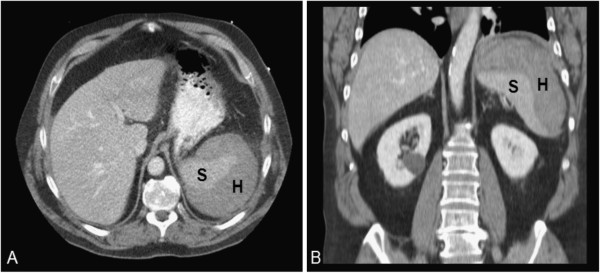
TC de abdomen de un paciente con rotura esplénica:
La rotura esplénica tiene múltiples etiologías posibles. El examen clínico y los antecedentes suelen ser insuficientes para el diagnóstico. Una TC de abdomen puede revelar áreas de hematoma (H) o sangre libre.
S: bazo

Imagen: “Spleen with large subcapsular hematoma” por de Kubber MM et al. Licencia: CC BY 2.0

Tratamiento

El tratamiento de la rotura esplénica depende del estado hemodinámico del paciente:

  • Hemodinámicamente estable:
    • Observación durante 10–14 días
    • Tratamiento no quirúrgico
    • Transfusión de sangre y control del dolor Dolor Inflammation según necesidad
    • Se aconseja a los LOS Neisseria pacientes que eviten la actividad intensa durante 6–8 semanas
    • Considerar la embolización angiográfica.
  • Hemodinámicamente inestable:
    • Laparotomía exploratoria de urgencia
    • La esplenectomía se considera para los LOS Neisseria pacientes con pérdidas de sangre continuas que requieren transfusión de ≥ 4 unidades de sangre.
Splenic artery embolization Rupture of Spleen

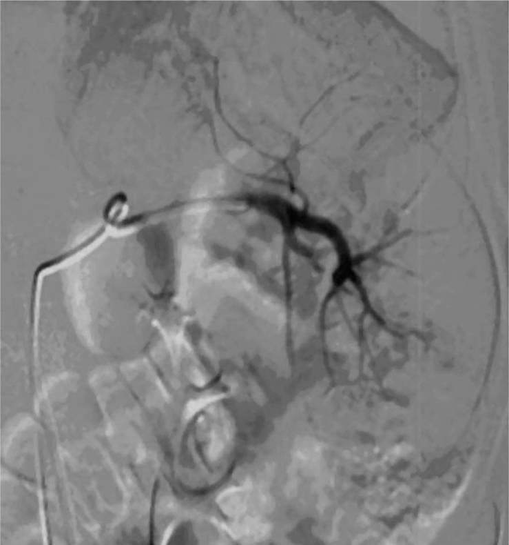
Embolización de la arteria esplénica:
Esta imagen muestra la embolización angiográfica en el marco de una lesión esplénica. El tratamiento no quirúrgico ha conseguido grandes mejoras en los resultados de los pacientes hemodinámicamente estables.

Imagen: “Splenic angiography obtained after the selective embolization” por Popovic P et al. Licencia: CC BY 3.0

Esplenectomia

  • Resección de las partes lesionadas del bazo
  • Consecuencias hematológicas e inmunológicas graves
  • El objetivo de la cirugía es preservar el bazo y realizar una resección parcial (especialmente importante en EN Erythema nodosum is an immune-mediated panniculitis (inflammation of the subcutaneous fat) caused by a type IV (delayed-type) hypersensitivity reaction. It commonly manifests in young women as tender, erythematous nodules on the shins. Erythema Nodosum los LOS Neisseria niños).
  • Complicaciones:
    • Hemorragia grave y shock Shock Shock is a life-threatening condition associated with impaired circulation that results in tissue hypoxia. The different types of shock are based on the underlying cause: distributive (↑ cardiac output (CO), ↓ systemic vascular resistance (SVR)), cardiogenic (↓ CO, ↑ SVR), hypovolemic (↓ CO, ↑ SVR), obstructive (↓ CO), and mixed. Types of Shock hipovolémico
    • Inmunosupresión: mayor riesgo de sepsis Sepsis Systemic inflammatory response syndrome with a proven or suspected infectious etiology. When sepsis is associated with organ dysfunction distant from the site of infection, it is called severe sepsis. When sepsis is accompanied by hypotension despite adequate fluid infusion, it is called septic shock. Sepsis and Septic Shock por infección bacteriana por polisacáridos
    • Complicación pancreática: lesión o infección

Post-esplenectomía:

Los LOS Neisseria pacientes asplénicos requieren vacunación contra bacterias encapsuladas:

  • Haemophilus influenzae Haemophilus Influenzae A species of Haemophilus found on the mucous membranes of humans and a variety of animals. The species is further divided into biotypes I through viii. Haemophilus
  • Streptococcus Streptococcus Streptococcus is one of the two medically important genera of gram-positive cocci, the other being Staphylococcus. Streptococci are identified as different species on blood agar on the basis of their hemolytic pattern and sensitivity to optochin and bacitracin. There are many pathogenic species of streptococci, including S. pyogenes, S. agalactiae, S. pneumoniae, and the viridans streptococci. Streptococcus pneumoniae
  • Neisseria meningitidis Neisseria meningitidis A species of gram-negative, aerobic bacteria. It is a commensal and pathogen only of humans, and can be carried asymptomatically in the nasopharynx. When found in cerebrospinal fluid it is the causative agent of cerebrospinal meningitis. It is also found in venereal discharges and blood. There are at least 13 serogroups based on antigenic differences in the capsular polysaccharides; the ones causing most meningitis infections being a, b, c, y, and w-135. Each serogroup can be further classified by serotype, serosubtype, and immunotype. Neisseria

Relevancia Clínica

  • Bazo: órgano sólido con forma de grano de café, que pesa unos 150 g. El bazo está situado en EN Erythema nodosum is an immune-mediated panniculitis (inflammation of the subcutaneous fat) caused by a type IV (delayed-type) hypersensitivity reaction. It commonly manifests in young women as tender, erythematous nodules on the shins. Erythema Nodosum la parte posterior izquierda del abdomen superior (hipocondrio izquierdo) y se encuentra junto a las costillas 9 a 11. El bazo tiene pulpa roja y blanca, que filtran las células sanguíneas y activan el sistema inmunitario, respectivamente. Este órgano está compuesto por un tejido muy blando y, como tal TAL Renal Sodium and Water Regulation, corre un alto riesgo de laceración y lesión tras un traumatismo abdominal.
  • Traumatismos abdominales y lesiones abdominales penetrantes: se clasifican según el mecanismo de la lesión como cerradas o penetrantes. El traumatismo abdominal cerrado es el tipo más común de traumatismo abdominal que se presenta en EN Erythema nodosum is an immune-mediated panniculitis (inflammation of the subcutaneous fat) caused by a type IV (delayed-type) hypersensitivity reaction. It commonly manifests in young women as tender, erythematous nodules on the shins. Erythema Nodosum urgencias. Las personas que han sufrido un accidente de tráfico pueden presentar un hallazgo clásico en EN Erythema nodosum is an immune-mediated panniculitis (inflammation of the subcutaneous fat) caused by a type IV (delayed-type) hypersensitivity reaction. It commonly manifests in young women as tender, erythematous nodules on the shins. Erythema Nodosum la exploración física denominado signo del cinturón de seguridad. Pueden lesionarse diferentes estructuras, como el duodeno, el bazo, el hígado, los LOS Neisseria riñones y los LOS Neisseria órganos pélvicos. La tomografía computarizada es la imagenología de elección para los LOS Neisseria pacientes que están hemodinámicamente estables.
  • Asplenia Asplenia Asplenia is the absence of splenic tissue or function and can stem from several factors ranging from congenital to iatrogenic. There is a distinction between anatomic asplenia, which is due to the surgical removal of the spleen, and functional asplenia, which is due to a condition that leads to splenic atrophy, infarct, congestion, or infiltrative disease. Asplenia: ausencia de bazo. La razón anatómica más común para la asplenia Asplenia Asplenia is the absence of splenic tissue or function and can stem from several factors ranging from congenital to iatrogenic. There is a distinction between anatomic asplenia, which is due to the surgical removal of the spleen, and functional asplenia, which is due to a condition that leads to splenic atrophy, infarct, congestion, or infiltrative disease. Asplenia es la extirpación quirúrgica. Solo en EN Erythema nodosum is an immune-mediated panniculitis (inflammation of the subcutaneous fat) caused by a type IV (delayed-type) hypersensitivity reaction. It commonly manifests in young women as tender, erythematous nodules on the shins. Erythema Nodosum raras ocasiones la asplenia Asplenia Asplenia is the absence of splenic tissue or function and can stem from several factors ranging from congenital to iatrogenic. There is a distinction between anatomic asplenia, which is due to the surgical removal of the spleen, and functional asplenia, which is due to a condition that leads to splenic atrophy, infarct, congestion, or infiltrative disease. Asplenia es congénita, y si es así, suele estar asociada a una malformación de los LOS Neisseria grandes vasos torácicos. La asplenia Asplenia Asplenia is the absence of splenic tissue or function and can stem from several factors ranging from congenital to iatrogenic. There is a distinction between anatomic asplenia, which is due to the surgical removal of the spleen, and functional asplenia, which is due to a condition that leads to splenic atrophy, infarct, congestion, or infiltrative disease. Asplenia se denomina “funcional” cuando el órgano sigue presente en EN Erythema nodosum is an immune-mediated panniculitis (inflammation of the subcutaneous fat) caused by a type IV (delayed-type) hypersensitivity reaction. It commonly manifests in young women as tender, erythematous nodules on the shins. Erythema Nodosum el paciente, pero no funciona debido a repetidas lesiones microscópicas. La enfermedad de células falciformes es una causa comúnmente probada de asplenia Asplenia Asplenia is the absence of splenic tissue or function and can stem from several factors ranging from congenital to iatrogenic. There is a distinction between anatomic asplenia, which is due to the surgical removal of the spleen, and functional asplenia, which is due to a condition that leads to splenic atrophy, infarct, congestion, or infiltrative disease. Asplenia funcional. Los LOS Neisseria pacientes deben ser vacunados adecuadamente para prevenir la sepsis Sepsis Systemic inflammatory response syndrome with a proven or suspected infectious etiology. When sepsis is associated with organ dysfunction distant from the site of infection, it is called severe sepsis. When sepsis is accompanied by hypotension despite adequate fluid infusion, it is called septic shock. Sepsis and Septic Shock por infecciones causadas por bacterias encapsuladas.
  • Mononucleosis Mononucleosis Infectious mononucleosis (IM), also known as “the kissing disease,” is a highly contagious viral infection caused by the Epstein-Barr virus. Its common name is derived from its main method of transmission: the spread of infected saliva via kissing. Clinical manifestations of IM include fever, tonsillar pharyngitis, and lymphadenopathy. Mononucleosis: mononucleosis Mononucleosis Infectious mononucleosis (IM), also known as “the kissing disease,” is a highly contagious viral infection caused by the Epstein-Barr virus. Its common name is derived from its main method of transmission: the spread of infected saliva via kissing. Clinical manifestations of IM include fever, tonsillar pharyngitis, and lymphadenopathy. Mononucleosis infecciosa, también conocida como “la enfermedad del beso”. La mononucleosis Mononucleosis Infectious mononucleosis (IM), also known as “the kissing disease,” is a highly contagious viral infection caused by the Epstein-Barr virus. Its common name is derived from its main method of transmission: the spread of infected saliva via kissing. Clinical manifestations of IM include fever, tonsillar pharyngitis, and lymphadenopathy. Mononucleosis es una enfermedad muy contagiosa causada por el VEB. El nombre común de la mononucleosis Mononucleosis Infectious mononucleosis (IM), also known as “the kissing disease,” is a highly contagious viral infection caused by the Epstein-Barr virus. Its common name is derived from its main method of transmission: the spread of infected saliva via kissing. Clinical manifestations of IM include fever, tonsillar pharyngitis, and lymphadenopathy. Mononucleosis proviene de su frecuente método de transmisión a través de la saliva Saliva The clear, viscous fluid secreted by the salivary glands and mucous glands of the mouth. It contains mucins, water, organic salts, and ptyalin. Salivary Glands: Anatomy. Una complicación bien conocida de la mononucleosis Mononucleosis Infectious mononucleosis (IM), also known as “the kissing disease,” is a highly contagious viral infection caused by the Epstein-Barr virus. Its common name is derived from its main method of transmission: the spread of infected saliva via kissing. Clinical manifestations of IM include fever, tonsillar pharyngitis, and lymphadenopathy. Mononucleosis es la esplenomegalia, que predispone a los LOS Neisseria pacientes a rotura esplénica.
  • Esplenomegalia: aumento masivo del tamaño del bazo. La esplenomegalia hace HACE Altitude Sickness que el bazo sea palpable bajo el reborde costal izquierdo. El ultrasonido muestra una forma abultada y un redondeo de los LOS Neisseria extremos normalmente puntiagudos del bazo. Cualquier tejido ectópico, como los LOS Neisseria bazos accesorios, también estaría hipertrofiado. La esplenomegalia puede estar causada por una obstrucción del flujo venoso, un infarto esplénico, una neoplasia y una infección en EN Erythema nodosum is an immune-mediated panniculitis (inflammation of the subcutaneous fat) caused by a type IV (delayed-type) hypersensitivity reaction. It commonly manifests in young women as tender, erythematous nodules on the shins. Erythema Nodosum curso. El tratamiento de 1ra línea es el de la causa subyacente. La esplenectomía se considera para pacientes con síntomas considerables que no se alivian con el tratamiento del trastorno subyacente.

Referencias

  1. Akoury, T., Whetstone, D. R. (2020) Splenic rupture. StatPearls. Obtenido el 20 de abril de 2021, de: https://www.ncbi.nlm.nih.gov/books/NBK525951/
  2. Coccolini, F., Montori, G., et al. (2017). Splenic trauma: WSES classification and guidelines for adult and pediatric patients. World Journal of Emergency Surgery 12(40).
  3. Lieberman, M. E., Levitt, M. A. (1989). Spontaneous rupture of the spleen: a case report and literature review. Am J Emerg Med 7:28-31.
  4. Maung, A. A., Kaplan, L. J. (2019). Management of splenic injury in the adult trauma patient. UpToDate. Obtenido el 22 de abril de 2021, de https://www.uptodate.com/contents/management-of-splenic-injury-in-the-adult-trauma-patient

¡Crea tu cuenta gratis o inicia una sesión para seguir leyendo!

Regístrate ahora y obtén acceso gratuito a Lecturio con páginas de concepto, videos médicos y cuestionarios para tu educación médica.

User Reviews

Que tengas una sesión de estudio alegre y navideña 🎁 Ahorra 50% en todos los planes >>

Details