La nefrolitiasis es la formación de un cálculo en EN Erythema nodosum is an immune-mediated panniculitis (inflammation of the subcutaneous fat) caused by a type IV (delayed-type) hypersensitivity reaction. It commonly manifests in young women as tender, erythematous nodules on the shins. Erythema Nodosum cualquier parte del tracto urinario causada por la precipitación de solutos en EN Erythema nodosum is an immune-mediated panniculitis (inflammation of the subcutaneous fat) caused by a type IV (delayed-type) hypersensitivity reaction. It commonly manifests in young women as tender, erythematous nodules on the shins. Erythema Nodosum la orina en EN Erythema nodosum is an immune-mediated panniculitis (inflammation of the subcutaneous fat) caused by a type IV (delayed-type) hypersensitivity reaction. It commonly manifests in young women as tender, erythematous nodules on the shins. Erythema Nodosum cualquier parte del tracto urinario. El tipo más común de cálculo renal es el oxalato cálcico, pero existen otros tipos como el fosfato cálcico, la estruvita (fosfato amónico magnésico), el ácido úrico y los LOS Neisseria cálculos de cistina. La nefrolitiasis cursa con dolor Dolor Inflammation intenso y cólico en EN Erythema nodosum is an immune-mediated panniculitis (inflammation of the subcutaneous fat) caused by a type IV (delayed-type) hypersensitivity reaction. It commonly manifests in young women as tender, erythematous nodules on the shins. Erythema Nodosum el costado, que a menudo se irradia a la ingle, y hematuria Hematuria Presence of blood in the urine. Renal Cell Carcinoma debida a lesiones ureterales. El diagnóstico se realiza mediante TC sin contraste del abdomen y la pelvis Pelvis The pelvis consists of the bony pelvic girdle, the muscular and ligamentous pelvic floor, and the pelvic cavity, which contains viscera, vessels, and multiple nerves and muscles. The pelvic girdle, composed of 2 "hip" bones and the sacrum, is a ring-like bony structure of the axial skeleton that links the vertebral column with the lower extremities. Pelvis: Anatomy o mediante ultrasonido renal, y es necesario un análisis de orina para excluir una infección del tracto urinario (ITU) concomitante. El tratamiento depende del tamaño del cálculo. Es probable que los LOS Neisseria cálculos pequeños (< 5 mm MM Multiple myeloma (MM) is a malignant condition of plasma cells (activated B lymphocytes) primarily seen in the elderly. Monoclonal proliferation of plasma cells results in cytokine-driven osteoclastic activity and excessive secretion of IgG antibodies. Multiple Myeloma) se eliminen por sí solos y se tratan de forma expectante con hidratación y analgésicos. Los LOS Neisseria cálculos grandes con pocas probabilidades de expulsión espontánea se tratan con litotricia extracorpórea por ondas de choque (LEOC), ureterorenoscopia o nefrolitotomía percutánea. La nefrolitiasis puede complicarse con hidronefrosis o pielonefritis aguda. La hidratación adecuada es la mejor intervención profiláctica para prevenir los LOS Neisseria cálculos renales.
Last updated: Dec 15, 2025
La nefrolitiasis (también conocida como cálculos renales, urolitiasis o cálculos urinarios) es la formación de piedras en EN Erythema nodosum is an immune-mediated panniculitis (inflammation of the subcutaneous fat) caused by a type IV (delayed-type) hypersensitivity reaction. It commonly manifests in young women as tender, erythematous nodules on the shins. Erythema Nodosum cualquier parte del tracto urinario.
Existen 5 tipos principales de cálculos renales:
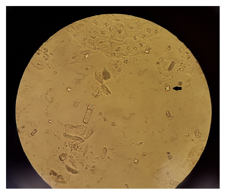
La flecha negra muestra un cristal de oxalato de calcio en forma de sobre o mancuerna bajo el microscopio
Imagen: “Ethylene Glycol Poisoning: An Unusual Cause of Altered Mental Status and the Lessons Learned from Management of the Disease in the Acute Setting” por Case Reports in Critical Care. Licencia: CC BY 4.0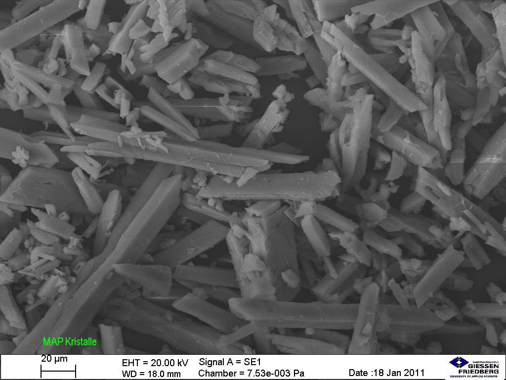
Cristales de cálculos de estruvita bajo el microscopio
Imagen: “Struvite under the microscope (5534899028)” por Sustainable Sanitation Alliance. Licencia: CC BY 2.0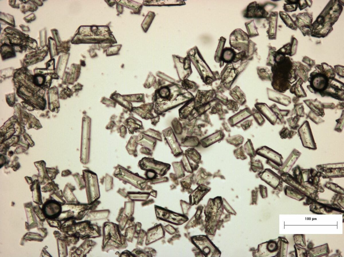
Cristales de estruvita y sus estructuras específicas al microscopio..
Imagen: “Struvite under the microscope (5267841405)” por SuSanA Secretariat. Licencia: CC BY 2.0El material normalmente soluble sobresatura la orina y da comienzo la formación de cristales.
Factores de riesgo:
| Tipo de cálculo | % | Causas | Cristales | pH pH The quantitative measurement of the acidity or basicity of a solution. Acid-Base Balance urinario |
|---|---|---|---|---|
| Oxalato de calcio | 75% |
|
En EN Erythema nodosum is an immune-mediated panniculitis (inflammation of the subcutaneous fat) caused by a type IV (delayed-type) hypersensitivity reaction. It commonly manifests in young women as tender, erythematous nodules on the shins. Erythema Nodosum forma de sobre o mancuerna | ↓ |
| Ácido úrico | 10% |
|
Forma romboidal o de roseta | ↓ |
| Estruvita (fosfato de amonio y magnesio) | 5%–10% | ITU con bacterias ureasa-positivas | Con forma de tapa de ataúd | ↑ |
| Fosfato de calcio | 5% | Aumento del pH pH The quantitative measurement of the acidity or basicity of a solution. Acid-Base Balance urinario | Prisma en EN Erythema nodosum is an immune-mediated panniculitis (inflammation of the subcutaneous fat) caused by a type IV (delayed-type) hypersensitivity reaction. It commonly manifests in young women as tender, erythematous nodules on the shins. Erythema Nodosum forma de cuña | ↑ |
| Cistina | 5% | Cistinuria | Hexagonal | ↓ |
Imagenología:
Estudios de laboratorio:
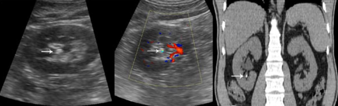
Izquierda: La flecha blanca señala un cálculo renal en el ultrasonido.
Derecha: La flecha blanca señala un cálculo renal en la TC.
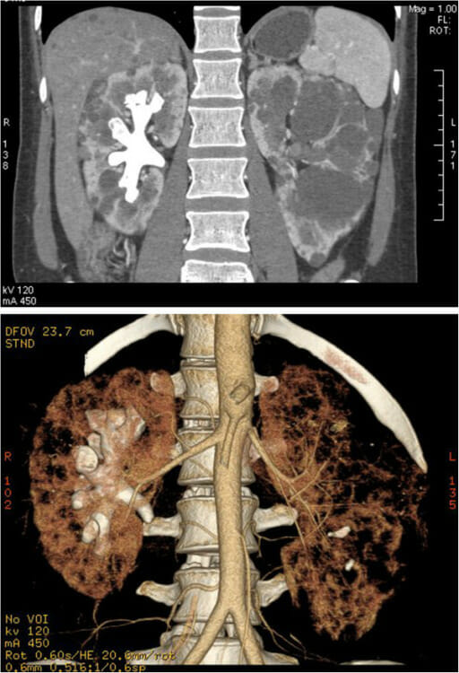
Imágenes de TC de un cálculo coraliforme en el riñón derecho de una mujer con poliquistosis renal autosómica dominante:
Arriba: cálculo coraliforme y riñón poliquístico en el plano coronal
Abajo: Imagen en 3D del cálculo y el riñón reconstruidos
Cuidados generales de soporte:
Tratamiento específico del cálculo: