El infarto de miocardio es la isquemia y muerte de un área de tejido miocárdico debido a un flujo sanguíneo y oxigenación insuficientes, generalmente debido a la formación de trombos en EN Erythema nodosum is an immune-mediated panniculitis (inflammation of the subcutaneous fat) caused by a type IV (delayed-type) hypersensitivity reaction. It commonly manifests in young women as tender, erythematous nodules on the shins. Erythema Nodosum una placa aterosclerótica rota en EN Erythema nodosum is an immune-mediated panniculitis (inflammation of the subcutaneous fat) caused by a type IV (delayed-type) hypersensitivity reaction. It commonly manifests in young women as tender, erythematous nodules on the shins. Erythema Nodosum las arterias epicárdicas. La presentación clínica es más comúnmente con dolor Dolor Inflammation torácico, pero las mujeres y los LOS Neisseria pacientes con diabetes Diabetes Diabetes mellitus (DM) is a metabolic disease characterized by hyperglycemia and dysfunction of the regulation of glucose metabolism by insulin. Type 1 DM is diagnosed mostly in children and young adults as the result of autoimmune destruction of β cells in the pancreas and the resulting lack of insulin. Type 2 DM has a significant association with obesity and is characterized by insulin resistance. Diabetes Mellitus pueden tener síntomas atípicos. El diagnóstico se basa en EN Erythema nodosum is an immune-mediated panniculitis (inflammation of the subcutaneous fat) caused by a type IV (delayed-type) hypersensitivity reaction. It commonly manifests in young women as tender, erythematous nodules on the shins. Erythema Nodosum los LOS Neisseria antecedentes clínicos, cambios en EN Erythema nodosum is an immune-mediated panniculitis (inflammation of the subcutaneous fat) caused by a type IV (delayed-type) hypersensitivity reaction. It commonly manifests in young women as tender, erythematous nodules on the shins. Erythema Nodosum el electrocardiograma ( ECG ECG An electrocardiogram (ECG) is a graphic representation of the electrical activity of the heart plotted against time. Adhesive electrodes are affixed to the skin surface allowing measurement of cardiac impulses from many angles. The ECG provides 3-dimensional information about the conduction system of the heart, the myocardium, and other cardiac structures. Electrocardiogram (ECG)), aumento de las enzimas cardíacas y la evidencia de anomalías en EN Erythema nodosum is an immune-mediated panniculitis (inflammation of the subcutaneous fat) caused by a type IV (delayed-type) hypersensitivity reaction. It commonly manifests in young women as tender, erythematous nodules on the shins. Erythema Nodosum el movimiento de la pared en EN Erythema nodosum is an immune-mediated panniculitis (inflammation of the subcutaneous fat) caused by a type IV (delayed-type) hypersensitivity reaction. It commonly manifests in young women as tender, erythematous nodules on the shins. Erythema Nodosum la imagenología. El tratamiento depende del momento de la presentación y de los LOS Neisseria recursos locales con respecto a la terapia trombolítica versus la intervención percutánea. Todos los LOS Neisseria pacientes reciben nitratos, control del dolor Dolor Inflammation, aspirina, anticoagulantes y betabloqueadores (a menos que estén contraindicados).
Last updated: Jan 18, 2026
El infarto de miocardio (IM), comúnmente conocido como “ataque cardíaco”, se define como una lesión miocárdica aguda y la muerte del tejido como resultado de la isquemia.
Los LOS Neisseria riesgos de IM aumentan proporcionalmente con el aumento de los LOS Neisseria factores de riesgo de aterosclerosis coronaria (también conocida como coronariopatía).
Clasificación del IM según la causa supuesta:
Clasificación del IM según los LOS Neisseria hallazgos del ECG ECG An electrocardiogram (ECG) is a graphic representation of the electrical activity of the heart plotted against time. Adhesive electrodes are affixed to the skin surface allowing measurement of cardiac impulses from many angles. The ECG provides 3-dimensional information about the conduction system of the heart, the myocardium, and other cardiac structures. Electrocardiogram (ECG) y la patología:
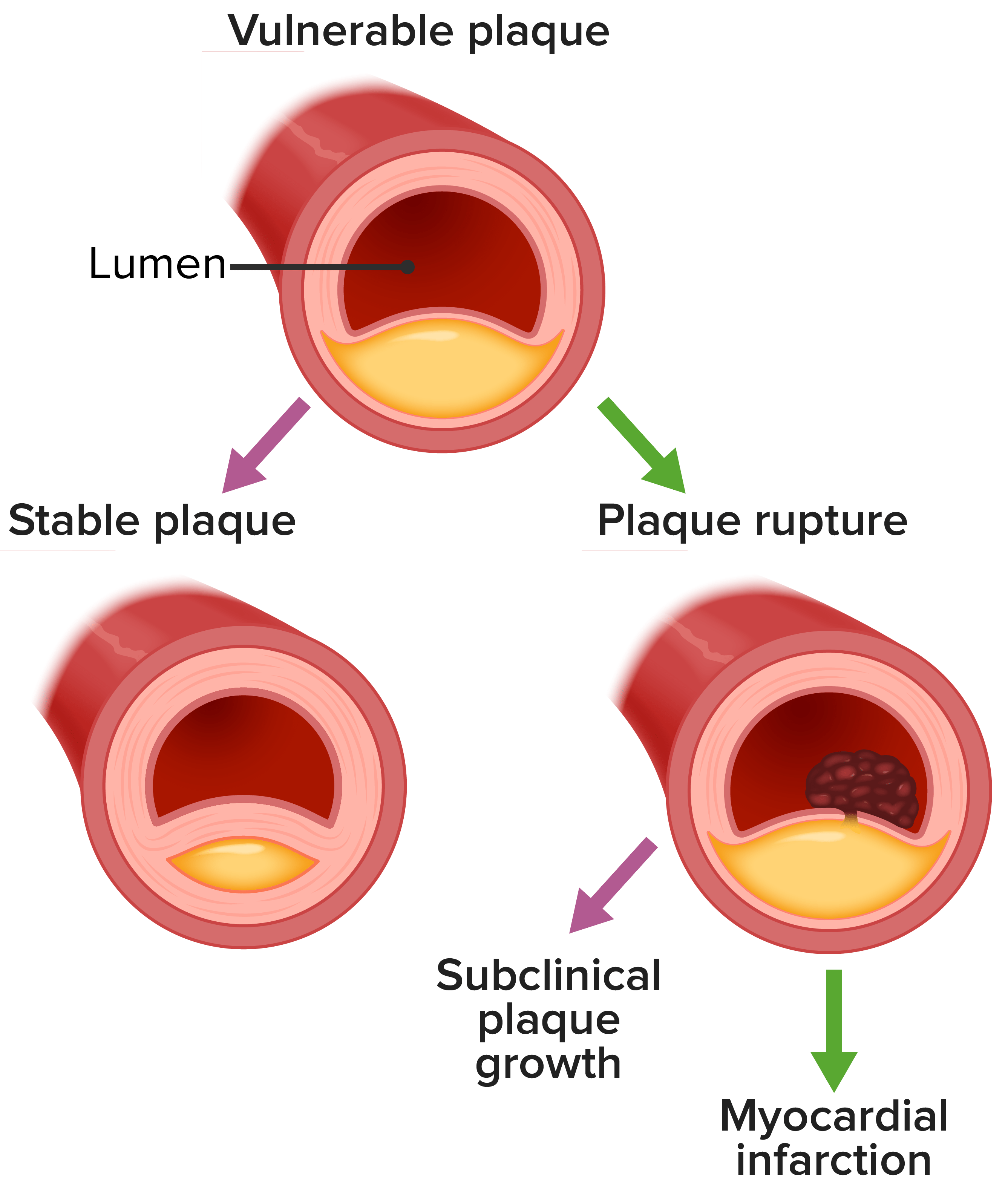
Historia natural de la placa vulnerable/inestable:
Se cree que las placas ateroscleróticas inestables representan la mayoría de los IM. Se caracterizan por inflamación por macrófagos, una cubierta fibrosa delgada, remodelación, microcalcificación y angiogénesis.
El síntoma clásico de IM en EN Erythema nodosum is an immune-mediated panniculitis (inflammation of the subcutaneous fat) caused by a type IV (delayed-type) hypersensitivity reaction. It commonly manifests in young women as tender, erythematous nodules on the shins. Erythema Nodosum la mayoría de los LOS Neisseria pacientes es el dolor Dolor Inflammation torácico agudo. Sin embargo, algunos pacientes pueden presentar síntomas más vagos.
Los LOS Neisseria pacientes que presentan antecedentes de dolor Dolor Inflammation torácico agudo o síntomas atípicos sospechosos se evalúan con ECG ECG An electrocardiogram (ECG) is a graphic representation of the electrical activity of the heart plotted against time. Adhesive electrodes are affixed to the skin surface allowing measurement of cardiac impulses from many angles. The ECG provides 3-dimensional information about the conduction system of the heart, the myocardium, and other cardiac structures. Electrocardiogram (ECG). Los LOS Neisseria niveles de troponina cardíaca deben obtenerse dentro de los LOS Neisseria 10 minutos posteriores a la llegada del paciente al AL Amyloidosis servicio de urgencias. Las anomalías en EN Erythema nodosum is an immune-mediated panniculitis (inflammation of the subcutaneous fat) caused by a type IV (delayed-type) hypersensitivity reaction. It commonly manifests in young women as tender, erythematous nodules on the shins. Erythema Nodosum ambos confirman el diagnóstico de IM.
| Arteria ocluida | Derivaciones con elevación ST | Localización del IM |
|---|---|---|
| LAD proximal | V1–V2 | Septal |
| LAD | V3–V4 | Anterior |
| LAD distal | V5–V6 | Apical |
| LCX o LAD | I, aVL | Lateral |
| RCA (más común) o LCX | II, III, aVF | Inferior |
| RCA o LCX | V7–V9 (depresiones ST en EN Erythema nodosum is an immune-mediated panniculitis (inflammation of the subcutaneous fat) caused by a type IV (delayed-type) hypersensitivity reaction. It commonly manifests in young women as tender, erythematous nodules on the shins. Erythema Nodosum V1–V3) | Posterolateral |
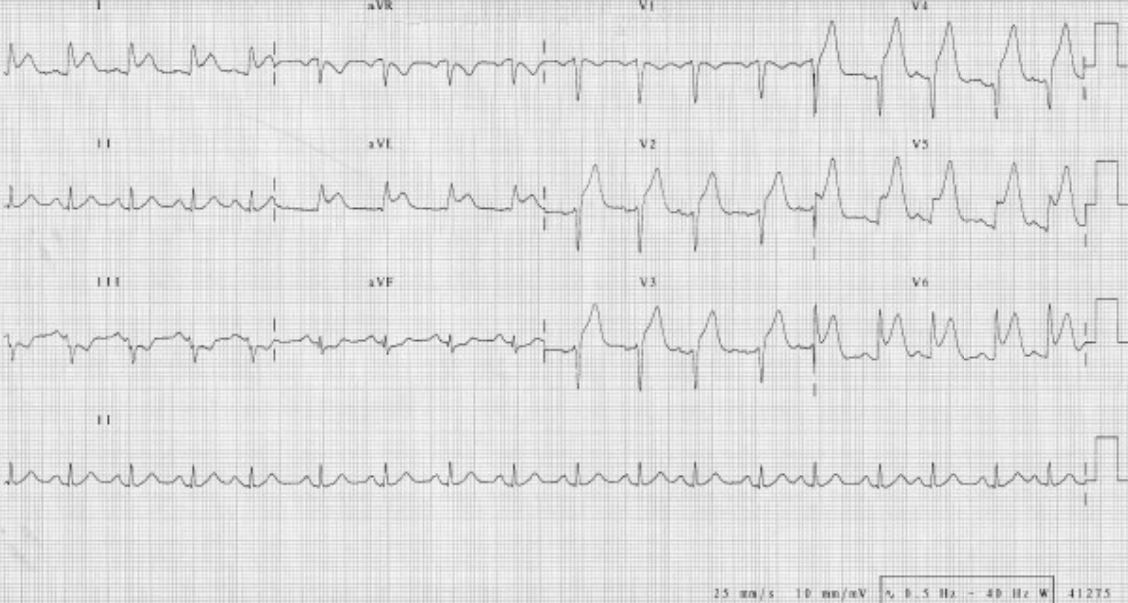
ECG que muestra un IM extendido de la pared anterior con elevaciones del segmento ST en V2–V6, I y aVL:
Obsérvense también las depresiones recíprocas del ST en III y aVF. La angiografía coronaria de este paciente mostró una oclusión total de la arteria descendente anterior izquierda.
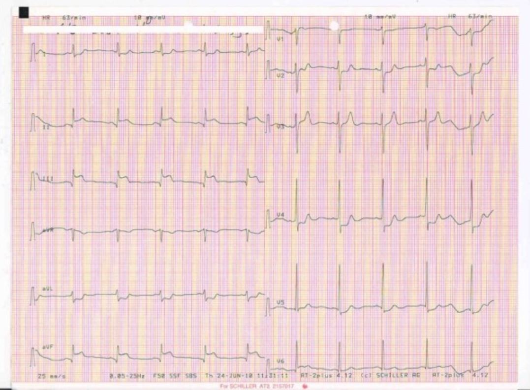
ECG que muestra un IM de la pared inferior con elevaciones del ST en las derivaciones II, III, aVF
Imagen: “The patient’s initial ECG, showing an acute inferior myocardial infarction” por Sogut O, Kaya H, Gokdemir MT, Sezen Y. Licencia: CC BY 2.0Enzimas cardíacas:
Laboratorios de soporte:
La siguiente tabla compara la angina inestable, IAMSEST e IAMCEST sobre la base de las características clínicas, ECG ECG An electrocardiogram (ECG) is a graphic representation of the electrical activity of the heart plotted against time. Adhesive electrodes are affixed to the skin surface allowing measurement of cardiac impulses from many angles. The ECG provides 3-dimensional information about the conduction system of the heart, the myocardium, and other cardiac structures. Electrocardiogram (ECG) y los LOS Neisseria hallazgos de laboratorio.
| Diagnóstico | Características clínicas | Hallazgos del ECG ECG An electrocardiogram (ECG) is a graphic representation of the electrical activity of the heart plotted against time. Adhesive electrodes are affixed to the skin surface allowing measurement of cardiac impulses from many angles. The ECG provides 3-dimensional information about the conduction system of the heart, the myocardium, and other cardiac structures. Electrocardiogram (ECG) | Hallazgos de laboratorio |
|---|---|---|---|
| Angina inestable | Dolor Dolor Inflammation torácico isquémico que ocurre en EN Erythema nodosum is an immune-mediated panniculitis (inflammation of the subcutaneous fat) caused by a type IV (delayed-type) hypersensitivity reaction. It commonly manifests in young women as tender, erythematous nodules on the shins. Erythema Nodosum reposo o con niveles de esfuerzo previamente tolerados |
|
Troponina normal |
| IAMSEST | Dolor Dolor Inflammation torácico isquémico prolongado en EN Erythema nodosum is an immune-mediated panniculitis (inflammation of the subcutaneous fat) caused by a type IV (delayed-type) hypersensitivity reaction. It commonly manifests in young women as tender, erythematous nodules on the shins. Erythema Nodosum cualquier situación |
|
Troponina elevada |
| IAMCEST | Dolor Dolor Inflammation torácico isquémico prolongado en EN Erythema nodosum is an immune-mediated panniculitis (inflammation of the subcutaneous fat) caused by a type IV (delayed-type) hypersensitivity reaction. It commonly manifests in young women as tender, erythematous nodules on the shins. Erythema Nodosum cualquier situación |
|
Troponina elevada |
El reconocimiento rápido del diagnóstico de IM agudo es imperativo para obtener el beneficio de la terapia de reperfusión.
Se observa un mejor pronóstico a largo plazo con:
Después de un IM, se desarrollan diferentes riesgos a medida que pasa el tiempo después del evento agudo. Los LOS Neisseria pacientes con IM tipo 2 tienen una mayor prevalencia de insuficiencia cardíaca, enfermedad renal como complicación del IM y fibrilación auricular.