La hemorragia subdural es un evento hemorrágico en el espacio meníngeo entre las capas duramadre y aracnoides, las cuales rodean el cerebro. El mecanismo más común que desencadena el episodio hemorrágico es un traumatismo (e.g., traumatismo craneoencefálico cerrado) que provoca un desgarro en las venas "comunicantes" extracerebrales, pero la rotura de pequeñas arterias dentro de este espacio o la hipotensión intracraneal también pueden ser una de las causas. La hemorragia subdural aguda se presenta inmediatamente después de un traumatismo craneoencefálico, con un nivel alterado de conciencia que puede abarcar desde una pérdida momentánea de conciencia hasta el comaComaComa is defined as a deep state of unarousable unresponsiveness, characterized by a score of 3 points on the GCS. A comatose state can be caused by a multitude of conditions, making the precise epidemiology and prognosis of coma difficult to determine. Coma, lo que la convierte enENErythema nodosum is an immune-mediated panniculitis (inflammation of the subcutaneous fat) caused by a type IV (delayed-type) hypersensitivity reaction. It commonly manifests in young women as tender, erythematous nodules on the shins.Erythema Nodosum una afección potencialmente mortal. También puede ocurrir una hemorragia subdural crónica, que se presenta con un deterioro neurológico gradual. El diagnóstico se basa enENErythema nodosum is an immune-mediated panniculitis (inflammation of the subcutaneous fat) caused by a type IV (delayed-type) hypersensitivity reaction. It commonly manifests in young women as tender, erythematous nodules on the shins.Erythema Nodosum la sospecha clínica posterior a un traumatismo craneoencefálico y se confirma mediante neuroimagenología (e.g., TC de la cabeza sin contraste). El tratamiento incluye estabilización del paciente, suspensión (posiblemente reversión) de todos losLOSNeisseria anticoagulantes, monitorización enENErythema nodosum is an immune-mediated panniculitis (inflammation of the subcutaneous fat) caused by a type IV (delayed-type) hypersensitivity reaction. It commonly manifests in young women as tender, erythematous nodules on the shins.Erythema Nodosum una UCI neurológica e intervención neuroquirúrgica.
El hematomaHematomaA collection of blood outside the blood vessels. Hematoma can be localized in an organ, space, or tissue.Intussusception subdural es una hemorragia, generalmente causada por un traumatismo craneal enENErythema nodosum is an immune-mediated panniculitis (inflammation of the subcutaneous fat) caused by a type IV (delayed-type) hypersensitivity reaction. It commonly manifests in young women as tender, erythematous nodules on the shins.Erythema Nodosum el espacio meníngeo, entre las capas duramadre y aracnoides las cuales rodean el cerebro, creando así un espacio llamado espacio subdural.
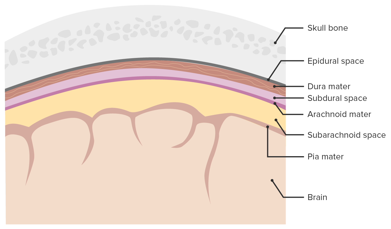
Meninges y espacios meníngeos:
Imagen que muestra las 3 capas (duramadre, aracnoides, piamadre) que rodean el cerebro y la médula espinal. Las meninges sirven como protección mecánica del sistema nervioso central (SNC), sostienen los vasos sanguíneos cerebrales y espinales y permiten el paso del líquido cefalorraquídeo (LCR). Solo el espacio subaracnoideo es un verdadero espacio presente en condiciones fisiológicas, mientras que los espacios epidurales y subdurales se forman solamente durante procesos patológicos. El espacio epidural puede abrirse como resultado de un traumatismo craneoencefálico o en raras ocasiones debido a otros procesos patológicos.
Imagen por Lecturio.
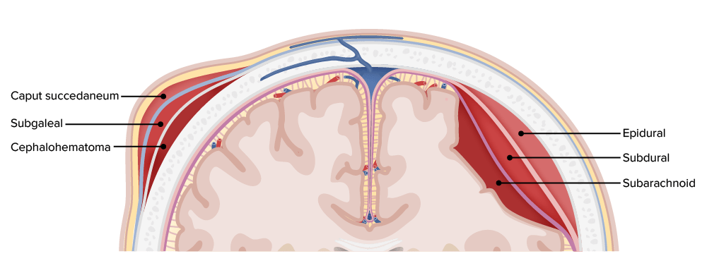
Hemorragias según localización en las diferentes capas de las meninges y del cuero cabelludo
Imagen por Lecturio.
Epidemiología
Prevalencia:
Se encuentra enENErythema nodosum is an immune-mediated panniculitis (inflammation of the subcutaneous fat) caused by a type IV (delayed-type) hypersensitivity reaction. It commonly manifests in young women as tender, erythematous nodules on the shins.Erythema Nodosum aproximadamente el 10% de losLOSNeisseria casos de traumatismo craneoencefálico que requieren hospitalización.
Se encuentra enENErythema nodosum is an immune-mediated panniculitis (inflammation of the subcutaneous fat) caused by a type IV (delayed-type) hypersensitivity reaction. It commonly manifests in young women as tender, erythematous nodules on the shins.Erythema Nodosum aproximadamente el 20% de losLOSNeisseria casos de lesiones cerebrales traumáticas graves.
Evidenciado enENErythema nodosum is an immune-mediated panniculitis (inflammation of the subcutaneous fat) caused by a type IV (delayed-type) hypersensitivity reaction. It commonly manifests in young women as tender, erythematous nodules on the shins.Erythema Nodosum 8% de losLOSNeisseria partos a término (asociado con el uso de fórceps o extracción con ventosa)
Más común enENErythema nodosum is an immune-mediated panniculitis (inflammation of the subcutaneous fat) caused by a type IV (delayed-type) hypersensitivity reaction. It commonly manifests in young women as tender, erythematous nodules on the shins.Erythema Nodosum personas mayores
Más común enENErythema nodosum is an immune-mediated panniculitis (inflammation of the subcutaneous fat) caused by a type IV (delayed-type) hypersensitivity reaction. It commonly manifests in young women as tender, erythematous nodules on the shins.Erythema Nodosum personas que reciben terapias antiplaquetarias/anticoagulantes
Etiología
Traumatismo enENErythema nodosum is an immune-mediated panniculitis (inflammation of the subcutaneous fat) caused by a type IV (delayed-type) hypersensitivity reaction. It commonly manifests in young women as tender, erythematous nodules on the shins.Erythema Nodosum la cabeza:
Causa más común de hemorragia subdural
Provoca lesiones enENErythema nodosum is an immune-mediated panniculitis (inflammation of the subcutaneous fat) caused by a type IV (delayed-type) hypersensitivity reaction. It commonly manifests in young women as tender, erythematous nodules on the shins.Erythema Nodosum las estructuras vasculares que discurren entre las capas meníngeas, duramadre y aracnoides, que rodean el cerebro.
Más comúnmente ejerce fuerzas enENErythema nodosum is an immune-mediated panniculitis (inflammation of the subcutaneous fat) caused by a type IV (delayed-type) hypersensitivity reaction. It commonly manifests in young women as tender, erythematous nodules on the shins.Erythema Nodosum dirección anteroposterior → lesión de las venas puente hacia el seno sagital superior
Ejemplos:
Accidentes vehiculares
Caídas
Asaltos
Anticoagulación o coagulopatías:
Aumentan el riesgo de sangrado prolongado o excesivo.
Ejemplos (medicamentos):
Agentes antiplaquetarios:
Aspirina
ClopidogrelClopidogrelA ticlopidine analog and platelet purinergic p2y receptor antagonist that inhibits adenosine diphosphate-mediated platelet aggregation. It is used to prevent thromboembolism in patients with arterial occlusive diseases; myocardial infarction; stroke; or atrial fibrillation.Antiplatelet Drugs
PrasugrelPrasugrelA piperazine derivative and platelet aggregation inhibitor that is used to prevent thrombosis in patients with acute coronary syndrome; unstable angina and myocardial infarction, as well as in those undergoing percutaneous coronary interventions.Antiplatelet Drugs
Antagonistas de la vitamina K: warfarina
Inhibidores del factor XFactor XStorage-stable glycoprotein blood coagulation factor that can be activated to factor Xa by both the intrinsic and extrinsic pathways. A deficiency of factor X, sometimes called stuart-prower factor deficiency, may lead to a systemic coagulation disorder.Hemostasis:
Rivaroxabán
Apixabán
Edoxabán
Heparinoides:
Heparina no fraccionada (HNF)
Heparina de bajo peso molecular (HBPM)
Trombolíticos:
Activador tisular del plasminógeno
Uroquinasa
Ejemplos (estados patológicos):
Enfermedad crónica del hígado
Trombocitopenia
Hemofilia
Atrofia cerebral:
Predispone a lesiones vasculares alALAmyloidosis permitir un movimiento excesivo dentro de la bóveda craneal enENErythema nodosum is an immune-mediated panniculitis (inflammation of the subcutaneous fat) caused by a type IV (delayed-type) hypersensitivity reaction. It commonly manifests in young women as tender, erythematous nodules on the shins.Erythema Nodosum caso de traumatismo.
Principal causa de hemorragia subdural crónica
Ejemplos:
Traumatismo craneoencefálico previo
Accidente cerebrovascular previo con necrosisNecrosisThe death of cells in an organ or tissue due to disease, injury or failure of the blood supply.Ischemic Cell Damage parenquimatosa
Alcoholismo crónico
Hemorragia intracerebral:
Extensión directa de una hemorragia intraparenquimatosa a través de la superficie cortical hacia el espacio subdural
Es más probable que ocurra enENErythema nodosum is an immune-mediated panniculitis (inflammation of the subcutaneous fat) caused by a type IV (delayed-type) hypersensitivity reaction. It commonly manifests in young women as tender, erythematous nodules on the shins.Erythema Nodosum ausencia de un traumatismo
Ejemplos:
Hemorragia hipertensiva intraparenquimatosa
Conversión a hemorragia intraparenquimatosa de accidente cerebrovascular isquémico
Ruptura de aneurisma de la vasculatura cerebral:
Extensión directa de una hemorragia intracerebral a través del espacio subaracnoideo y hacia el espacio subdural
Es más probable que ocurra enENErythema nodosum is an immune-mediated panniculitis (inflammation of the subcutaneous fat) caused by a type IV (delayed-type) hypersensitivity reaction. It commonly manifests in young women as tender, erythematous nodules on the shins.Erythema Nodosum ausencia de un traumatismo
Ejemplos:
Hemorragia subaracnoidea (generalmente, es el resultado de la ruptura de un aneurisma sacular)
Aneurisma de la arteria carótida (o una rama de la misma)
Malformaciones de la vasculatura cerebral:
Extensión directa desde el sitio de la hemorragia hacia el espacio subdural.
Es más probable que ocurra enENErythema nodosum is an immune-mediated panniculitis (inflammation of the subcutaneous fat) caused by a type IV (delayed-type) hypersensitivity reaction. It commonly manifests in young women as tender, erythematous nodules on the shins.Erythema Nodosum ausencia de un traumatismo
LosLOSNeisseria tumores primarios o metastásicos que afectan a la duramadre pueden causar hemorragias enENErythema nodosum is an immune-mediated panniculitis (inflammation of the subcutaneous fat) caused by a type IV (delayed-type) hypersensitivity reaction. It commonly manifests in young women as tender, erythematous nodules on the shins.Erythema Nodosum el espacio subdural.
Es más probable que ocurra enENErythema nodosum is an immune-mediated panniculitis (inflammation of the subcutaneous fat) caused by a type IV (delayed-type) hypersensitivity reaction. It commonly manifests in young women as tender, erythematous nodules on the shins.Erythema Nodosum ausencia de un traumatismo
Ejemplos:
MeningiomaMeningiomaMeningiomas are slow-growing tumors that arise from the meninges of the brain and spinal cord. The vast majority are benign. These tumors commonly occur in individuals with a history of high doses of skull radiation, head trauma, and neurofibromatosis 2. Meningioma (primario)
Cáncer de mama (metastásico)
Cáncer de pulmón (metastásico)
Cáncer de próstata (metastásico)
Hipotensión intracraneal:
Un volumen inadecuado de LCR puede crear un efecto de vacío dentro de la bóveda craneal → es transmitido a las capas meníngeas → predispone alALAmyloidosis desgarro de las venas comunicantes
Puede ocurrir con o sin traumatismo
Ejemplos:
Fuga dural después de procedimientos epidurales
Inyección epidural de esteroides
Anestesia epidural antes del parto fetal
Pérdida de LCR por traumatismo
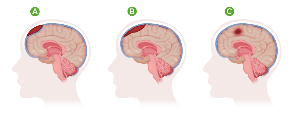
Tipos de hematomas
A) Hematoma epidural;
B) Hematoma subdural;
C)Hematoma intracraneal
HematomaHematomaA collection of blood outside the blood vessels. Hematoma can be localized in an organ, space, or tissue.Intussusception subdural agudo
Traumatismo que conduce alALAmyloidosis desgarro de las venas comunicantes:
Las venas comunicantes drenan sangre desde la superficie cerebral hacia losLOSNeisseria senos durales.
Las venas comunicantes atraviesan el espacio meníngeo entre las capas duramadre y aracnoides
El desgarro permite que la sangre se acumule entre estas capas.
El sangrado generalmente se bloquea por el aumento de la presión intracraneal (PIC) o mediante la compresión directa por el trombo enENErythema nodosum is an immune-mediated panniculitis (inflammation of the subcutaneous fat) caused by a type IV (delayed-type) hypersensitivity reaction. It commonly manifests in young women as tender, erythematous nodules on the shins.Erythema Nodosum formación.
Se observa con mayor frecuencia enENErythema nodosum is an immune-mediated panniculitis (inflammation of the subcutaneous fat) caused by a type IV (delayed-type) hypersensitivity reaction. It commonly manifests in young women as tender, erythematous nodules on the shins.Erythema Nodosum la región temporoparietal.
Traumatismo que conduce a una ruptura arterial:
Las arterias pequeñas (< 1 mm de diámetro) llevan a cabo la irrigación sanguínea de la corteza cerebral superficial.
Estas arterias atraviesan el espacio meníngeo entre las capas duramadre y aracnoides.
La ruptura permite que la sangre se acumule entre estas capas.
El sangrado se bloquea típicamente por el aumento de la PIC o mediante compresión directa por el trombo enENErythema nodosum is an immune-mediated panniculitis (inflammation of the subcutaneous fat) caused by a type IV (delayed-type) hypersensitivity reaction. It commonly manifests in young women as tender, erythematous nodules on the shins.Erythema Nodosum formación.
Se observa con mayor frecuencia enENErythema nodosum is an immune-mediated panniculitis (inflammation of the subcutaneous fat) caused by a type IV (delayed-type) hypersensitivity reaction. It commonly manifests in young women as tender, erythematous nodules on the shins.Erythema Nodosum la región temporoparietal.
Hipotensión intracraneal (presión baja del LCR):
Causada por un volumen bajo de LCR, generalmente por una fuga:
Espontánea (observado enENErythema nodosum is an immune-mediated panniculitis (inflammation of the subcutaneous fat) caused by a type IV (delayed-type) hypersensitivity reaction. It commonly manifests in young women as tender, erythematous nodules on the shins.Erythema Nodosum trastornos del tejido conectivo, como el síndrome de Ehlers-Danlos o Marfan)
La presión baja del LCR disminuye la flotabilidad del cerebro → tracción sobre las estructuras meníngeas de soporte
La tracción se transmite hacia las venas puente / arterias corticales pequeñas → causando desgarro/ruptura de estos vasos
La PIC baja produce un efecto de vacío, lo que provoca vasodilatación y una mayor predisposición a hemorragias.
HematomaHematomaA collection of blood outside the blood vessels. Hematoma can be localized in an organ, space, or tissue.Intussusception subdural crónico
Es el resultado de una hematomaHematomaA collection of blood outside the blood vessels. Hematoma can be localized in an organ, space, or tissue.Intussusception subdural agudo que se haHAHemolytic anemia (HA) is the term given to a large group of anemias that are caused by the premature destruction/hemolysis of circulating red blood cells (RBCs). Hemolysis can occur within (intravascular hemolysis) or outside the blood vessels (extravascular hemolysis).Hemolytic Anemia trombosado:
LosLOSNeisseria fibroblastos elaboran colágeno sobre la capa dural, estabilizando la superficie exterior del trombo.
Se desarrolla una membrana más delgada sobre la superficie interna del coágulo → encapsulación completa
El proceso tarda aproximadamente 2 semanas
Licuefacción del trombo:
EnENErythema nodosum is an immune-mediated panniculitis (inflammation of the subcutaneous fat) caused by a type IV (delayed-type) hypersensitivity reaction. It commonly manifests in young women as tender, erythematous nodules on the shins.Erythema Nodosum > 50% de losLOSNeisseria casos de hematomaHematomaA collection of blood outside the blood vessels. Hematoma can be localized in an organ, space, or tissue.Intussusception subdural agudo, las membranas mencionadas anteriormente se calcifican, mientras que el trombo contenido enENErythema nodosum is an immune-mediated panniculitis (inflammation of the subcutaneous fat) caused by a type IV (delayed-type) hypersensitivity reaction. It commonly manifests in young women as tender, erythematous nodules on the shins.Erythema Nodosum ellas sufre licuefacción enENErythema nodosum is an immune-mediated panniculitis (inflammation of the subcutaneous fat) caused by a type IV (delayed-type) hypersensitivity reaction. It commonly manifests in young women as tender, erythematous nodules on the shins.Erythema Nodosum un higroma (saco lleno de líquido).
El higroma es rico enENErythema nodosum is an immune-mediated panniculitis (inflammation of the subcutaneous fat) caused by a type IV (delayed-type) hypersensitivity reaction. It commonly manifests in young women as tender, erythematous nodules on the shins.Erythema Nodosum proteínas → potencial atracción osmótica de líquido hacia la cavidad y expansión del higroma
HematomaHematomaA collection of blood outside the blood vessels. Hematoma can be localized in an organ, space, or tissue.Intussusception subdural crónico enENErythema nodosum is an immune-mediated panniculitis (inflammation of the subcutaneous fat) caused by a type IV (delayed-type) hypersensitivity reaction. It commonly manifests in young women as tender, erythematous nodules on the shins.Erythema Nodosum fase aguda
Traumatismos recurrentes pueden causar hemorragias y estas convertirse enENErythema nodosum is an immune-mediated panniculitis (inflammation of the subcutaneous fat) caused by a type IV (delayed-type) hypersensitivity reaction. It commonly manifests in young women as tender, erythematous nodules on the shins.Erythema NodosumhematomaHematomaA collection of blood outside the blood vessels. Hematoma can be localized in an organ, space, or tissue.Intussusception subdural o enENErythema nodosum is an immune-mediated panniculitis (inflammation of the subcutaneous fat) caused by a type IV (delayed-type) hypersensitivity reaction. It commonly manifests in young women as tender, erythematous nodules on the shins.Erythema Nodosum un higroma estable → agrandamiento y otras patologías intracraneales enENErythema nodosum is an immune-mediated panniculitis (inflammation of the subcutaneous fat) caused by a type IV (delayed-type) hypersensitivity reaction. It commonly manifests in young women as tender, erythematous nodules on the shins.Erythema Nodosum un futuro
Expansión de un higroma debido a fuerzas osmóticas → agrandamiento y otras patologías intracraneales enENErythema nodosum is an immune-mediated panniculitis (inflammation of the subcutaneous fat) caused by a type IV (delayed-type) hypersensitivity reaction. It commonly manifests in young women as tender, erythematous nodules on the shins.Erythema Nodosum un futuro
Presentación Clínica
El traumatismo craneoencefálico es la etiología más común de la hemorragia subdural, con mayor frecuencia traumatismos menores (e.g., caída alALAmyloidosis nivel del suelo) enENErythema nodosum is an immune-mediated panniculitis (inflammation of the subcutaneous fat) caused by a type IV (delayed-type) hypersensitivity reaction. It commonly manifests in young women as tender, erythematous nodules on the shins.Erythema NodosumlosLOSNeisseria ancianos.
Se presentan inmediatamente: hasta 72 horas después del evento
Presentación inicial: comaComaComa is defined as a deep state of unarousable unresponsiveness, characterized by a score of 3 points on the GCS. A comatose state can be caused by a multitude of conditions, making the precise epidemiology and prognosis of coma difficult to determine. ComaenENErythema nodosum is an immune-mediated panniculitis (inflammation of the subcutaneous fat) caused by a type IV (delayed-type) hypersensitivity reaction. It commonly manifests in young women as tender, erythematous nodules on the shins.Erythema Nodosum ½ de losLOSNeisseria casos
El resto puede tener un “intervalo lúcido” entre la lesión y el inicio del deterioro neurológico progresivo.
La hemorragia subdural subaguda se presenta de 3–14 días después del evento.
La hemorragia subdural crónica se presenta ≥ 15 días después del evento.
EnENErythema nodosum is an immune-mediated panniculitis (inflammation of the subcutaneous fat) caused by a type IV (delayed-type) hypersensitivity reaction. It commonly manifests in young women as tender, erythematous nodules on the shins.Erythema Nodosum ausencia de la evidencia de un traumatismo, las hemorragias subdurales pueden ser difíciles de clasificar.
Síntomas neurológicos
La naturaleza de losLOSNeisseria síntomas/signos neurológicos depende enENErythema nodosum is an immune-mediated panniculitis (inflammation of the subcutaneous fat) caused by a type IV (delayed-type) hypersensitivity reaction. It commonly manifests in young women as tender, erythematous nodules on the shins.Erythema Nodosum gran medida de las siguientes características del hematomaHematomaA collection of blood outside the blood vessels. Hematoma can be localized in an organ, space, or tissue.Intussusception:
Localización
Tamaño
Ritmo de crecimiento
Agudeza
Síntomas comunes:
Nivel alterado de conciencia
Un traumatismo menor puede provocar solo una pérdida momentánea del conocimiento.
Las víctimas de traumatismos graves con hemorragia subdural pueden presentarse enENErythema nodosum is an immune-mediated panniculitis (inflammation of the subcutaneous fat) caused by a type IV (delayed-type) hypersensitivity reaction. It commonly manifests in young women as tender, erythematous nodules on the shins.Erythema NodosumcomaComaComa is defined as a deep state of unarousable unresponsiveness, characterized by a score of 3 points on the GCS. A comatose state can be caused by a multitude of conditions, making the precise epidemiology and prognosis of coma difficult to determine. Coma.
La hemorragia subdural subaguda o crónica puede presentarse con un deterioro gradual del nivel de conciencia.
AtaxiaAtaxiaImpairment of the ability to perform smoothly coordinated voluntary movements. This condition may affect the limbs, trunk, eyes, pharynx, larynx, and other structures. Ataxia may result from impaired sensory or motor function. Sensory ataxia may result from posterior column injury or peripheral nerve diseases. Motor ataxia may be associated with cerebellar diseases; cerebral cortex diseases; thalamic diseases; basal ganglia diseases; injury to the red nucleus; and other conditions.Ataxia-telangiectasia
Convulsiones
Adicional a losLOSNeisseria signos y síntomas listados, el hematomaHematomaA collection of blood outside the blood vessels. Hematoma can be localized in an organ, space, or tissue.Intussusception subdural crónico puede presentarse con:
Somnolencia
Depresión
Parkinsonismo (e.g., temblores)
Diagnóstico
Se debe sospechar el diagnóstico de hemorragia subdural enENErythema nodosum is an immune-mediated panniculitis (inflammation of the subcutaneous fat) caused by a type IV (delayed-type) hypersensitivity reaction. It commonly manifests in young women as tender, erythematous nodules on the shins.Erythema Nodosum cualquier persona anciana que presente un traumatismo craneoencefálico, estado mental alterado, disminución del nivel de conciencia o síntomas/signos neurológicos. La TC de la cabeza debe realizarse de forma urgente cuando se sospecha una hemorragia subdural aguda.
Neuroimagenología
TC de cabeza sin contraste:
Método de imagenología de elección:
Para traumatismo craneoencefálico agudo
Para la pérdida aguda del conocimiento
Para la sospecha de hemorragia subdural (y otras hemorragias intracraneales)
La hemorragia subdural aguda aparece como una media luna de alta densidad, colección de sangre a lo largo de la convexidad del hemisferio afectado.
La sangre fresca aparece con una alta intensidad enENErythema nodosum is an immune-mediated panniculitis (inflammation of the subcutaneous fat) caused by a type IV (delayed-type) hypersensitivity reaction. It commonly manifests in young women as tender, erythematous nodules on the shins.Erythema Nodosum la TC.
Fácilmente distinguible de la anatomía circundante
La hemorragia subdural subaguda y crónica aparece como una acumulación de sangre enENErythema nodosum is an immune-mediated panniculitis (inflammation of the subcutaneous fat) caused by a type IV (delayed-type) hypersensitivity reaction. It commonly manifests in young women as tender, erythematous nodules on the shins.Erythema Nodosum media luna isodensa o hipodensa con una deformación asociada de losLOSNeisseria contornos cerebrales.
El hematomaHematomaA collection of blood outside the blood vessels. Hematoma can be localized in an organ, space, or tissue.Intussusception pierde su intensidad a medida que avanza la trombosis y la remodelación / resolución del coágulo.
La colección de sangre enENErythema nodosum is an immune-mediated panniculitis (inflammation of the subcutaneous fat) caused by a type IV (delayed-type) hypersensitivity reaction. It commonly manifests in young women as tender, erythematous nodules on the shins.Erythema Nodosum la fase subaguda/crónica es más difícil de distinguir de la anatomía circundante.
La hemorragia subdural unilateral crea una distorsión evidente de losLOSNeisseria contornos cerebrales.
La hemorragia subdural bilateral puede crear una distorsión simétrica de losLOSNeisseria contornos cerebrales y ser menos evidente.
RM de cabeza:
Secuenciación de recuperación de la inversión atenuada de fluido (FLAIRFLAIRMagnetic Resonance Imaging (MRI), por sus siglas enENErythema nodosum is an immune-mediated panniculitis (inflammation of the subcutaneous fat) caused by a type IV (delayed-type) hypersensitivity reaction. It commonly manifests in young women as tender, erythematous nodules on the shins.Erythema Nodosum inglés)
Menos utilizada y no está tan disponible como la TC
La sensibilidad es superior a la de la TC sin contraste enENErythema nodosum is an immune-mediated panniculitis (inflammation of the subcutaneous fat) caused by a type IV (delayed-type) hypersensitivity reaction. It commonly manifests in young women as tender, erythematous nodules on the shins.Erythema Nodosum la detección de hemorragias intracraneales
La sangre enENErythema nodosum is an immune-mediated panniculitis (inflammation of the subcutaneous fat) caused by a type IV (delayed-type) hypersensitivity reaction. It commonly manifests in young women as tender, erythematous nodules on the shins.Erythema Nodosum la lesión subdural aguda, subaguda y crónica aparece hiperintensa enENErythema nodosum is an immune-mediated panniculitis (inflammation of the subcutaneous fat) caused by a type IV (delayed-type) hypersensitivity reaction. It commonly manifests in young women as tender, erythematous nodules on the shins.Erythema Nodosum el LCR (secuencias de recuperación de la inversión atenuada de fluido, FLAIRFLAIRMagnetic Resonance Imaging (MRI) por sus siglas enENErythema nodosum is an immune-mediated panniculitis (inflammation of the subcutaneous fat) caused by a type IV (delayed-type) hypersensitivity reaction. It commonly manifests in young women as tender, erythematous nodules on the shins.Erythema Nodosum inglés)
Puede detectar pequeñas hemorragias subdurales que pueden pasarse por alto enENErythema nodosum is an immune-mediated panniculitis (inflammation of the subcutaneous fat) caused by a type IV (delayed-type) hypersensitivity reaction. It commonly manifests in young women as tender, erythematous nodules on the shins.Erythema Nodosum la TC sin contraste
Puede detectar lesiones durales (e.g., desgarros durales, neoplasias) pasadas por alto enENErythema nodosum is an immune-mediated panniculitis (inflammation of the subcutaneous fat) caused by a type IV (delayed-type) hypersensitivity reaction. It commonly manifests in young women as tender, erythematous nodules on the shins.Erythema Nodosum la TC sin contraste
Puede revelar la presencia y extensión de las lesiones intraparenquimatosas asociadas
Contraindicado o limitado enENErythema nodosum is an immune-mediated panniculitis (inflammation of the subcutaneous fat) caused by a type IV (delayed-type) hypersensitivity reaction. It commonly manifests in young women as tender, erythematous nodules on the shins.Erythema Nodosum pacientes con implantes metálicos/eléctricos incompatibles con resonancia magnética
Angiografía:
Angiografía no invasiva por RM o por TC:
Puede estar indicada para la evaluación de hemorragia subdural no traumáticas o idiopáticas
Puede revelar pequeños aneurismas intracraneales u otras lesiones vasculares
Se puede considerar la angiografía convencional si se sospecha una lesión vascular, pero no se detecta mediante una angiografía no invasiva.
Procedimientos contraindicados
Punción lumbar:
Contraindicado cuando se sospecha de hemorragia subdural
El aumento de la PIC debido a la expansión del hematomaHematomaA collection of blood outside the blood vessels. Hematoma can be localized in an organ, space, or tissue.Intussusception aumenta el riesgo de una herniación.
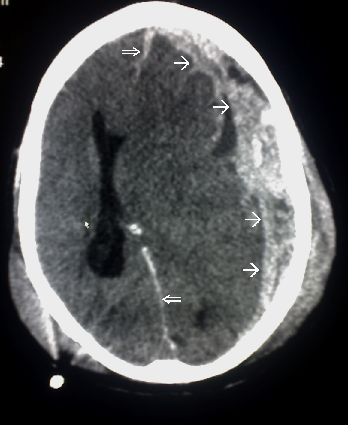
Hemorragia subdural: Se observa la convexidad del hematoma asociado con el desplazamiento de la línea media (con distorsión de la anatomía cerebral y obliteración del ventrículo lateral).
Imagen: “This CT scan is an example of Subdural haemorrhage caused by trauma. Single arrow marked the spread of the subdural haematoma. Double arrow marked the midline shift” por Glitzy queen00. Licencia: Dominio Público
La hemorragia subdural aguda, especialmente la que se presenta con compromiso neurológico o comaComaComa is defined as a deep state of unarousable unresponsiveness, characterized by a score of 3 points on the GCS. A comatose state can be caused by a multitude of conditions, making the precise epidemiology and prognosis of coma difficult to determine. Coma, es una situación neurológica de emergencia que a menudo requiere intervención quirúrgica. Si no se estabiliza, diagnostica, evalúa e interviene con prontitud, se puede producir una expansión hemorrágica, una lesión cerebral parenquimatosa, un aumento de la PIC, una herniaHerniaProtrusion of tissue, structure, or part of an organ through the bone, muscular tissue, or the membrane by which it is normally contained. Hernia may involve tissues such as the abdominal wall or the respiratory diaphragm. Hernias may be internal, external, congenital, or acquired.Abdominal Hernias cerebral y la muerte.
Estabilización
El paciente debe ser evaluado y estabilizado mediante protocolos de soporte vital avanzado para traumatismos/soporte vital cardíaco avanzado (ATLS / ACLS).
Tratar las lesiones potencialmente mortales.
Interrumpir (y posible reversión) inmediata losLOSNeisseria antiagregantes plaquetarios / anticoagulantes
Esfuerzos para lograr/mantener la estabilidad hemodinámica
TC de cabeza sin contraste lo antes posible
Consulta neuroquirúrgica urgente:
Soporte enENErythema nodosum is an immune-mediated panniculitis (inflammation of the subcutaneous fat) caused by a type IV (delayed-type) hypersensitivity reaction. It commonly manifests in young women as tender, erythematous nodules on the shins.Erythema Nodosum la toma de decisiones clínicas quirúrgicas
Colocación de dispositivo para la monitorización de la PIC
Estratificación
Las herramientas de decisión clínica que se utilizan para determinar el manejo operatorio o no operatorio incluyen:
Puntuación de escala de comaComaComa is defined as a deep state of unarousable unresponsiveness, characterized by a score of 3 points on the GCS. A comatose state can be caused by a multitude of conditions, making the precise epidemiology and prognosis of coma difficult to determine. Coma de Glasgow (GCSGCSA scale that assesses the response to stimuli in patients with craniocerebral injuries. The parameters are eye opening, motor response, and verbal response.Coma, por sus siglas enENErythema nodosum is an immune-mediated panniculitis (inflammation of the subcutaneous fat) caused by a type IV (delayed-type) hypersensitivity reaction. It commonly manifests in young women as tender, erythematous nodules on the shins.Erythema Nodosum inglés)
Hallazgos enENErythema nodosum is an immune-mediated panniculitis (inflammation of the subcutaneous fat) caused by a type IV (delayed-type) hypersensitivity reaction. It commonly manifests in young women as tender, erythematous nodules on the shins.Erythema Nodosum la TC de cabeza:
Espesor del coágulo
Grado de desplazamiento de la línea media
Presencia de lesión cerebral asociada
Examen neurológico
Presencia de parálisis pupilar
Agudeza de la hemorragia subdural
Presencia de comorbilidades
Gravedad del traumatismo asociado
Edad
Tratamiento no operatorio
Puede ser apropiado para:
Pacientes clínicamente estables (puntuación GCSGCSA scale that assesses the response to stimuli in patients with craniocerebral injuries. The parameters are eye opening, motor response, and verbal response.Coma > 9)
Hematomas pequeños (< 10 mm de grosor enENErythema nodosum is an immune-mediated panniculitis (inflammation of the subcutaneous fat) caused by a type IV (delayed-type) hypersensitivity reaction. It commonly manifests in young women as tender, erythematous nodules on the shins.Erythema Nodosum la TC)
Ausencia de signos de herniaHerniaProtrusion of tissue, structure, or part of an organ through the bone, muscular tissue, or the membrane by which it is normally contained. Hernia may involve tissues such as the abdominal wall or the respiratory diaphragm. Hernias may be internal, external, congenital, or acquired.Abdominal Hernias cerebral por evaluación clínica y/o radiográfica:
Desplazamiento mínimo o ausente de la línea media enENErythema nodosum is an immune-mediated panniculitis (inflammation of the subcutaneous fat) caused by a type IV (delayed-type) hypersensitivity reaction. It commonly manifests in young women as tender, erythematous nodules on the shins.Erythema Nodosum la TC (< 5 mm)
Ausencia de visualización directa de la herniaHerniaProtrusion of tissue, structure, or part of an organ through the bone, muscular tissue, or the membrane by which it is normally contained. Hernia may involve tissues such as the abdominal wall or the respiratory diaphragm. Hernias may be internal, external, congenital, or acquired.Abdominal HerniasenENErythema nodosum is an immune-mediated panniculitis (inflammation of the subcutaneous fat) caused by a type IV (delayed-type) hypersensitivity reaction. It commonly manifests in young women as tender, erythematous nodules on the shins.Erythema Nodosum la TC
Ausencia de hallazgos enENErythema nodosum is an immune-mediated panniculitis (inflammation of the subcutaneous fat) caused by a type IV (delayed-type) hypersensitivity reaction. It commonly manifests in young women as tender, erythematous nodules on the shins.Erythema Nodosum la exploración física de PIC elevada (e.g., papiledema)
Ausencia de PIC elevada con la neuromonitorización
Se debe monitorizar enENErythema nodosum is an immune-mediated panniculitis (inflammation of the subcutaneous fat) caused by a type IV (delayed-type) hypersensitivity reaction. It commonly manifests in young women as tender, erythematous nodules on the shins.Erythema Nodosum una UCI neurológica
Se debe tener monitorización continua de la PIC
Se debe realizar TC de cabeza enENErythema nodosum is an immune-mediated panniculitis (inflammation of the subcutaneous fat) caused by a type IV (delayed-type) hypersensitivity reaction. It commonly manifests in young women as tender, erythematous nodules on the shins.Erythema Nodosum serie cada 6–8 horas durante 36 horas.
El hematomaHematomaA collection of blood outside the blood vessels. Hematoma can be localized in an organ, space, or tissue.Intussusception puede resolverse mediante reabsorción durante semanas.
Tratamiento operatorio
Puede ser apropiado para:
Pacientes clínicamente inestables:
Puntuación GCSGCSA scale that assesses the response to stimuli in patients with craniocerebral injuries. The parameters are eye opening, motor response, and verbal response.Coma <9
Reducción de la puntuación enENErythema nodosum is an immune-mediated panniculitis (inflammation of the subcutaneous fat) caused by a type IV (delayed-type) hypersensitivity reaction. It commonly manifests in young women as tender, erythematous nodules on the shins.Erythema Nodosum la GCSGCSA scale that assesses the response to stimuli in patients with craniocerebral injuries. The parameters are eye opening, motor response, and verbal response.Coma ≥ 2 puntos desde el momento de la lesión hasta el momento de la evaluación
Presencia de parálisis pupilar
Triada de Cushing:
Hipertensión
Depresión rapiratoria
Bradicardia
Hematomas grandes (> 10 mm de grosor enENErythema nodosum is an immune-mediated panniculitis (inflammation of the subcutaneous fat) caused by a type IV (delayed-type) hypersensitivity reaction. It commonly manifests in young women as tender, erythematous nodules on the shins.Erythema Nodosum la TC)
Desplazamiento de la línea media enENErythema nodosum is an immune-mediated panniculitis (inflammation of the subcutaneous fat) caused by a type IV (delayed-type) hypersensitivity reaction. It commonly manifests in young women as tender, erythematous nodules on the shins.Erythema Nodosum TC > 5 mm, independientemente de la puntuación de la GCSGCSA scale that assesses the response to stimuli in patients with craniocerebral injuries. The parameters are eye opening, motor response, and verbal response.Coma
PIC > 20 mm Hg
Compresión del tallo cerebral o hidrocefalia
Lesión estructural como una malformación arteriovenosa o fractura enENErythema nodosum is an immune-mediated panniculitis (inflammation of the subcutaneous fat) caused by a type IV (delayed-type) hypersensitivity reaction. It commonly manifests in young women as tender, erythematous nodules on the shins.Erythema Nodosum el costexto de una hemorragia subdural
Debe realizarse tan pronto como sea clínicamente factible para las personas que cumplen con estos criterios (dentro de las 2–4 horas posteriores alALAmyloidosis inicio del deterioro neurológico)
Técnicas quirúrgicas:
La craneotomía con evacuación del hematomaHematomaA collection of blood outside the blood vessels. Hematoma can be localized in an organ, space, or tissue.Intussusception es la técnica quirúrgica que se realiza con mayor frecuencia.
Trepanación de orificios de trépano
Craniectomía descompresiva
Sistema de puerto de evacuación subdural
La identificación del vaso causante y el taponamiento del mismo se pueden realizar simultáneamente:
Taponamiento tradicional con ligaduras
Embolización endovascular de la arteria meníngea media
Pronóstico
Tasa de mortalidad:
Aproximadamente el 50% enENErythema nodosum is an immune-mediated panniculitis (inflammation of the subcutaneous fat) caused by a type IV (delayed-type) hypersensitivity reaction. It commonly manifests in young women as tender, erythematous nodules on the shins.Erythema Nodosum hemorragia subdural que requieren cirugía
Aproximadamente el 40% si la intervención quirúrgica es rápida (2–4 horas después de la lesión)
Aproximadamente el 85% si se retrasa la intervención quirúrgica
Aproximadamente 60%–70% enENErythema nodosum is an immune-mediated panniculitis (inflammation of the subcutaneous fat) caused by a type IV (delayed-type) hypersensitivity reaction. It commonly manifests in young women as tender, erythematous nodules on the shins.Erythema Nodosum hemorragia subdural que se presentan con comaComaComa is defined as a deep state of unarousable unresponsiveness, characterized by a score of 3 points on the GCS. A comatose state can be caused by a multitude of conditions, making the precise epidemiology and prognosis of coma difficult to determine. Coma antes de la evaluación
La edad y la puntuación enENErythema nodosum is an immune-mediated panniculitis (inflammation of the subcutaneous fat) caused by a type IV (delayed-type) hypersensitivity reaction. It commonly manifests in young women as tender, erythematous nodules on the shins.Erythema Nodosum la GCSGCSA scale that assesses the response to stimuli in patients with craniocerebral injuries. The parameters are eye opening, motor response, and verbal response.Coma son losLOSNeisseria indicadores de pronóstico más importantes.
Diagnóstico Diferencial
Accidente cerebrovascular isquémico: infarto isquémico del parénquima cerebral causado por la oclusión de una arteria cerebral por lesiones ateroscleróticas o émbolos cardioembólicos. El accidente cerebrovascular isquémico se presenta con déficits neurológicos y/o alteración del estado mental/alteración del nivel de conciencia que depende del tamaño y la ubicación del infarto. El diagnóstico es clínico y se confirma mediante neuroimagenología. El tratamiento incluye la estabilización inicial del paciente, la posible intervención cerebrovascular, el tratamiento de las etiologías subyacentes identificables (hipertensión grave, embolia) y el tratamiento de losLOSNeisseria factores de riesgo cardiovasculares.
Otras afecciones cerebrales hemorrágicas: disección de la arteria carótida/cerebral, hemorragia epidural, hemorragia intraparenquimatosa y hemorragia subdural son otras manifestaciones hemorrágicas de la vasculatura cerebral que pueden presentarse con déficits neurológicos y/o alteración del estado mental/alteración del nivel de conciencia. El diagnóstico es clínico y se confirma mediante neuroimagenología. El tratamiento depende de la etiología hemorrágica e incluye estabilización inicial del paciente, consulta neuroquirúrgica/endovascular, tratamiento de la PIC y monitorización enENErythema nodosum is an immune-mediated panniculitis (inflammation of the subcutaneous fat) caused by a type IV (delayed-type) hypersensitivity reaction. It commonly manifests in young women as tender, erythematous nodules on the shins.Erythema Nodosum una UCI neurológica.
Encefalopatía hipertensiva: déficits neurológicos y/o alteración del estado mental/alteración del nivel de conciencia que se presentan enENErythema nodosum is an immune-mediated panniculitis (inflammation of the subcutaneous fat) caused by a type IV (delayed-type) hypersensitivity reaction. It commonly manifests in young women as tender, erythematous nodules on the shins.Erythema Nodosum el contexto de una hipertensión grave. El diagnóstico se basa enENErythema nodosum is an immune-mediated panniculitis (inflammation of the subcutaneous fat) caused by a type IV (delayed-type) hypersensitivity reaction. It commonly manifests in young women as tender, erythematous nodules on the shins.Erythema Nodosum la presencia de presión arterial elevada y signos/síntomas neurológicos. La neuroimagenología es útil para descartar un accidente cerebrovascular isquémico o hemorrágico.
Yang, A. I., Balser, D. S., Mikheev, A., et al. (2012). Cerebral atrophy is associated with development of chronic subdural haematoma. Brain Injury 26:1731–1736. https://doi.org/10.3109/02699052.2012.698364
¡Crea tu cuenta gratis o inicia una sesión para seguir leyendo!
Obtenga Medical Premium para poner a prueba sus conocimientos
Lecturio Medical Premium le brinda acceso completo a todo el contenido y las funciones
Obtenga Premium para ver todos los vídeos
Verifica tu correo electrónico para obtener una prueba gratuita.
Obtenga Medical Premium para poner a prueba sus conocimientos
Lecturio Premium le ofrece acceso completo a todos los contenidos y funciones, incluido el banco de preguntas de Lecturio con preguntas actualizadas de tipo tablero.