Chlamydiae son bacterias gram-negativas intracelulares obligadas. Carecen de una capa de peptidoglucana y se visualizan mejor con la tinción de Giemsa. Las especies de Chlamydiae tienen un ciclo de replicación complejo que consta de 2 formas morfológicas: cuerpos elementales y cuerpos reticulados. La familia Chlamydiaceae comprende 3 patógenos que pueden infectar a los LOS Neisseria humanos: Chlamydia Chlamydia Chlamydiae are obligate intracellular gram-negative bacteria. They lack a peptidoglycan layer and are best visualized using Giemsa stain. The family of Chlamydiaceae comprises 3 pathogens that can infect humans: Chlamydia trachomatis, Chlamydia psittaci, and Chlamydia pneumoniae. Chlamydia trachomatis, Chlamydia psittaci Chlamydia psittaci A genus of chlamydophila infecting primarily birds. It contains eight known serovars, some of which infect more than one type of host, including humans. Chlamydia y Chlamydia pneumoniae Chlamydia pneumoniae A species of chlamydophila that causes acute respiratory infection, especially atypical pneumonia, in humans, horses, and koalas. Chlamydia. C. trachomatis es la bacteria Bacteria Bacteria are prokaryotic single-celled microorganisms that are metabolically active and divide by binary fission. Some of these organisms play a significant role in the pathogenesis of diseases. Bacteriology más común responsable de causar enfermedades de transmisión sexual en EN Erythema nodosum is an immune-mediated panniculitis (inflammation of the subcutaneous fat) caused by a type IV (delayed-type) hypersensitivity reaction. It commonly manifests in young women as tender, erythematous nodules on the shins. Erythema Nodosum Estados Unidos y está asociada con infecciones urogenitales, linfogranuloma venéreo, conjuntivitis neonatal y tracoma. C. psittaci causa psitacosis (fiebre del loro), mientras que C. pneumoniae causa neumonía atípica.
Last updated: Dec 15, 2025
Mnemotécnia
Para ayudar a recordar las características del ciclo de vida, recordar las 3 es y las 2 eres:
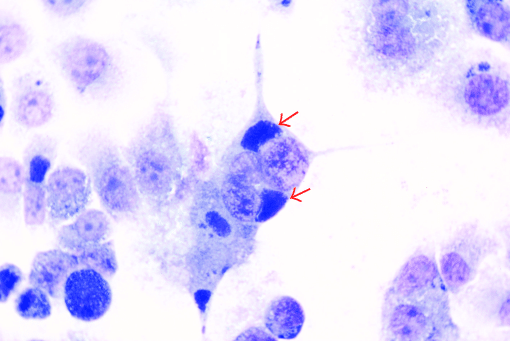
C. trachomatis: Se observan inclusiones perinucleares de clamidias (flechas) en un cultivo de tejido de células McCoy (una línea celular derivada originalmente del líquido sinovial humano) en una tinción de Giemsa.
Imagen: “Perinuclear chlamydial inclusions (arrows) in McCoy cells when infected with the isolated strain” por Zhaocai Li, Xiaoan Cao, Baoquan Fu, Jizhang Zhou. Licencia: CC BY 3.0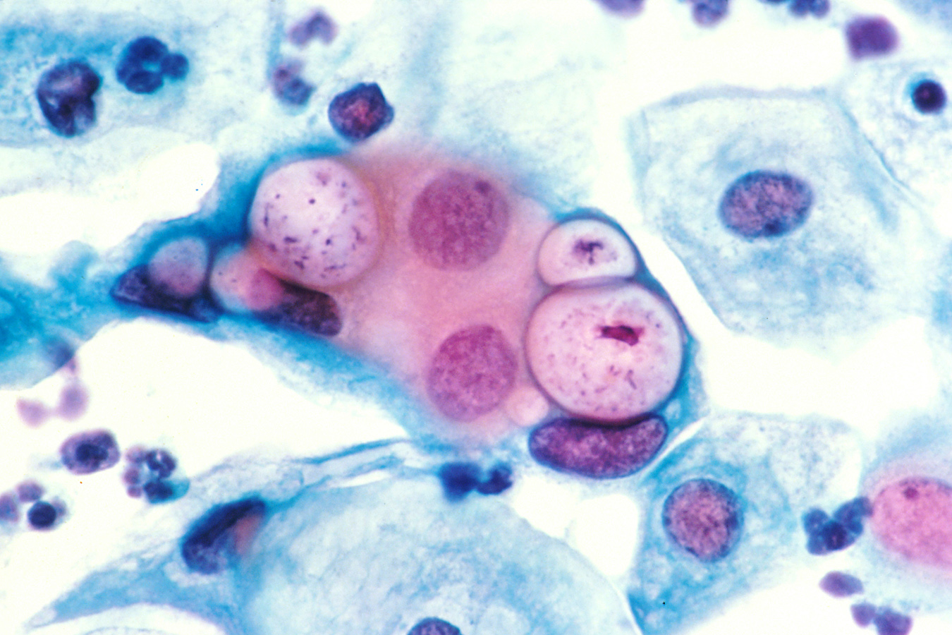
Tinción de Papanicolaou de una preparación de citología cervicovaginal que muestra tres inclusiones intranucleares de clamidias dentro de células epiteliales escamosas que se encuentran junto a varias células epiteliales normales
Imagen : “Pap smear showing chlamydia in the vacuoles 500x H&E” por el Dr. Lance Liotta Laboratory. Licencia: Dominio Público| C. trachomatis | C. psittaci | C. pneumoniae | |
|---|---|---|---|
| Rango de huéspedes | Principalmente humanos |
|
Solo humanos |
| Transmisión |
|
Inhalación de heces de aves secas y contaminadas | Gotitas en EN Erythema nodosum is an immune-mediated panniculitis (inflammation of the subcutaneous fat) caused by a type IV (delayed-type) hypersensitivity reaction. It commonly manifests in young women as tender, erythematous nodules on the shins. Erythema Nodosum aerosol |
| Factores de riesgo |
|
Exposición a las aves | Entornos concurridos (escuelas, hogares de ancianos); los LOS Neisseria ancianos tienen un mayor riesgo de enfermedad grave. |
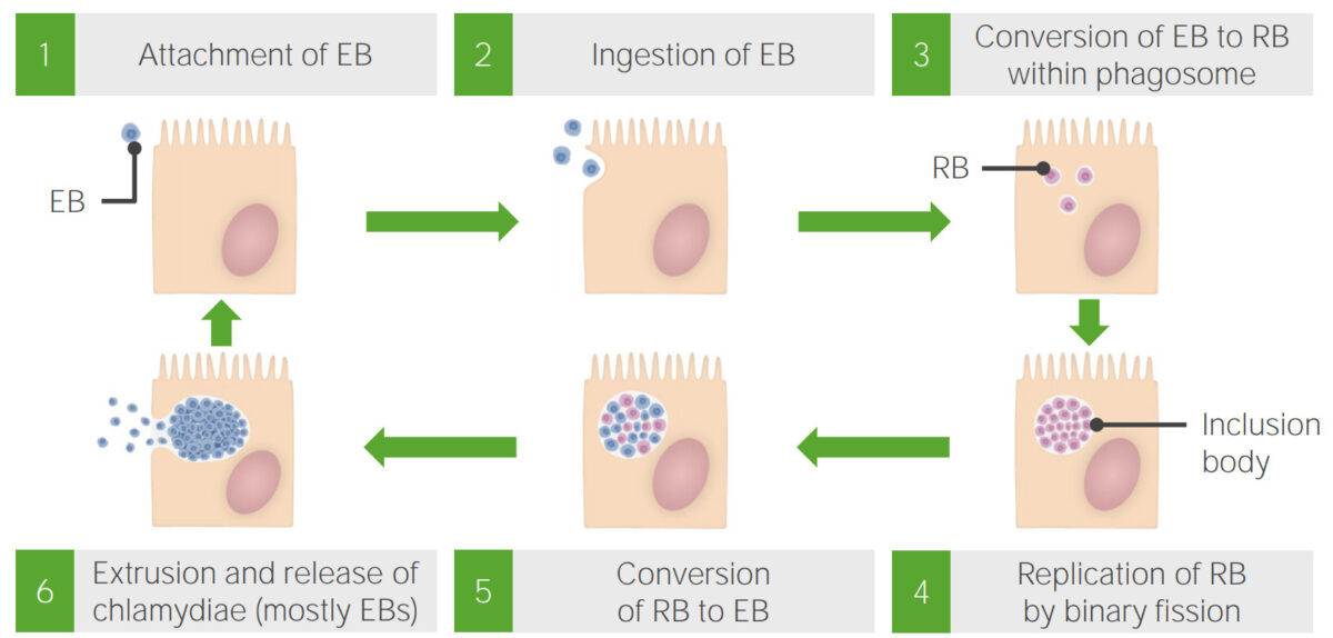
Ciclo del crecimiento de la clamidia:
Los cuerpos elementales (EB, por sus siglas en inglés) ingresan a la célula después de adherirse a su superficie y luego se convierten en la forma de cuerpos reticulados (RB, por sus siglas en inglés). A medida que los RB comienzan a replicarse, crean un grupo conocido como cuerpo de inclusión. Una vez que termina la replicación, los cuerpos de inclusión liberan formas EB, que abandonan la célula.
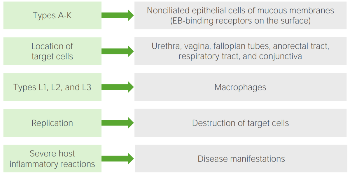
Patogénesis de Chlamydia trachomatis
Imagen por Lecturio.Transmisión sexual:
Infecciones neonatales (serovares D–K):
Tracoma (serovares A–C):
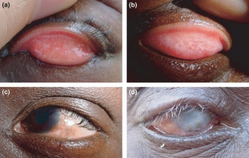
Tracoma
Los episodios recurrentes de infección con los serovares A–C de Chlamydia trachomatis causan inflamación conjuntival en los niños, que eventualmente desarrollan cicatrices y ceguera en la edad adulta.
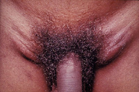
Linfogranuloma venéreo causado por los serovares invasivos L1, L2 o L3 de Chlamydia trachomatis. Este joven adulto experimentó una aparición aguda de ganglios linfáticos sensibles y agrandados en ambas ingles.
Imagen: “Lymphogranuloma venerum – lymph nodes” por Herbert L. Fred, Hendrik A. van Dijk. Licencia: CC BY 2.0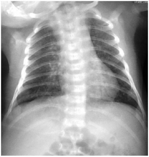
Neumonía neonatal por Chlamydia trachomatis: manchas difusas en una radiografía de tórax
Imagen: “Chlamydial pneumonitis: a creepy neonatal disease” por Hon KL, Leung AK. Licencia: CC BY 3.0