La catarata es una afección que se define como una nubosidad indolora del cristalino. Las cataratas provocan una alteración de la visión, ya que el cristalino proporciona parte del poder de refracción del ojo. Esta afección es una de las causas más comunes de ceguera pediátrica. Las cataratas pueden estar presentes desde el nacimiento (congénitas) o desarrollarse después de la infancia (adquiridas). Las mutaciones genéticas, las enfermedades sistémicas, los LOS Neisseria traumatismos y los LOS Neisseria medicamentos pueden provocar el desarrollo de cataratas. Los LOS Neisseria niños presentan un reflejo rojo anormal, leucocoria o disminución de la agudeza visual. Un examen oftalmológico revela la morfología y la localización de la catarata. El tratamiento depende de la edad de presentación y los LOS Neisseria defectos visuales. Cuando la opacidad es de cierto tamaño o está causando problemas de visión, estrabismo y nistagmo, se recomienda la cirugía de cataratas.
Last updated: May 3, 2022
La catarata es una opacidad indolora del cristalino que interrumpe la luz que se proyecta sobre la retina Retina The ten-layered nervous tissue membrane of the eye. It is continuous with the optic nerve and receives images of external objects and transmits visual impulses to the brain. Its outer surface is in contact with the choroid and the inner surface with the vitreous body. The outermost layer is pigmented, whereas the inner nine layers are transparent. Eye: Anatomy, lo que provoca una nubosidad en EN Erythema nodosum is an immune-mediated panniculitis (inflammation of the subcutaneous fat) caused by a type IV (delayed-type) hypersensitivity reaction. It commonly manifests in young women as tender, erythematous nodules on the shins. Erythema Nodosum la visión. Las cataratas pueden causar ceguera parcial o total.
Cristalino:
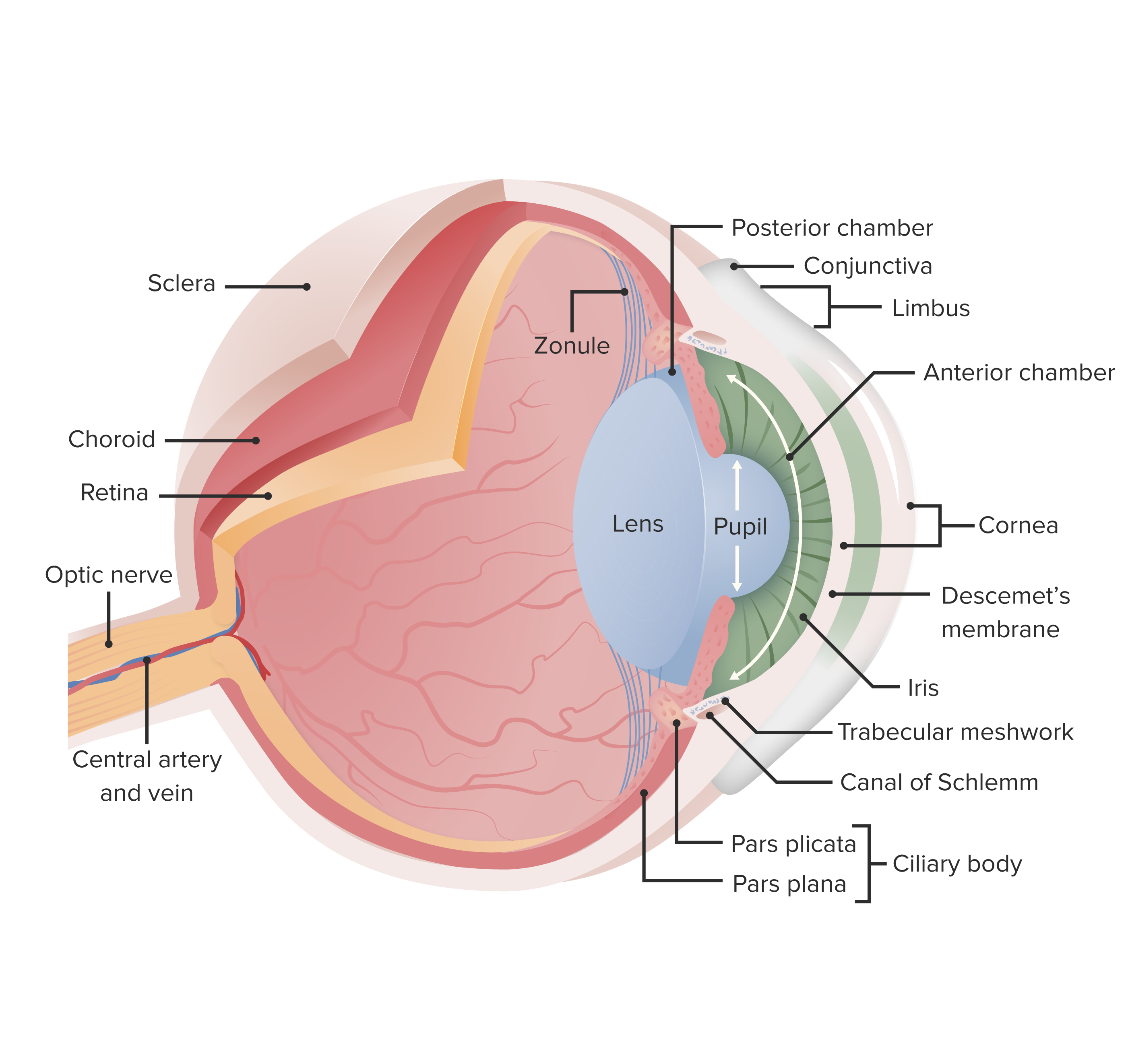
Esta imagen muestra la anatomía esencial del ojo. En las cataratas, se produce una nubosidad del cristalino, que opacifica la luz cuando se proyecta a la retina; esto provoca una reducción de la visión, sobre todo por la noche, cuando los niveles de luz son bajos.
Imagen por Lecturio.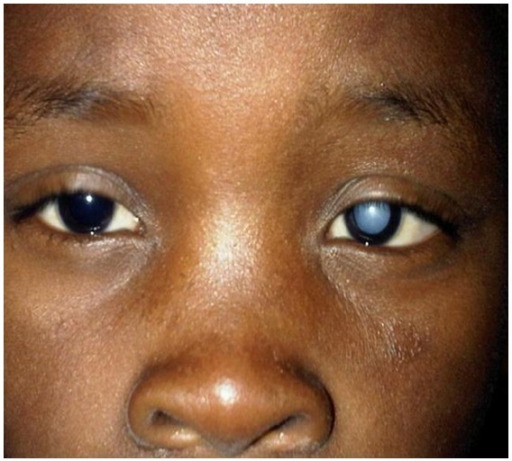
Niño que desarrolló una catarata en el ojo izquierdo tras sufrir una lesión por rayo
Imagen: “A dense left cataract” por Division of Ophthalmology, Red Cross War Memorial Children’s Hospital and the University of Cape Town, Klipfontein Road, Rondebosch, Cape Town 7700, South Africa. Licencia: CC BY 3.0
El reflejo rojo es un reflejo rojo-anaranjado de la parte posterior del ojo. La ausencia de este color es anormal y podría indicar una afección grave. El niño de esta imagen tiene una catarata en el ojo izquierdo.
Imagen: “How to test for the red reflex” por US National Library of Medicine. Licencia: CC BY 2.0
Catarata congénita: Las imágenes (B, C, D) de un ojo dilatado muestran una catarata subcapsular posterior.
Imagen: “Clinical features of the family” por Eye Center of the 2nd Affiliated Hospital, Medical College of Zhejiang University, Hangzhou, China. Licencia: CC BY 3.0, editada por Lecturio.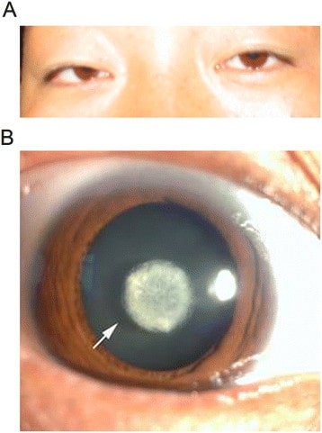
A. Examen ocular sin dilatación de un paciente con catarata congénita.
B. El examen dilatado mostró una catarata nuclear con córnea e iris normales.