


O descolamento da retina Retina The ten-layered nervous tissue membrane of the eye. It is continuous with the optic nerve and receives images of external objects and transmits visual impulses to the brain. Its outer surface is in contact with the choroid and the inner surface with the vitreous body. The outermost layer is pigmented, whereas the inner nine layers are transparent. Eye: Anatomy consiste na separação da retina Retina The ten-layered nervous tissue membrane of the eye. It is continuous with the optic nerve and receives images of external objects and transmits visual impulses to the brain. Its outer surface is in contact with the choroid and the inner surface with the vitreous body. The outermost layer is pigmented, whereas the inner nine layers are transparent. Eye: Anatomy neurossensorial do epitélio pigmentar da retina Retina The ten-layered nervous tissue membrane of the eye. It is continuous with the optic nerve and receives images of external objects and transmits visual impulses to the brain. Its outer surface is in contact with the choroid and the inner surface with the vitreous body. The outermost layer is pigmented, whereas the inner nine layers are transparent. Eye: Anatomy e da coroide. O descolamento de retina Retina The ten-layered nervous tissue membrane of the eye. It is continuous with the optic nerve and receives images of external objects and transmits visual impulses to the brain. Its outer surface is in contact with the choroid and the inner surface with the vitreous body. The outermost layer is pigmented, whereas the inner nine layers are transparent. Eye: Anatomy regmatogénico, o tipo mais MAIS Androgen Insensitivity Syndrome comum, decorre de uma rotura na retina Retina The ten-layered nervous tissue membrane of the eye. It is continuous with the optic nerve and receives images of external objects and transmits visual impulses to the brain. Its outer surface is in contact with the choroid and the inner surface with the vitreous body. The outermost layer is pigmented, whereas the inner nine layers are transparent. Eye: Anatomy, que permite a acumulação de líquido no espaço sub-retiniano. Na presença de uma retina Retina The ten-layered nervous tissue membrane of the eye. It is continuous with the optic nerve and receives images of external objects and transmits visual impulses to the brain. Its outer surface is in contact with the choroid and the inner surface with the vitreous body. The outermost layer is pigmented, whereas the inner nine layers are transparent. Eye: Anatomy intacta, o descolamento ocorre quando o vítreo puxa a retina Retina The ten-layered nervous tissue membrane of the eye. It is continuous with the optic nerve and receives images of external objects and transmits visual impulses to the brain. Its outer surface is in contact with the choroid and the inner surface with the vitreous body. The outermost layer is pigmented, whereas the inner nine layers are transparent. Eye: Anatomy (tração) ou quando uma doença subjacente leva ao aumento do extravazamento de fluido (exsudativo). Os sintomas como fotopsia, moscas volantes e defeitos visuais podem apresentar-se em horas ou gradualmente ao longo de semanas. O descolamento da retina Retina The ten-layered nervous tissue membrane of the eye. It is continuous with the optic nerve and receives images of external objects and transmits visual impulses to the brain. Its outer surface is in contact with the choroid and the inner surface with the vitreous body. The outermost layer is pigmented, whereas the inner nine layers are transparent. Eye: Anatomy com perda visual é uma emergência. Assim que ocorre o descolamento macular, o prognóstico visual é mau. O descolamento regmatogénico da retina Retina The ten-layered nervous tissue membrane of the eye. It is continuous with the optic nerve and receives images of external objects and transmits visual impulses to the brain. Its outer surface is in contact with the choroid and the inner surface with the vitreous body. The outermost layer is pigmented, whereas the inner nine layers are transparent. Eye: Anatomy sintomático com acuidade visual central intacta implica uma cirurgia de urgência. Nos descolamentos não regmatogénicos da retina Retina The ten-layered nervous tissue membrane of the eye. It is continuous with the optic nerve and receives images of external objects and transmits visual impulses to the brain. Its outer surface is in contact with the choroid and the inner surface with the vitreous body. The outermost layer is pigmented, whereas the inner nine layers are transparent. Eye: Anatomy, o tratamento deve ser dirigido ao processo patológico primário.
Last updated: Dec 15, 2025
O descolamento de retina Retina The ten-layered nervous tissue membrane of the eye. It is continuous with the optic nerve and receives images of external objects and transmits visual impulses to the brain. Its outer surface is in contact with the choroid and the inner surface with the vitreous body. The outermost layer is pigmented, whereas the inner nine layers are transparent. Eye: Anatomy consiste na separação da retina Retina The ten-layered nervous tissue membrane of the eye. It is continuous with the optic nerve and receives images of external objects and transmits visual impulses to the brain. Its outer surface is in contact with the choroid and the inner surface with the vitreous body. The outermost layer is pigmented, whereas the inner nine layers are transparent. Eye: Anatomy do epitélio pigmentar da retina Retina The ten-layered nervous tissue membrane of the eye. It is continuous with the optic nerve and receives images of external objects and transmits visual impulses to the brain. Its outer surface is in contact with the choroid and the inner surface with the vitreous body. The outermost layer is pigmented, whereas the inner nine layers are transparent. Eye: Anatomy e da coroide subjacentes.
Retina Retina The ten-layered nervous tissue membrane of the eye. It is continuous with the optic nerve and receives images of external objects and transmits visual impulses to the brain. Its outer surface is in contact with the choroid and the inner surface with the vitreous body. The outermost layer is pigmented, whereas the inner nine layers are transparent. Eye: Anatomy:
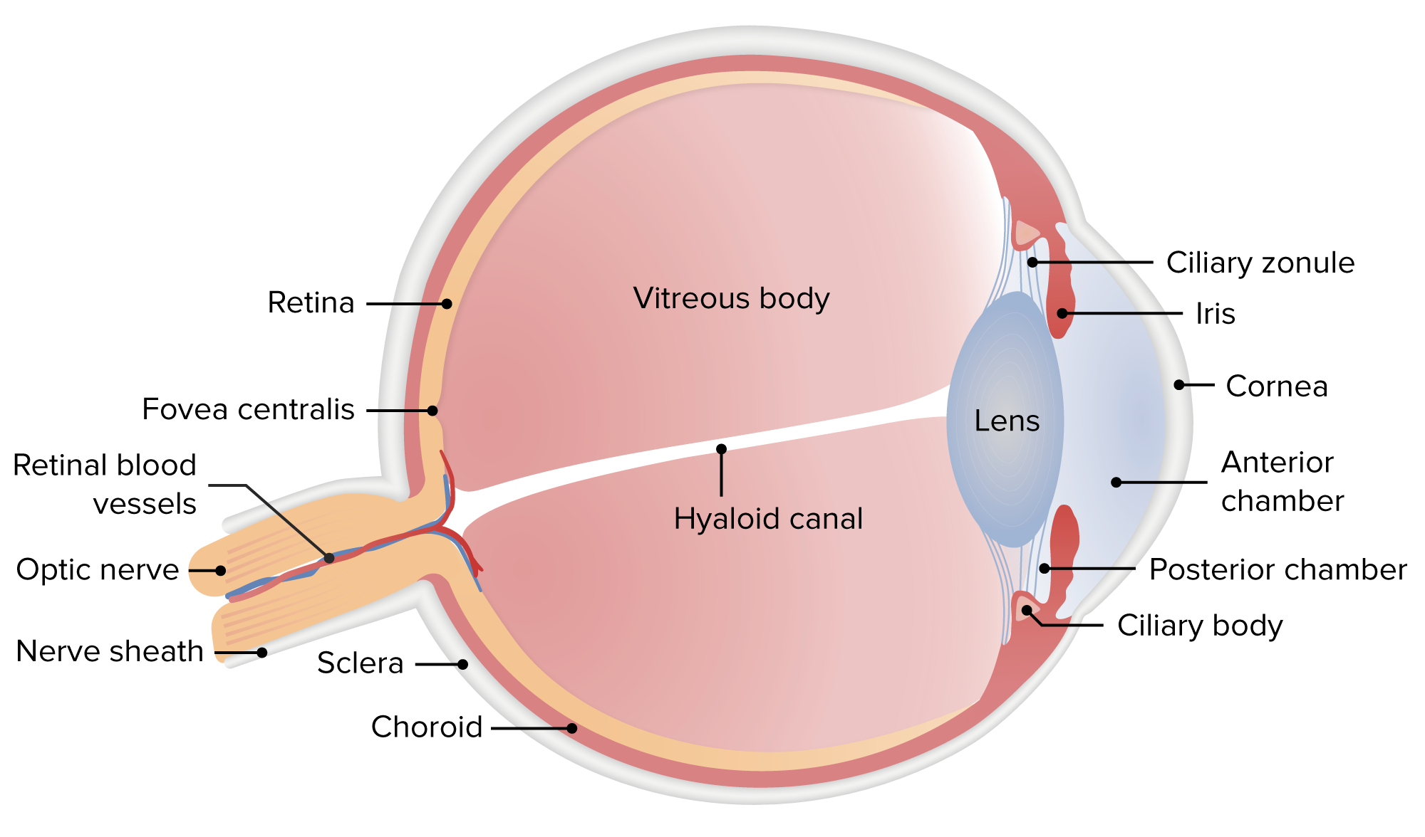
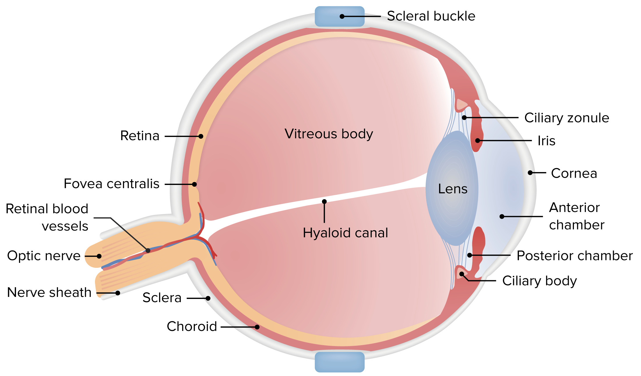
Anatomia do olho humano
Imagem de Lecturio.
Esquerda: componentes das camadas mais interna, neuronal e sensorial do olho
Direita: estrutura geral do olho
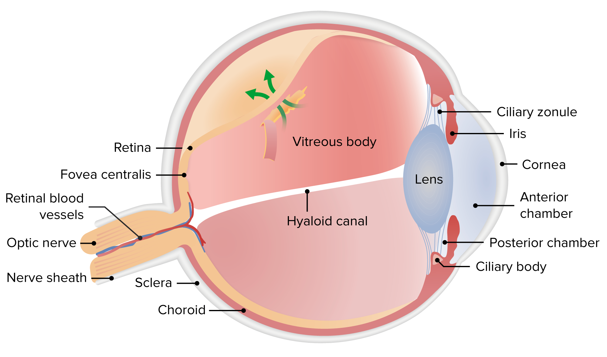
Esta imagem mostra uma rotura da retina (rasgo) que permite a entrada do fluido no espaço sub-retiniano.
Imagem de Lecturio.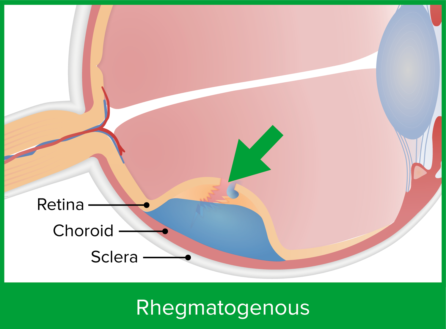
Esta imagem mostra uma rotura retiniana, que permite a entrada e acumulação de fluido no espaço sub-retiniano. Isto predispõe o doente a um descolamento de retina do tipo regmatogénico.
Imagem de Lecturio.
Um vítreo com uma adesão forte ou uma membrana proliferativa puxa a retina, causando um descolamento por tração.
Imagem de Lecturio.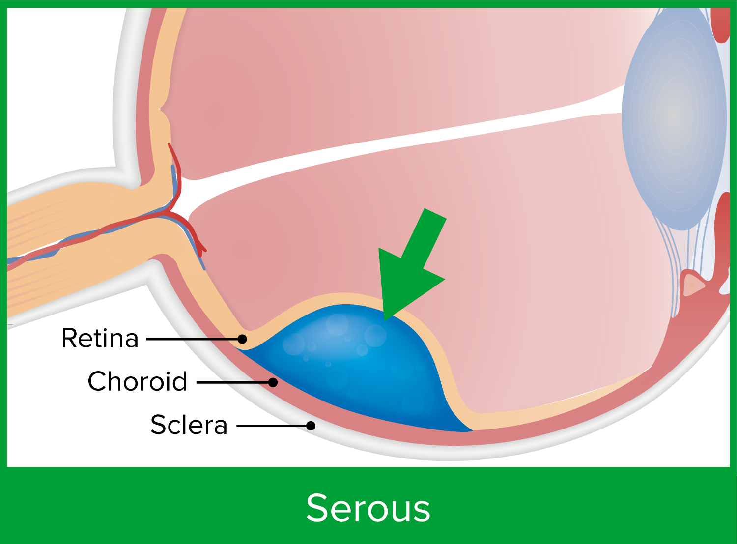
O descolamento exsudativo ou seroso da retina resulta do fluxo disfuncional de fluido ou do aumento da permeabilidade vascular devido a uma doença subjacente.
Imagem de Lecturio.| Sintoma | Causa |
|---|---|
| Fotopsia | Tração vitreorretiniana |
| Moscas volantes | Células vítreas, sangue |
| Defeito do campo visual | Descolamento periférico |
| Visão turva ou perda da visão central | Descolamento macular |
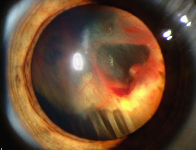
Fotografia de lâmpada de fenda que mostra um descolamento da retina com hemorragia vítrea
Imagem: “Slit lamp photograph showing retinal detachment” do National Eye Institute/National Institutes of Health. Licença: Domínio Público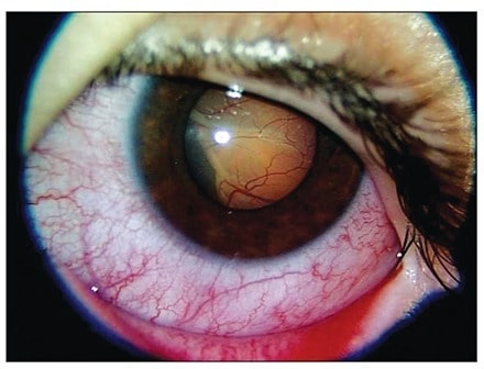
Fotografia de lâmpada de fenda do olho esquerdo que mostra um descolamento exsudativo da retina na área nasal inferior. O descolamento de retina é secundário a uma leucemia linfoblástica aguda.
Imagem: “Slit-lamp photograph of the left eye at presentation” do Diskapi Children’s Hospital, Department of Pediatric Hematology, Ankara, Turkey. Licença: CC BY 2.5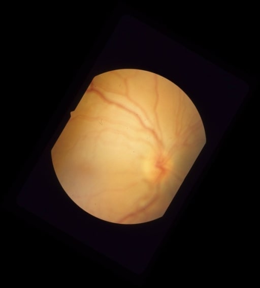
Fotografia colorida do fundo do olho que mostra um descolamento da retina regmatogénico sem afetar a mácula devido a um buraco necrótico na retin por uma lesão plana da coróide inferior à arcada inferotemporal
Imagem: “Pre-operative color fundus photograph of the right eye” da University of Southern California and Doheny Eye Institute, Los Angeles, California 90033, USA. Licença: CC BY 2.0
Descolamento da retina exsudativo bolhoso unilateral na doença de Behçet. Fundoscopia e angiofluoresceinografia:
(a) Extenso revestimento perivascular com infiltrados retinianos amarelados dispersos e hemorragias em ambos os olhos, com descolamento da retina exsudativo bolhoso nos quadrantes superior e inferior da retina temporal no olho direito (setas brancas)
(b) Angiografia de fluoresceína que mostra coloração tardia dos vasos retinianos com extravazamento difuso de corante em ambos os olhos e acumulação de fluoresceína no espaço sub-retiniano no olho direito (setas pretas)
Esta imagem ilustra a introflexão escleral, a colocação de uma estrutura em silicone flexível suturada à esclera. A fivela escleral empurra a esclera para dentro, mantendo assim as estruturas separadas mais próximas e permitindo que a retina se fixe novamente.
Imagem de Lecturio.