Domina Conceptos Médicos

Estudia para la escuela de medicina y tus examenes con Lecturio.

Virus JC y Virus BK

Los LOS Neisseria virus Virus Viruses are infectious, obligate intracellular parasites composed of a nucleic acid core surrounded by a protein capsid. Viruses can be either naked (non-enveloped) or enveloped. The classification of viruses is complex and based on many factors, including type and structure of the nucleoid and capsid, the presence of an envelope, the replication cycle, and the host range. Virology BK (BKV, por sus siglas en EN Erythema nodosum is an immune-mediated panniculitis (inflammation of the subcutaneous fat) caused by a type IV (delayed-type) hypersensitivity reaction. It commonly manifests in young women as tender, erythematous nodules on the shins. Erythema Nodosum inglés) y JC (JCV, por sus siglas en EN Erythema nodosum is an immune-mediated panniculitis (inflammation of the subcutaneous fat) caused by a type IV (delayed-type) hypersensitivity reaction. It commonly manifests in young women as tender, erythematous nodules on the shins. Erythema Nodosum inglés) son pequeños virus Virus Viruses are infectious, obligate intracellular parasites composed of a nucleic acid core surrounded by a protein capsid. Viruses can be either naked (non-enveloped) or enveloped. The classification of viruses is complex and based on many factors, including type and structure of the nucleoid and capsid, the presence of an envelope, the replication cycle, and the host range. Virology de ácido desoxirribonucleico (ADN) de doble cadena sin envoltura, pertenecientes a la familia Polyomaviridae Polyomaviridae A family of small, non-enveloped DNA viruses, infecting mainly mammals. JC Virus and BK Virus, que son ubicuos en EN Erythema nodosum is an immune-mediated panniculitis (inflammation of the subcutaneous fat) caused by a type IV (delayed-type) hypersensitivity reaction. It commonly manifests in young women as tender, erythematous nodules on the shins. Erythema Nodosum la población humana. Mientras que la infección primaria suele ser asintomática, la infección se mantiene latente de por vida en EN Erythema nodosum is an immune-mediated panniculitis (inflammation of the subcutaneous fat) caused by a type IV (delayed-type) hypersensitivity reaction. It commonly manifests in young women as tender, erythematous nodules on the shins. Erythema Nodosum los LOS Neisseria riñones y órganos linfoides. Las infecciones latentes pueden activarse en EN Erythema nodosum is an immune-mediated panniculitis (inflammation of the subcutaneous fat) caused by a type IV (delayed-type) hypersensitivity reaction. It commonly manifests in young women as tender, erythematous nodules on the shins. Erythema Nodosum pacientes inmunodeprimidos. La reactivación del BKV se observa con mayor frecuencia en EN Erythema nodosum is an immune-mediated panniculitis (inflammation of the subcutaneous fat) caused by a type IV (delayed-type) hypersensitivity reaction. It commonly manifests in young women as tender, erythematous nodules on the shins. Erythema Nodosum pacientes trasplantados y puede provocar nefropatía, pérdida de aloinjertos, cistitis hemorrágica y estenosis uretral. La reactivación del JCV causa la leucoencefalopatía multifocal Multifocal Retinoblastoma progresiva, que se considera una enfermedad definitoria del síndrome de inmunodeficiencia adquirida (SIDA). El diagnóstico puede confirmarse mediante reacción en EN Erythema nodosum is an immune-mediated panniculitis (inflammation of the subcutaneous fat) caused by a type IV (delayed-type) hypersensitivity reaction. It commonly manifests in young women as tender, erythematous nodules on the shins. Erythema Nodosum cadena de polimerasa ( PCR PCR Polymerase chain reaction (PCR) is a technique that amplifies DNA fragments exponentially for analysis. The process is highly specific, allowing for the targeting of specific genomic sequences, even with minuscule sample amounts. The PCR cycles multiple times through 3 phases: denaturation of the template DNA, annealing of a specific primer to the individual DNA strands, and synthesis/elongation of new DNA molecules. Polymerase Chain Reaction (PCR), por sus siglas en EN Erythema nodosum is an immune-mediated panniculitis (inflammation of the subcutaneous fat) caused by a type IV (delayed-type) hypersensitivity reaction. It commonly manifests in young women as tender, erythematous nodules on the shins. Erythema Nodosum inglés) o biopsia de tejido. No hay disponible una terapia específica. El objetivo del tratamiento es reducir la inmunosupresión.

Last updated: Dec 15, 2025

Editorial responsibility: Stanley Oiseth, Lindsay Jones, Evelin Maza

Clasificación

Diagrama de flujo de la clasificación de los virus del ADN

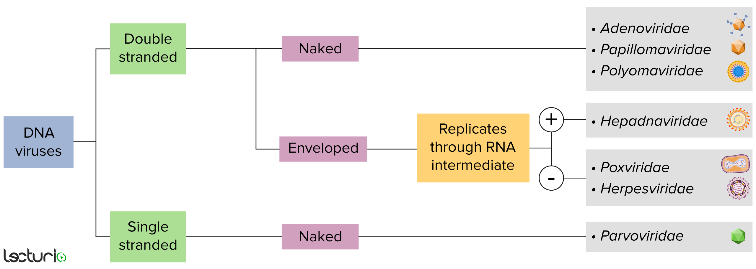
Identificación de virus de ADN:
Los virus pueden clasificarse de muchas maneras. Sin embargo, la mayoría de los virus tienen un genoma formado por ADN o ácido ribonucleico (ARN). Los virus con un genoma de ADN pueden caracterizarse además como de cadena simple o doble. Los virus “envueltos” están cubiertos por una fina capa de membrana celular, que suele tomarse de la célula huésped. Sin embargo, si la capa está ausente, los virus se denominan “desnudos”. Algunos virus con envoltura traducen el ADN en ARN antes de incorporarse al genoma de la célula huésped.

Imagen por Lecturio. Licencia: CC BY-NC-SA 4.0

Características Generales y Epidemiología

Taxonomía

  • Familia: Polyomaviridae Polyomaviridae A family of small, non-enveloped DNA viruses, infecting mainly mammals. JC Virus and BK Virus
  • Género: Betapolyomavirus Betapolyomavirus JC Virus and BK Virus
  • Especies:
    • Poliomavirus humano 1: virus Virus Viruses are infectious, obligate intracellular parasites composed of a nucleic acid core surrounded by a protein capsid. Viruses can be either naked (non-enveloped) or enveloped. The classification of viruses is complex and based on many factors, including type and structure of the nucleoid and capsid, the presence of an envelope, the replication cycle, and the host range. Virology BK (BKV)
    • Poliomavirus humano 2: virus Virus Viruses are infectious, obligate intracellular parasites composed of a nucleic acid core surrounded by a protein capsid. Viruses can be either naked (non-enveloped) or enveloped. The classification of viruses is complex and based on many factors, including type and structure of the nucleoid and capsid, the presence of an envelope, the replication cycle, and the host range. Virology JC (JCV)

Características generales

El BKV y el JCV comparten características similares:

  • Virus Virus Viruses are infectious, obligate intracellular parasites composed of a nucleic acid core surrounded by a protein capsid. Viruses can be either naked (non-enveloped) or enveloped. The classification of viruses is complex and based on many factors, including type and structure of the nucleoid and capsid, the presence of an envelope, the replication cycle, and the host range. Virology de ADN:
    • Circulares
    • Doble cadena
  • Estructura:
    • Pequeños
    • No envueltos
    • Icosaédricos
Micrografía electrónica que muestra el virus BK virus jc y el virus bk

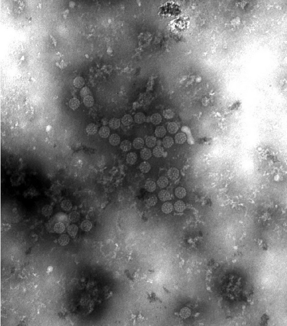
Micrografía electrónica que muestra los viriones del virus BK (BKV):
Obsérvese el pequeño tamaño y la simetría icosaédrica.

Imagen: “Viral inclusions seen on electron microscopy of urine epithelial cells” por Perram J et al. Licencia: CC BY 3.0

Enfermedades asociadas

  • BKV:
    • Nefropatía asociada al AL Amyloidosis BKV
    • Estenosis ureteral
    • Cistitis hemorrágica
  • JCV:
    • Leucoencefalopatía multifocal Multifocal Retinoblastoma progresiva
    • Encefalopatía y meningitis Meningitis Meningitis is inflammation of the meninges, the protective membranes of the brain, and spinal cord. The causes of meningitis are varied, with the most common being bacterial or viral infection. The classic presentation of meningitis is a triad of fever, altered mental status, and nuchal rigidity. Meningitis (raro)

Epidemiología

  • Se desconoce gran parte de la epidemiología del BKV y el JCV en EN Erythema nodosum is an immune-mediated panniculitis (inflammation of the subcutaneous fat) caused by a type IV (delayed-type) hypersensitivity reaction. It commonly manifests in young women as tender, erythematous nodules on the shins. Erythema Nodosum la población general es desconocida.
  • Seroprevalencia:
    • En EN Erythema nodosum is an immune-mediated panniculitis (inflammation of the subcutaneous fat) caused by a type IV (delayed-type) hypersensitivity reaction. It commonly manifests in young women as tender, erythematous nodules on the shins. Erythema Nodosum Estados Unidos y Europa: 30%–90%
    • Aumenta con la edad
  • La viruria Viruria JC Virus and BK Virus asintomática es detectable en EN Erythema nodosum is an immune-mediated panniculitis (inflammation of the subcutaneous fat) caused by a type IV (delayed-type) hypersensitivity reaction. It commonly manifests in young women as tender, erythematous nodules on the shins. Erythema Nodosum 10%–30% de los LOS Neisseria individuos sanos.
  • Las manifestaciones clínicas son raras.

Patogénesis

Reservorio

El ser humano es el huésped natural del JCV y del BKV (no reservorios animales)

Transmisión

  • Los LOS Neisseria métodos de transmisión son poco conocidos tanto para el BKV como para el JCV.
  • Posibles rutas:
    • Oral
    • Respiratorias
    • Perinatal
    • Tejido de donante

Factores de riesgo del huésped

La enfermedad clínica suele surgir por la reactivación del virus Virus Viruses are infectious, obligate intracellular parasites composed of a nucleic acid core surrounded by a protein capsid. Viruses can be either naked (non-enveloped) or enveloped. The classification of viruses is complex and based on many factors, including type and structure of the nucleoid and capsid, the presence of an envelope, the replication cycle, and the host range. Virology en EN Erythema nodosum is an immune-mediated panniculitis (inflammation of the subcutaneous fat) caused by a type IV (delayed-type) hypersensitivity reaction. It commonly manifests in young women as tender, erythematous nodules on the shins. Erythema Nodosum pacientes inmunodeprimidos:

  • Virus Virus Viruses are infectious, obligate intracellular parasites composed of a nucleic acid core surrounded by a protein capsid. Viruses can be either naked (non-enveloped) or enveloped. The classification of viruses is complex and based on many factors, including type and structure of the nucleoid and capsid, the presence of an envelope, the replication cycle, and the host range. Virology de inmunodeficiencia humana (VIH)/SIDA (la leucoencefalopatía multifocal Multifocal Retinoblastoma progresiva se considera una enfermedad definitoria del SIDA)
  • Trasplante de órganos
  • Trasplante de células hematopoyéticas
  • Enfermedades hematológicas malignas

Fisiopatología

  • La infección primaria da lugar a una persistencia subclínica y de por vida en EN Erythema nodosum is an immune-mediated panniculitis (inflammation of the subcutaneous fat) caused by a type IV (delayed-type) hypersensitivity reaction. It commonly manifests in young women as tender, erythematous nodules on the shins. Erythema Nodosum el tejido del huésped.
  • Una vez dentro de la célula, el virus Virus Viruses are infectious, obligate intracellular parasites composed of a nucleic acid core surrounded by a protein capsid. Viruses can be either naked (non-enveloped) or enveloped. The classification of viruses is complex and based on many factors, including type and structure of the nucleoid and capsid, the presence of an envelope, the replication cycle, and the host range. Virology se desplaza hasta el núcleo y establece una infección latente o lítica.
  • La infección se controla mediante anticuerpos y células T citotóxicas.
  • Fallo de la respuesta inmunitaria → reactivación
    • JCV:
      • Establece la latencia (principalmente en EN Erythema nodosum is an immune-mediated panniculitis (inflammation of the subcutaneous fat) caused by a type IV (delayed-type) hypersensitivity reaction. It commonly manifests in young women as tender, erythematous nodules on the shins. Erythema Nodosum los LOS Neisseria riñones y el tejido linfático)
      • Reactivación → replicación y producción de variantes neurotrópicas
      • Puede atravesar la barrera hematoencefálica → replicación lítica en EN Erythema nodosum is an immune-mediated panniculitis (inflammation of the subcutaneous fat) caused by a type IV (delayed-type) hypersensitivity reaction. It commonly manifests in young women as tender, erythematous nodules on the shins. Erythema Nodosum los LOS Neisseria oligodendrocitos → desmielinización → leucoencefalopatía multifocal Multifocal Retinoblastoma progresiva
    • BKV:
      • Latencia en EN Erythema nodosum is an immune-mediated panniculitis (inflammation of the subcutaneous fat) caused by a type IV (delayed-type) hypersensitivity reaction. It commonly manifests in young women as tender, erythematous nodules on the shins. Erythema Nodosum los LOS Neisseria riñones
      • Reactivación → ↑ replicación → daño a las células tubulares y uroepiteliales renales e infiltración inflamatoria
      • Inflamación intersticial → fibrosis Fibrosis Any pathological condition where fibrous connective tissue invades any organ, usually as a consequence of inflammation or other injury. Bronchiolitis Obliterans y lesión tubular

Presentación Clínica

BKV

Infección primaria:

  • La mayoría de los LOS Neisseria pacientes son asintomáticos.
  • Otros pueden tener una enfermedad respiratoria leve.

Infección secundaria:

  • Ocurre en EN Erythema nodosum is an immune-mediated panniculitis (inflammation of the subcutaneous fat) caused by a type IV (delayed-type) hypersensitivity reaction. It commonly manifests in young women as tender, erythematous nodules on the shins. Erythema Nodosum pacientes inmunodeprimidos
  • Nefropatía asociada al AL Amyloidosis BKV (más frecuente en EN Erythema nodosum is an immune-mediated panniculitis (inflammation of the subcutaneous fat) caused by a type IV (delayed-type) hypersensitivity reaction. It commonly manifests in young women as tender, erythematous nodules on the shins. Erythema Nodosum pacientes con trasplante renal):
    • Lesión renal aguda
    • Hematuria Hematuria Presence of blood in the urine. Renal Cell Carcinoma, piuria y cilindros celulares
    • Puede dar lugar a una disfunción o pérdida de aloinjerto renal
  • Estenosis ureteral:
    • Obstrucción de las vías urinarias
    • Lesión renal aguda
  • Cistitis hemorrágica (más frecuente en EN Erythema nodosum is an immune-mediated panniculitis (inflammation of the subcutaneous fat) caused by a type IV (delayed-type) hypersensitivity reaction. It commonly manifests in young women as tender, erythematous nodules on the shins. Erythema Nodosum receptores de trasplante de células hematopoyéticas):

JCV

La leucoencefalopatía multifocal Multifocal Retinoblastoma progresiva es una enfermedad progresiva (y a menudo mortal) que se observa con mayor frecuencia en EN Erythema nodosum is an immune-mediated panniculitis (inflammation of the subcutaneous fat) caused by a type IV (delayed-type) hypersensitivity reaction. It commonly manifests in young women as tender, erythematous nodules on the shins. Erythema Nodosum pacientes con SIDA (considerada una condición definidora de SIDA) con un recuento de CD4 < 200 células/µl.

  • Torpeza
  • Hemiparesia (hallazgo más común)
  • Afasia y disartria
  • Hemianopsia o diplopía
  • Ataxia Ataxia Impairment of the ability to perform smoothly coordinated voluntary movements. This condition may affect the limbs, trunk, eyes, pharynx, larynx, and other structures. Ataxia may result from impaired sensory or motor function. Sensory ataxia may result from posterior column injury or peripheral nerve diseases. Motor ataxia may be associated with cerebellar diseases; cerebral cortex diseases; thalamic diseases; basal ganglia diseases; injury to the red nucleus; and other conditions. Ataxia-telangiectasia de la marcha
  • Deterioro cognitivo
  • Deterioro de la vigilancia
  • Cefalea
  • Convulsiones (raras)

Diagnóstico y Tratamiento

Diagnóstico

BKV:

  • PCR PCR Polymerase chain reaction (PCR) is a technique that amplifies DNA fragments exponentially for analysis. The process is highly specific, allowing for the targeting of specific genomic sequences, even with minuscule sample amounts. The PCR cycles multiple times through 3 phases: denaturation of the template DNA, annealing of a specific primer to the individual DNA strands, and synthesis/elongation of new DNA molecules. Polymerase Chain Reaction (PCR) cuantitativa para el ADN del BKV en EN Erythema nodosum is an immune-mediated panniculitis (inflammation of the subcutaneous fat) caused by a type IV (delayed-type) hypersensitivity reaction. It commonly manifests in young women as tender, erythematous nodules on the shins. Erythema Nodosum plasma Plasma The residual portion of blood that is left after removal of blood cells by centrifugation without prior blood coagulation. Transfusion Products:
    • Prueba de tamizaje preferida
    • Realizar en EN Erythema nodosum is an immune-mediated panniculitis (inflammation of the subcutaneous fat) caused by a type IV (delayed-type) hypersensitivity reaction. It commonly manifests in young women as tender, erythematous nodules on the shins. Erythema Nodosum todos los LOS Neisseria pacientes de trasplante renal.
  • Citología de orina para la identificación de células infectadas
  • Biopsia de aloinjerto renal:
    • El estándar de oro para el diagnóstico
    • Se puede utilizar la inmunohistoquímica para detectar los LOS Neisseria antígenos del BKV.
    • Hallazgos:
      • Inclusiones virales intranucleares y basófilas
      • Variación del tamaño de los LOS Neisseria núcleos celulares o hipercromasia (indica lesión nuclear)
      • Lesión tubular con infiltrados celulares intersticiales e inflamatorios
      • Tubulitis (linfocitos en EN Erythema nodosum is an immune-mediated panniculitis (inflammation of the subcutaneous fat) caused by a type IV (delayed-type) hypersensitivity reaction. It commonly manifests in young women as tender, erythematous nodules on the shins. Erythema Nodosum la membrana basal tubular)

JCV:

  • Tomografía computarizada (TC) o resonancia magnética (RM) del cerebro:
    • Lesiones diseminadas, sin realce, en EN Erythema nodosum is an immune-mediated panniculitis (inflammation of the subcutaneous fat) caused by a type IV (delayed-type) hypersensitivity reaction. It commonly manifests in young women as tender, erythematous nodules on the shins. Erythema Nodosum la sustancia blanca (indica desmielinización)
    • No se ajustan a los LOS Neisseria territorios cerebrovasculares
    • Sin efecto de masa
  • Punción lumbar con análisis del LCR: PCR PCR Polymerase chain reaction (PCR) is a technique that amplifies DNA fragments exponentially for analysis. The process is highly specific, allowing for the targeting of specific genomic sequences, even with minuscule sample amounts. The PCR cycles multiple times through 3 phases: denaturation of the template DNA, annealing of a specific primer to the individual DNA strands, and synthesis/elongation of new DNA molecules. Polymerase Chain Reaction (PCR) para el ADN del JCV (preferido)
  • Biopsia cerebral:
    • Se considera si la PCR PCR Polymerase chain reaction (PCR) is a technique that amplifies DNA fragments exponentially for analysis. The process is highly specific, allowing for the targeting of specific genomic sequences, even with minuscule sample amounts. The PCR cycles multiple times through 3 phases: denaturation of the template DNA, annealing of a specific primer to the individual DNA strands, and synthesis/elongation of new DNA molecules. Polymerase Chain Reaction (PCR) es negativa, pero la sospecha sigue siendo alta
    • Hallazgos:
      • Desmielinización
      • Astrocitos anormales
      • Núcleos oligodendrogliales agrandados
      • Por lo general, no hay infiltrados inflamatorios
Imágenes de resonancia magnética de una leucoencefalopatía multifocal

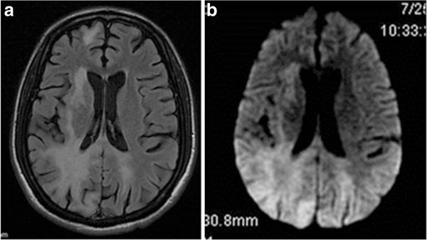
Resonancia magnética (RM) de un paciente con leucoencefalopatía multifocal progresiva:
a: RM axial en T2-flair del cerebro que muestra una hiperintensidad de señal bilateral en la sustancia blanca parietooccipital
b: imagen axial ponderada en T2-flair de las mismas lesiones

Imagen: “Axial T2-Flair MRI and axial T2-Flair diffuse-weighted image” por Jeyaraman VA et al. Licencia: CC BY 2.0

Tratamiento

No hay disponible terapias específicas para el BKV o el JCV. El tratamiento suele estar dirigido a reducir la inmunosupresión.

  • Reducir la terapia inmunosupresora (cuando sea posible)
  • Terapia antirretroviral para el VIH/SIDA
  • Medidas de soporte

Diagnóstico Diferencial

  • Linfoma del SNC: una neoplasia asociada a la infección por Epstein-Barr, que puede aparecer en EN Erythema nodosum is an immune-mediated panniculitis (inflammation of the subcutaneous fat) caused by a type IV (delayed-type) hypersensitivity reaction. It commonly manifests in young women as tender, erythematous nodules on the shins. Erythema Nodosum pacientes con SIDA con un recuento de CD4 < 50 células/µL. Los LOS Neisseria pacientes pueden tener cefalea, confusión, déficit neurológicos focales, convulsiones y síntomas constitucionales. La imagenología cerebral, los LOS Neisseria estudios de LCR para citología y PCR PCR Polymerase chain reaction (PCR) is a technique that amplifies DNA fragments exponentially for analysis. The process is highly specific, allowing for the targeting of specific genomic sequences, even with minuscule sample amounts. The PCR cycles multiple times through 3 phases: denaturation of the template DNA, annealing of a specific primer to the individual DNA strands, and synthesis/elongation of new DNA molecules. Polymerase Chain Reaction (PCR), y la biopsia cerebral pueden proporcionar el diagnóstico. El tratamiento incluye terapia antirretroviral y quimioterapia.
  • Demencia asociada al AL Amyloidosis VIH: una condición en EN Erythema nodosum is an immune-mediated panniculitis (inflammation of the subcutaneous fat) caused by a type IV (delayed-type) hypersensitivity reaction. It commonly manifests in young women as tender, erythematous nodules on the shins. Erythema Nodosum pacientes con SIDA con un recuento de CD4 < 200 células/µL. Los LOS Neisseria pacientes presentan pérdida de memoria, mal funcionamiento ejecutivo, cambios de comportamiento y de humor Humor Defense Mechanisms, y pérdida gradual de las habilidades motoras. La RM puede mostrar hiperintensidades difusas irregulares en EN Erythema nodosum is an immune-mediated panniculitis (inflammation of the subcutaneous fat) caused by a type IV (delayed-type) hypersensitivity reaction. It commonly manifests in young women as tender, erythematous nodules on the shins. Erythema Nodosum la materia blanca. El análisis del LCR debe descartar otras causas, como la leucoencefalopatía multifocal Multifocal Retinoblastoma progresiva y el linfoma. El tratamiento incluye terapia antirretroviral y medidas de soporte.
  • Esclerosis múltiple: enfermedad autoinmune crónica e inflamatoria que conduce a la desmielinización del SNC. La presentación clínica puede incluir síntomas neurológicos que afectan a la visión, funciones motoras, sensibilidad y función autonómica. La presentación varía en EN Erythema nodosum is an immune-mediated panniculitis (inflammation of the subcutaneous fat) caused by a type IV (delayed-type) hypersensitivity reaction. It commonly manifests in young women as tender, erythematous nodules on the shins. Erythema Nodosum función de la localización de las lesiones. El diagnóstico se realiza con imagenología RM del cerebro y la columna vertebral, así como con el examen del LCR. El tratamiento incluye corticosteroides para las exacerbaciones agudas y agentes modificadores de la enfermedad para frenar su progresión.
  • Citomegalovirus: un virus Virus Viruses are infectious, obligate intracellular parasites composed of a nucleic acid core surrounded by a protein capsid. Viruses can be either naked (non-enveloped) or enveloped. The classification of viruses is complex and based on many factors, including type and structure of the nucleoid and capsid, the presence of an envelope, the replication cycle, and the host range. Virology ubicuo de ADN de doble cadena perteneciente a la familia Herpesviridae Herpesviridae A family of enveloped, linear, double-stranded DNA viruses infecting a wide variety of animals. Subfamilies, based on biological characteristics, include: alphaherpesvirinae; betaherpesvirinae; and gammaherpesvirinae. Herpes Simplex Virus 1 and 2. La infección inicial suele ser asintomática en EN Erythema nodosum is an immune-mediated panniculitis (inflammation of the subcutaneous fat) caused by a type IV (delayed-type) hypersensitivity reaction. It commonly manifests in young women as tender, erythematous nodules on the shins. Erythema Nodosum el huésped inmunocompetente. Sin embargo, la reactivación puede ocurrir en EN Erythema nodosum is an immune-mediated panniculitis (inflammation of the subcutaneous fat) caused by a type IV (delayed-type) hypersensitivity reaction. It commonly manifests in young women as tender, erythematous nodules on the shins. Erythema Nodosum pacientes inmunodeprimidos, causando una serie de manifestaciones como colitis Colitis Inflammation of the colon section of the large intestine, usually with symptoms such as diarrhea (often with blood and mucus), abdominal pain, and fever. Pseudomembranous Colitis, hepatitis, neumonitis, meningoencefalitis y nefritis. El diagnóstico puede confirmarse mediante PCR PCR Polymerase chain reaction (PCR) is a technique that amplifies DNA fragments exponentially for analysis. The process is highly specific, allowing for the targeting of specific genomic sequences, even with minuscule sample amounts. The PCR cycles multiple times through 3 phases: denaturation of the template DNA, annealing of a specific primer to the individual DNA strands, and synthesis/elongation of new DNA molecules. Polymerase Chain Reaction (PCR) o biopsia. El tratamiento incluye antivirales y una disminución de la terapia inmunosupresora.

Referencias

  1. Zanotto E, Allesina A, Barreca A, Sidoti F, Gallo E, Bottino P, Iannaccone M, Bianco G, Biancone L, Cavallo R, Costa C. (2020). Renal Allograft Biopsies with Polyomavirus BK Nephropathy: Turin Transplant Center, 2015–19. Viruses; 12(9):1047. https://www.mdpi.com/1999-4915/12/9/1047
  2. Tan CS, Koralnik IJ. (2010). Progressive multifocal leukoencephalopathy and other disorders caused by JC virus: clinical features and pathogenesis. Lancet Neurol; 9(4):425–37. https://pubmed.ncbi.nlm.nih.gov/20298966/
  3. Iacobaeus E, Burkill S, Bahmanyar S, Hakim R, Byström C, Fored M. Olsson T, Brundin L, Montgomery S. (2018). The national incidence of PML in Sweden, 1988-2013. Neurology; 90(6):e498–e506. https://pubmed.ncbi.nlm.nih.gov/29321229/
  4. Tan K, Roda R, Ostrow L, McArthur J, Nath A. (2009). PML-IRIS in patients with HIV infection: clinical manifestations and treatment with steroids. Neurology; 72(17):1458–64. https://pubmed.ncbi.nlm.nih.gov/19129505/
  5. Kleinschmidt-DeMasters BK, Tyler KL. (2005). Progressive multifocal leukoencephalopathy complicating treatment with natalizumab and interferon beta-1a for multiple sclerosis. N Engl J Med; 353(4):369–74. https://pubmed.ncbi.nlm.nih.gov/15947079/
  6. Carson KR, Evens AM, Richey EA, Habermann TM, Focosi D, Seymour JF, Laubach J, Bawn SD, Gordon LI, Winter JN, Furman RR, Vose JM, Zelenetz AD, Mamtani R, Raisch DW, Dorshimer GW, Rosen ST, Muro K, Gottardi-Littell NR, Talley R., Sartor O, Green D, Major EO, Bennett CL. (2009). Progressive multifocal leukoencephalopathy after rituximab therapy in HIV-negative patients: a report of 57 cases from the Research on Adverse Drug Events and Reports project. Blood; 113(20):4834–40. https://pubmed.ncbi.nlm.nih.gov/19264918/
  7. Gómez-Cibeira E, Ivanovic-Barbeito Y, Gutiérrez-Martínez E, Morales E, Abradelo M, Hilario A, Ramos A, Ruiz-Morales J, Villarejo-Galende A. (2016). Eculizumab-related progressive multifocal leukoencephalopathy. Neurology; 86(4):399–400. https://pubmed.ncbi.nlm.nih.gov/26718572/
  8. Harypursat V, Zhou Y, Tang S, Chen Y. (2020). JC Polyomavirus, progressive multifocal leukoencephalopathy, and immune reconstitution inflammatory syndrome: a review. AIDS Res Ther; 17(1):37. https://pubmed.ncbi.nlm.nih.gov/32631361/
  9. Bohra C, Sokol L, Dalia S. (2017). Progressive Multifocal Leukoencephalopathy and Monoclonal Antibodies: A Review. Cancer Control. 24 (4): 1073274817729901. https://pubmed.ncbi.nlm.nih.gov/28975841/
  10. Kartau M, Sipilä JO, Auvinen E, Palomäki M, Verkkoniemi-Ahola A. (2019). Progressive Multifocal Leukoencephalopathy: Current Insights. Degenerative Neurological and Neuromuscular Disease. 9: 109–121. https://www.ncbi.nlm.nih.gov/pmc/articles/PMC6896915/
  11. Greenlee, JE. (2024). Progressive multifocal leukoencephalopathy (PML). MSD Manual Professional Version. Retrieved February 18, 2025, from https://www.msdmanuals.com/professional/neurologic-disorders/brain-infections/progressive-multifocal-leukoencephalopathy-pml
  12. Hirsch, HH. (2025). Overview of JC polyomavirus, BK polyomavirus, and other polyovavirus infections. In Bond, S. (Ed.), UpToDate. Retrieved February 18, 2025, from https://www.uptodate.com/contents/overview-of-jc-polyomavirus-bk-polyomavirus-and-other-polyomavirus-infections
  13. Koralnik, IJ. (2025). Progressive multifocal leukoencephalopathy (PML): Epidemiology, clinical manifestations, and diagnosis. In Dasche, JF. (Ed.), UpToDate. Retrieved February 18, 2025, from https://www.uptodate.com/contents/progressive-multifocal-leukoencephalopathy-pml-epidemiology-clinical-manifestations-and-diagnosis

¡Crea tu cuenta gratis o inicia una sesión para seguir leyendo!

Regístrate ahora y obtén acceso gratuito a Lecturio con páginas de concepto, videos médicos y cuestionarios para tu educación médica.

User Reviews

Que tengas una sesión de estudio alegre y navideña 🎁 Ahorra 50% en todos los planes >>

Details