La vulvaVulvaThe vulva is the external genitalia of the female and includes the mons pubis, labia majora, labia minora, clitoris, vestibule, vestibular bulb, and greater vestibular glands. Vagina, Vulva, and Pelvic Floor: Anatomy es el órgano genital externo de la mujer e incluye el monte de Venus, losLOSNeisseria labios mayores, losLOSNeisseria labios menores, el clítoris, el vestíbulo, el bulbo vestibular y las glándulas vestibulares mayores. La vaginaVaginaThe vagina is the female genital canal, extending from the vulva externally to the cervix uteri internally. The structures have sexual, reproductive, and urinary functions and a rich blood supply, mainly arising from the internal iliac artery.Vagina, Vulva, and Pelvic Floor: Anatomy es el canal genital femenino, que se extiende desde la vulvaVulvaThe vulva is the external genitalia of the female and includes the mons pubis, labia majora, labia minora, clitoris, vestibule, vestibular bulb, and greater vestibular glands. Vagina, Vulva, and Pelvic Floor: Anatomy por fuera hasta el cuello uterino por dentro. Estas estructuras tienen funciones sexuales, reproductivas y urinarias y una rica irrigación, que llega principalmente a través de la arteria ilíaca interna.
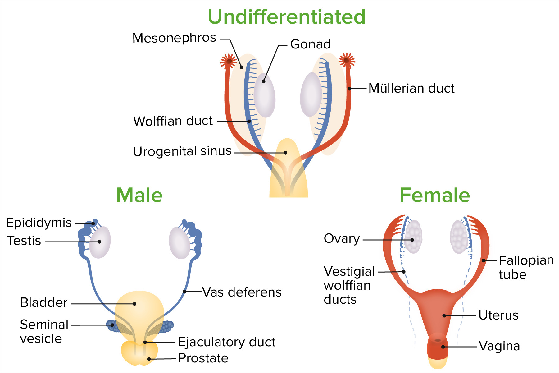
La diferenciación sexual comienza con el desarrollo de las gónadas.
LosLOSNeisseriagenesGenesA category of nucleic acid sequences that function as units of heredity and which code for the basic instructions for the development, reproduction, and maintenance of organisms.DNA Types and Structure presentes enENErythema nodosum is an immune-mediated panniculitis (inflammation of the subcutaneous fat) caused by a type IV (delayed-type) hypersensitivity reaction. It commonly manifests in young women as tender, erythematous nodules on the shins.Erythema Nodosum la fecundación determinan si las gónadas bipotentes enENErythema nodosum is an immune-mediated panniculitis (inflammation of the subcutaneous fat) caused by a type IV (delayed-type) hypersensitivity reaction. It commonly manifests in young women as tender, erythematous nodules on the shins.Erythema Nodosum desarrollo se diferencian enENErythema nodosum is an immune-mediated panniculitis (inflammation of the subcutaneous fat) caused by a type IV (delayed-type) hypersensitivity reaction. It commonly manifests in young women as tender, erythematous nodules on the shins.Erythema Nodosum testículos u ovarios.
La presencia y/o ausencia de hormonas específicas secretadas por las gónadas determinan la diferenciación del resto de estructuras.
Semana 20: Se completa la diferenciación fenotípica.
Diferenciación femenina del embrión
El desarrollo masculino se desencadena por la presencia de ciertas hormonas; el desarrollo femenino se desencadena principalmente por la ausencia de hormonas.
El desarrollo masculino está impulsado por 2 hormonas principales producidas enENErythema nodosum is an immune-mediated panniculitis (inflammation of the subcutaneous fat) caused by a type IV (delayed-type) hypersensitivity reaction. It commonly manifests in young women as tender, erythematous nodules on the shins.Erythema NodosumlosLOSNeisseria testículos:
Testosterona: estimula la diferenciación de losLOSNeisseria conductos wolffianos enENErythema nodosum is an immune-mediated panniculitis (inflammation of the subcutaneous fat) caused by a type IV (delayed-type) hypersensitivity reaction. It commonly manifests in young women as tender, erythematous nodules on the shins.Erythema Nodosum las estructuras masculinas (e.g., epidídimo, conductos deferentes, vesículas seminales y conductos eyaculatorios)
Hormona antimülleriana: provoca la degeneración de losLOSNeisseria conductos müllerianos
LosLOSNeisseria ovarios enENErythema nodosum is an immune-mediated panniculitis (inflammation of the subcutaneous fat) caused by a type IV (delayed-type) hypersensitivity reaction. It commonly manifests in young women as tender, erythematous nodules on the shins.Erythema Nodosum desarrollo no secretan testosterona ni hormona antimülleriana.
EnENErythema nodosum is an immune-mediated panniculitis (inflammation of the subcutaneous fat) caused by a type IV (delayed-type) hypersensitivity reaction. It commonly manifests in young women as tender, erythematous nodules on the shins.Erythema Nodosumausencia de hormona antimülleriana, losLOSNeisseria conductos paramesonéfricos/müllerianos persisten para formar losLOSNeisseria órganos sexuales femeninos internos:
Trompas de Falopio
Útero
⅓ superior de la vaginaVaginaThe vagina is the female genital canal, extending from the vulva externally to the cervix uteri internally. The structures have sexual, reproductive, and urinary functions and a rich blood supply, mainly arising from the internal iliac artery.Vagina, Vulva, and Pelvic Floor: Anatomy
EnENErythema nodosum is an immune-mediated panniculitis (inflammation of the subcutaneous fat) caused by a type IV (delayed-type) hypersensitivity reaction. It commonly manifests in young women as tender, erythematous nodules on the shins.Erythema Nodosumausencia de testosterona:
El seno urogenital, el tubérculo genital, losLOSNeisseria pliegues y la hinchazón se diferencian enENErythema nodosum is an immune-mediated panniculitis (inflammation of the subcutaneous fat) caused by a type IV (delayed-type) hypersensitivity reaction. It commonly manifests in young women as tender, erythematous nodules on the shins.Erythema NodosumlosLOSNeisseria genitales externos femeninos.
Nota: Todos losLOSNeisseria lactantes (hombres y mujeres) están expuestos a losLOSNeisseria altos niveles de estrógeno de la madre in utero; por lo tanto, no está claro el papel que desempeña el estrógeno enENErythema nodosum is an immune-mediated panniculitis (inflammation of the subcutaneous fat) caused by a type IV (delayed-type) hypersensitivity reaction. It commonly manifests in young women as tender, erythematous nodules on the shins.Erythema Nodosum el desarrollo sexual femenino.
El seno urogenital forma:
⅔ inferiores de la vaginaVaginaThe vagina is the female genital canal, extending from the vulva externally to the cervix uteri internally. The structures have sexual, reproductive, and urinary functions and a rich blood supply, mainly arising from the internal iliac artery.Vagina, Vulva, and Pelvic Floor: Anatomy
Glándulas vestibulares mayores (también conocidas como glándulas de Bartolino)
Glándulas uretrales y parauretrales (también conocidas como glándulas de Skene)
El tubérculo genital forma:
Glande del clítoris
Bulbos vestibulares
El pliegue urogenital forma: labios menores
Hinchazón labioescrotal: labios mayores
Diferenciación de sexos
Imagen por Lecturio.
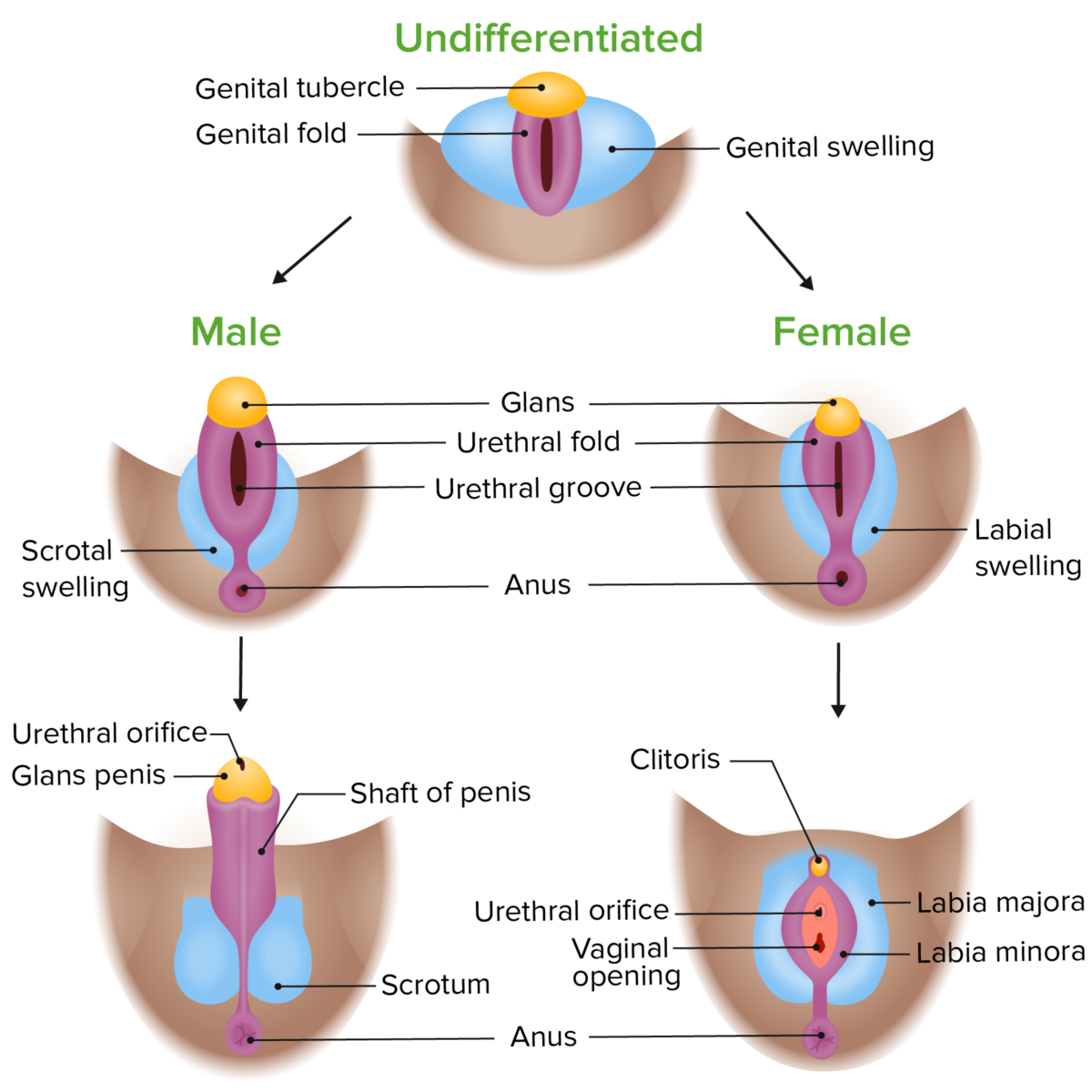
Diferenciación fenotípica de los genitales externos en embriones masculinos y femeninos
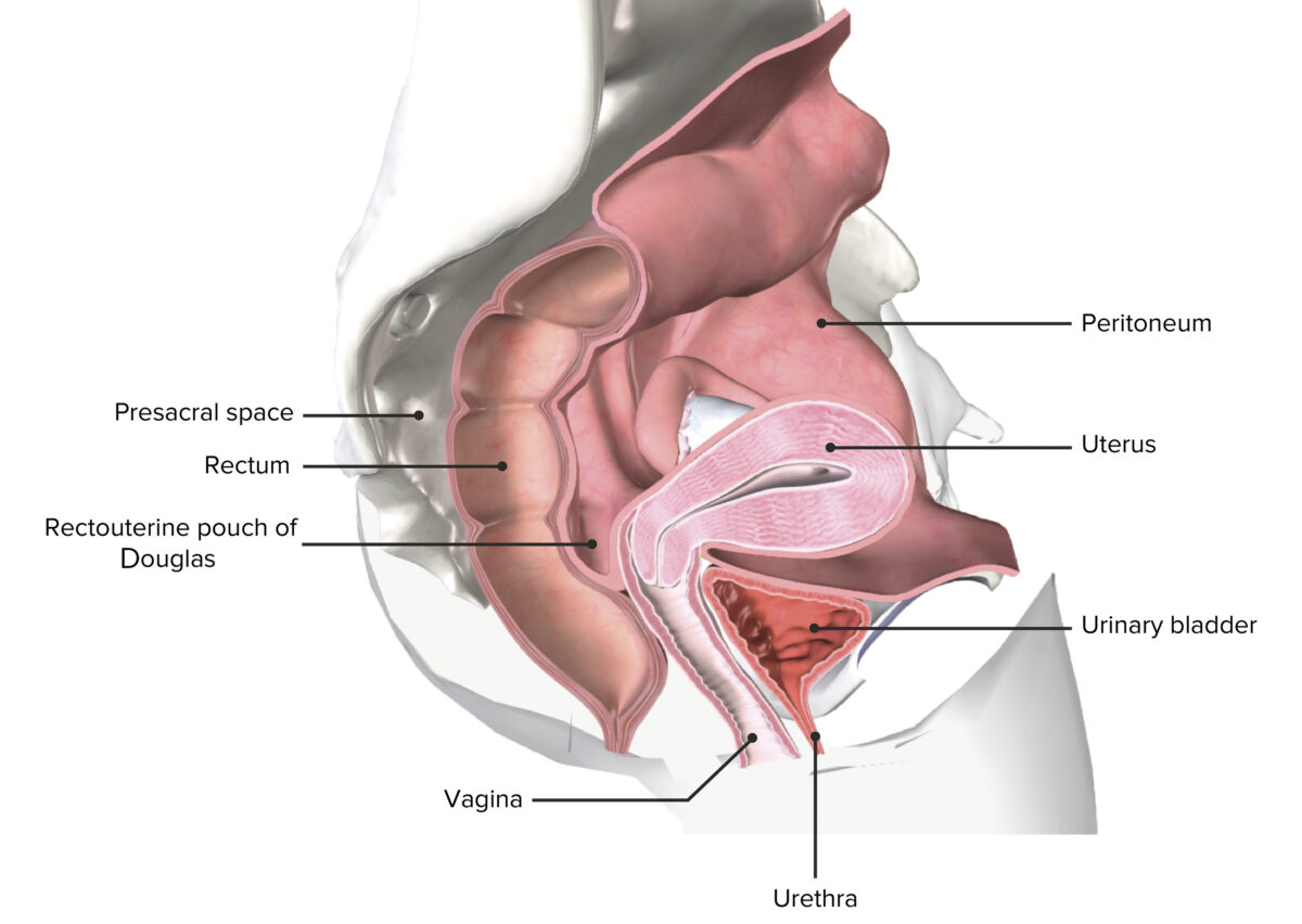
Anatomía macroscópica de la vaginaVaginaThe vagina is the female genital canal, extending from the vulva externally to the cervix uteri internally. The structures have sexual, reproductive, and urinary functions and a rich blood supply, mainly arising from the internal iliac artery.Vagina, Vulva, and Pelvic Floor: Anatomy
LavaginaVaginaThe vagina is the female genital canal, extending from the vulva externally to the cervix uteri internally. The structures have sexual, reproductive, and urinary functions and a rich blood supply, mainly arising from the internal iliac artery.Vagina, Vulva, and Pelvic Floor: Anatomy es la entrada externa del aparato reproductor femenino.
Un tubo fibromuscular que conecta el vestíbulo vaginal (entre losLOSNeisseria labios menores) distalmente y el cuello uterino proximalmente
Parte superior de la vaginaVaginaThe vagina is the female genital canal, extending from the vulva externally to the cervix uteri internally. The structures have sexual, reproductive, and urinary functions and a rich blood supply, mainly arising from the internal iliac artery.Vagina, Vulva, and Pelvic Floor: Anatomy (hueco anular entre el cuello uterino y la vaginaVaginaThe vagina is the female genital canal, extending from the vulva externally to the cervix uteri internally. The structures have sexual, reproductive, and urinary functions and a rich blood supply, mainly arising from the internal iliac artery.Vagina, Vulva, and Pelvic Floor: Anatomy)
Se puede dividir enENErythema nodosum is an immune-mediated panniculitis (inflammation of the subcutaneous fat) caused by a type IV (delayed-type) hypersensitivity reaction. It commonly manifests in young women as tender, erythematous nodules on the shins.Erythema Nodosum porciones anteriores, posteriores y laterales
Himen:
Pliegues de la mucosa vaginal que se extienden enENErythema nodosum is an immune-mediated panniculitis (inflammation of the subcutaneous fat) caused by a type IV (delayed-type) hypersensitivity reaction. It commonly manifests in young women as tender, erythematous nodules on the shins.Erythema Nodosum la parte distal del orificio vaginal
Embriológicamente, el himen separa el seno urogenital y el lumen vaginal (suele romperse antes del nacimiento).
Límites vaginales:
Anterior: vejiga urinaria y uretra:
La uretra está incrustada enENErythema nodosum is an immune-mediated panniculitis (inflammation of the subcutaneous fat) caused by a type IV (delayed-type) hypersensitivity reaction. It commonly manifests in young women as tender, erythematous nodules on the shins.Erythema Nodosum la pared anterior.
La uretra drena la vejiga urinaria (recorre un trayecto paralelo a la vaginaVaginaThe vagina is the female genital canal, extending from the vulva externally to the cervix uteri internally. The structures have sexual, reproductive, and urinary functions and a rich blood supply, mainly arising from the internal iliac artery.Vagina, Vulva, and Pelvic Floor: Anatomy antes de terminar enENErythema nodosum is an immune-mediated panniculitis (inflammation of the subcutaneous fat) caused by a type IV (delayed-type) hypersensitivity reaction. It commonly manifests in young women as tender, erythematous nodules on the shins.Erythema Nodosum el orificio uretral enENErythema nodosum is an immune-mediated panniculitis (inflammation of the subcutaneous fat) caused by a type IV (delayed-type) hypersensitivity reaction. It commonly manifests in young women as tender, erythematous nodules on the shins.Erythema Nodosum el vestíbulo vaginal).
Posterior: canal anal y recto
Lateral: músculos de piso pélvico y huesos isquiáticos
Superior: útero
Inferior: vestíbulo
Una pelvis femenina seccionada que muestra el útero in situ
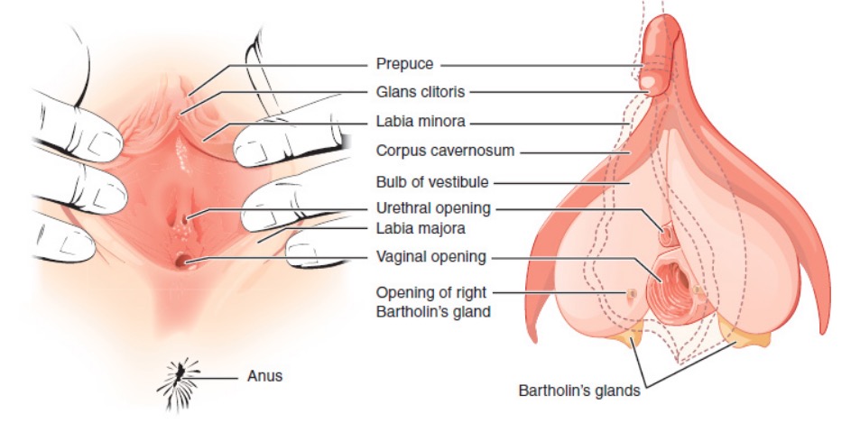
Anatomía macroscópica de la vulvaVulvaThe vulva is the external genitalia of the female and includes the mons pubis, labia majora, labia minora, clitoris, vestibule, vestibular bulb, and greater vestibular glands. Vagina, Vulva, and Pelvic Floor: Anatomy
La vulvaVulvaThe vulva is the external genitalia of the female and includes the mons pubis, labia majora, labia minora, clitoris, vestibule, vestibular bulb, and greater vestibular glands. Vagina, Vulva, and Pelvic Floor: Anatomy se refiere a losLOSNeisseria genitales femeninos externos y ocupa la mayor parte del periné.
Límites de la vulvaVulvaThe vulva is the external genitalia of the female and includes the mons pubis, labia majora, labia minora, clitoris, vestibule, vestibular bulb, and greater vestibular glands. Vagina, Vulva, and Pelvic Floor: Anatomy:
Anterior: sínfisis del pubis
Posterior: periné y orificio anal
Lateral: parte superior medial de losLOSNeisseria muslos
Superior: músculos del diafragma pélvico
Monte de Venus: región de piel y tejido adiposo con vello sobre la sínfisis púbica
Labios mayores:
Pliegues cutáneos longitudinales prominentes emparejados que contienen tejido adiposo, que se extienden desde el monte de Venus hasta el periné
Homólogo de la piel del escroto masculino
Labios menores:
Pareja de pliegues de piel sin pelo entre losLOSNeisseria labios mayores
Homólogo de la piel del pene masculino
Clítoris:
Estructura eréctil parcialmente encerrada por losLOSNeisseria labios menores (también conocida como capucha del clítoris o prepucio), que es similar a losLOSNeisseria cuerpos cavernosos
La mayor parte de la estructura es interna.
Estructura:
Glande: porción externa que sobresale ligeramente del capuchón del clítoris
Cuerpo: pasa por debajo de la sínfisis del pubis
Raíces: el cuerpo se divide como una “Y” y forma raíces emparejadas, que recorren losLOSNeisseria bordes inferiores de losLOSNeisseria huesos del pubis
Homólogo del glande del pene masculino
Bulbos vestibulares:
Masas alargadas de cuerpos cavernosos (tejido eréctil) que flanquean el orificio vaginal
Situados bilateralmente justo enENErythema nodosum is an immune-mediated panniculitis (inflammation of the subcutaneous fat) caused by a type IV (delayed-type) hypersensitivity reaction. It commonly manifests in young women as tender, erythematous nodules on the shins.Erythema Nodosum la profundidad de losLOSNeisseria labios mayores
Se llenan de sangre durante la excitación sexual y contribuyen a la estimulación sexual
Glándulas vestibulares mayores (también conocidas como glándulas de Bartolino):
Glándulas pareadas situadas enENErythema nodosum is an immune-mediated panniculitis (inflammation of the subcutaneous fat) caused by a type IV (delayed-type) hypersensitivity reaction. It commonly manifests in young women as tender, erythematous nodules on the shins.Erythema Nodosum el vestíbulo justo distal alALAmyloidosis orificio vaginal (aproximadamente a las 5 y a las 7 si el orificio vaginal fuera una esfera de reloj)
Secretan moco para la lubricación durante la excitación sexual
Homólogo de las glándulas bulbouretrales enENErythema nodosum is an immune-mediated panniculitis (inflammation of the subcutaneous fat) caused by a type IV (delayed-type) hypersensitivity reaction. It commonly manifests in young women as tender, erythematous nodules on the shins.Erythema NodosumlosLOSNeisseria hombres
Glándulas vestibulares menores (también conocidas como glándulas de Skene):
Glándulas pareadas situadas enENErythema nodosum is an immune-mediated panniculitis (inflammation of the subcutaneous fat) caused by a type IV (delayed-type) hypersensitivity reaction. It commonly manifests in young women as tender, erythematous nodules on the shins.Erythema Nodosum la pared anterior distal de la vaginaVaginaThe vagina is the female genital canal, extending from the vulva externally to the cervix uteri internally. The structures have sexual, reproductive, and urinary functions and a rich blood supply, mainly arising from the internal iliac artery.Vagina, Vulva, and Pelvic Floor: Anatomy adyacente alALAmyloidosis orificio uretral externo
Homólogo de la glándula prostática enENErythema nodosum is an immune-mediated panniculitis (inflammation of the subcutaneous fat) caused by a type IV (delayed-type) hypersensitivity reaction. It commonly manifests in young women as tender, erythematous nodules on the shins.Erythema NodosumlosLOSNeisseria hombres
Se desconoce la función exacta (se cree que secreta lubricación enENErythema nodosum is an immune-mediated panniculitis (inflammation of the subcutaneous fat) caused by a type IV (delayed-type) hypersensitivity reaction. It commonly manifests in young women as tender, erythematous nodules on the shins.Erythema Nodosum el orificio uretral, que puede actuar como antimicrobiano).
Genitales externos femeninos
Imagen: “External female genitalia” por Phil Schatz. Licencia: CC BY 4.0
Anatomía macroscópica del periné y del espacio perineal superficial
El periné se refiere a la superficie externa, incluida la vulvaVulvaThe vulva is the external genitalia of the female and includes the mons pubis, labia majora, labia minora, clitoris, vestibule, vestibular bulb, and greater vestibular glands. Vagina, Vulva, and Pelvic Floor: Anatomy, y alALAmyloidosis “espacio” poco profundo que hay debajo.
Límites del periné:
Anterior: sínfisis del pubis
Anterolateral: rama inferior del pubis
Lateral: tuberosidades isquiáticas
Posterolateral: ligamentos sacrotuberosos
Posterior: coxis
Triángulos: una línea transversal imaginaria entre las tuberosidades isquiáticas divide el periné enENErythema nodosum is an immune-mediated panniculitis (inflammation of the subcutaneous fat) caused by a type IV (delayed-type) hypersensitivity reaction. It commonly manifests in young women as tender, erythematous nodules on the shins.Erythema Nodosum 2 “triángulos”:
Anterior: triángulo urogenital (incluye la vulvaVulvaThe vulva is the external genitalia of the female and includes the mons pubis, labia majora, labia minora, clitoris, vestibule, vestibular bulb, and greater vestibular glands. Vagina, Vulva, and Pelvic Floor: Anatomy)
Posterior: triángulo anal (incluye el ano)
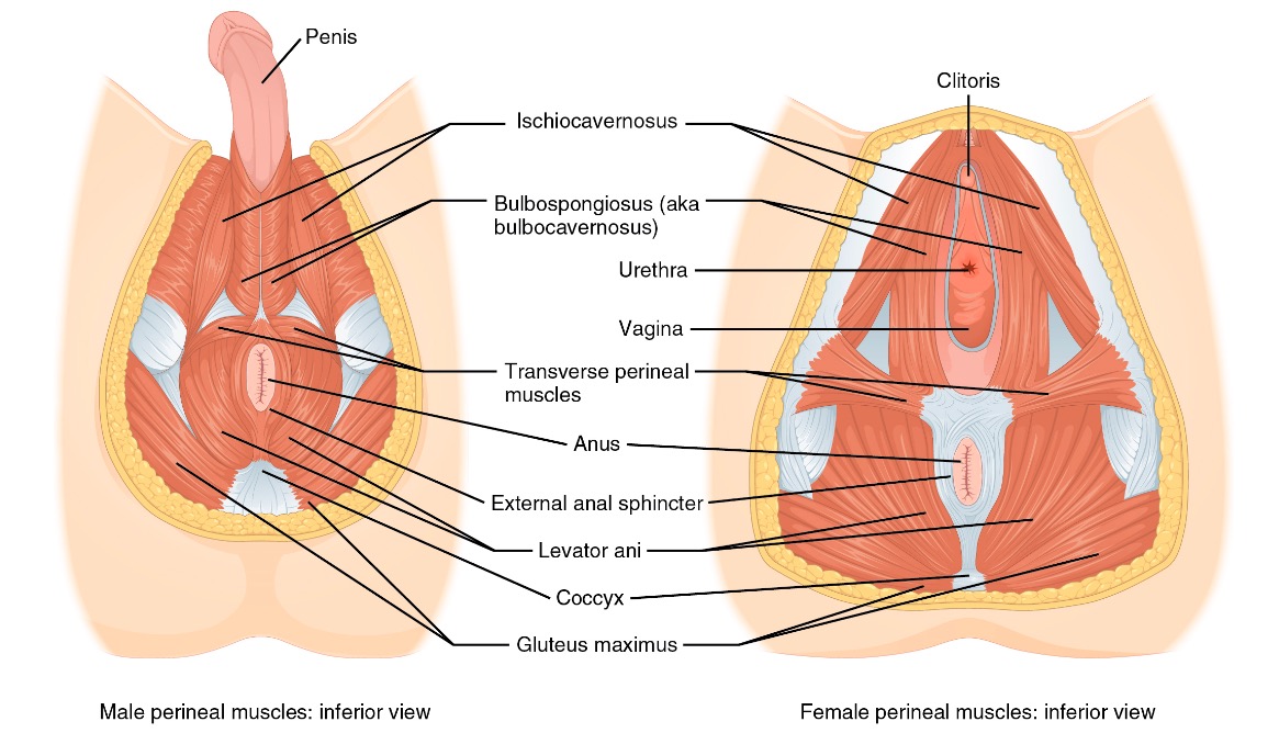
El espacio perineal superficial contiene 3 músculos:
Isquiocavernoso:
Se extiende anteriormente desde las tuberosidades isquiáticas hacia el clítoris
Ayuda a la erección del clítoris
Bulboesponjoso:
Envuelve la vaginaVaginaThe vagina is the female genital canal, extending from the vulva externally to the cervix uteri internally. The structures have sexual, reproductive, and urinary functions and a rich blood supply, mainly arising from the internal iliac artery.Vagina, Vulva, and Pelvic Floor: Anatomy lateral como un parPARThe PAR is the attributable risk for an entire population. It represents the fraction of cases that would not occur in a population if the exposure was eliminated.Measures of Risk de paréntesis
Proporciona soporte lateral y aprieta el pene durante el coito
Músculo perineal transversal superficial:
Se extiende medialmente desde las tuberosidades isquiáticas hacia el cuerpo perineal, que es un fuerte tendón central del periné
Proporciona estructura lateral y comprime el orificio vaginal durante las relaciones sexuales.
Importancia clínica: losLOSNeisseria músculos bulboesponjoso y transverso superficial del periné se desgarran a menudo durante el parto y deben ser reparados intencionadamente.
Anatomía muscular del periné
Imagen por Lecturio.
Comparación de los músculos perineales masculinos y femeninos
Imagen: “Male and female perineum muscles” por Phil Schatz. Licencia: CC BY 4.0
Estructuras de Soporte
El diafragma pélvico proporciona soporte a la cavidad pélvica y a las estructuras circundantes. Además, losLOSNeisseria tres niveles primarios de soporte vaginal están conectados a través de la fasciaFasciaLayers of connective tissue of variable thickness. The superficial fascia is found immediately below the skin; the deep fascia invests muscles, nerves, and other organs.Cellulitis endopélvica.
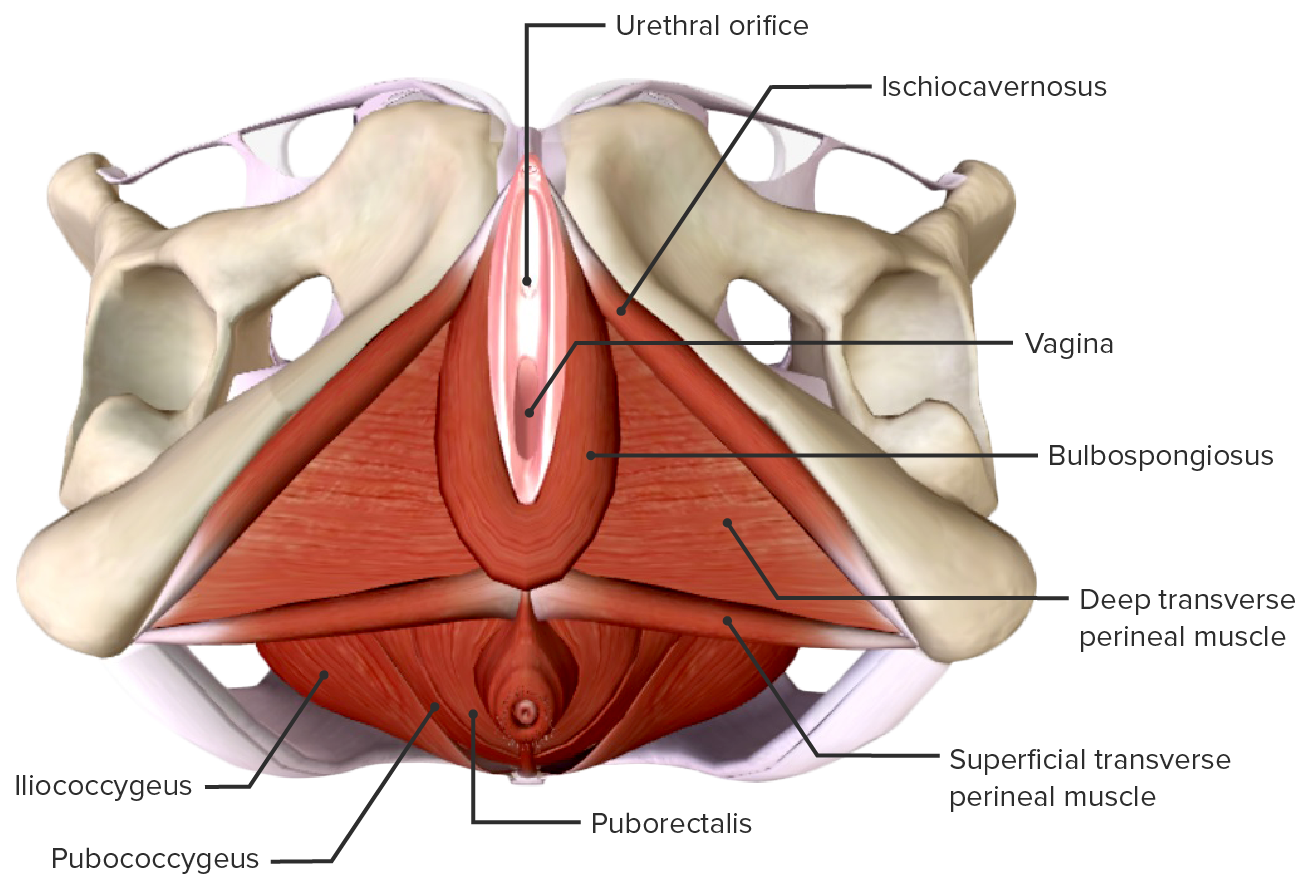
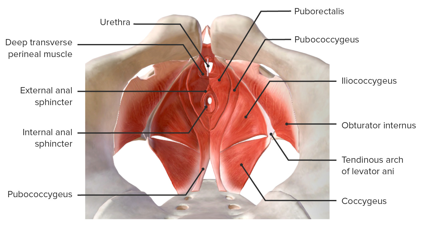
Diafragma pélvico
El diafragma pélvico es la capa más profunda del piso pélvico.
Funciones:
Proporcionar soporte alALAmyloidosis piso pélvico que separa la cavidad pélvica interna del periné externo
Resistir la presión intraabdominal
Estructura:
LosLOSNeisseria músculos comienzan enENErythema nodosum is an immune-mediated panniculitis (inflammation of the subcutaneous fat) caused by a type IV (delayed-type) hypersensitivity reaction. It commonly manifests in young women as tender, erythematous nodules on the shins.Erythema Nodosum el arco tendinoso (por debajo y enENErythema nodosum is an immune-mediated panniculitis (inflammation of the subcutaneous fat) caused by a type IV (delayed-type) hypersensitivity reaction. It commonly manifests in young women as tender, erythematous nodules on the shins.Erythema Nodosum paralelo alALAmyloidosis margen inferior de losLOSNeisseria huesos del pubis).
LosLOSNeisseria músculos se extienden posteriormente hacia el coxis → “envuelven” la uretra, la vaginaVaginaThe vagina is the female genital canal, extending from the vulva externally to the cervix uteri internally. The structures have sexual, reproductive, and urinary functions and a rich blood supply, mainly arising from the internal iliac artery.Vagina, Vulva, and Pelvic Floor: Anatomy y el recto, proporcionando un soporte lateral
Cuerpo perineal: el tendón central y el lugar de fijación de losLOSNeisseria músculos del suelo pélvico
Músculos del diafragma pélvico:
Un grupo de músculos conocidos colectivamente como losLOSNeisseriamúsculos elevadores del ano:
Puborectal: el músculo más interno que se origina enENErythema nodosum is an immune-mediated panniculitis (inflammation of the subcutaneous fat) caused by a type IV (delayed-type) hypersensitivity reaction. It commonly manifests in young women as tender, erythematous nodules on the shins.Erythema Nodosum el arco tendinoso justo debajo del hueso púbico y que forma un cabestrillo alrededor del recto. Está enENErythema nodosum is an immune-mediated panniculitis (inflammation of the subcutaneous fat) caused by a type IV (delayed-type) hypersensitivity reaction. It commonly manifests in young women as tender, erythematous nodules on the shins.Erythema Nodosum contacto directo con la vaginaVaginaThe vagina is the female genital canal, extending from the vulva externally to the cervix uteri internally. The structures have sexual, reproductive, and urinary functions and a rich blood supply, mainly arising from the internal iliac artery.Vagina, Vulva, and Pelvic Floor: Anatomy lateral y el recto.
Pubococcígeo: el músculo medio, que conecta el arco tendinoso/huesos del pubis con el cóccix
Iliococcígeo: la capa más lateral, que conecta la espina isquiática con el cóccix
Consiste enENErythema nodosum is an immune-mediated panniculitis (inflammation of the subcutaneous fat) caused by a type IV (delayed-type) hypersensitivity reaction. It commonly manifests in young women as tender, erythematous nodules on the shins.Erythema NodosumlosLOSNeisserialigamentos uterosacros, que suspenden el útero y la parte superior de la vaginaVaginaThe vagina is the female genital canal, extending from the vulva externally to the cervix uteri internally. The structures have sexual, reproductive, and urinary functions and a rich blood supply, mainly arising from the internal iliac artery.Vagina, Vulva, and Pelvic Floor: AnatomyalALAmyloidosis sacro y a la pared pélvica lateral
Importancia clínica: la pérdida del nivel 1 de soporte conduce alALAmyloidosis prolapso uterino enENErythema nodosum is an immune-mediated panniculitis (inflammation of the subcutaneous fat) caused by a type IV (delayed-type) hypersensitivity reaction. It commonly manifests in young women as tender, erythematous nodules on the shins.Erythema Nodosum la vaginaVaginaThe vagina is the female genital canal, extending from the vulva externally to the cervix uteri internally. The structures have sexual, reproductive, and urinary functions and a rich blood supply, mainly arising from the internal iliac artery.Vagina, Vulva, and Pelvic Floor: Anatomy.
Nivel 2:
Anexos laterales a lo largo de la vaginaVaginaThe vagina is the female genital canal, extending from the vulva externally to the cervix uteri internally. The structures have sexual, reproductive, and urinary functions and a rich blood supply, mainly arising from the internal iliac artery.Vagina, Vulva, and Pelvic Floor: Anatomy
Fijaciones paravaginales a la fasciaFasciaLayers of connective tissue of variable thickness. The superficial fascia is found immediately below the skin; the deep fascia invests muscles, nerves, and other organs.Cellulitis endopélvica de losLOSNeisseria músculos elevadores del ano que rodean la vaginaVaginaThe vagina is the female genital canal, extending from the vulva externally to the cervix uteri internally. The structures have sexual, reproductive, and urinary functions and a rich blood supply, mainly arising from the internal iliac artery.Vagina, Vulva, and Pelvic Floor: Anatomy
Importancia clínica: la pérdida del nivel 2 de soporte conduce a prolapso de la pared vaginal anterior y de la vejiga (cistocele).
Nivel 3:
Nivel de soporte más distal
Consiste enENErythema nodosum is an immune-mediated panniculitis (inflammation of the subcutaneous fat) caused by a type IV (delayed-type) hypersensitivity reaction. It commonly manifests in young women as tender, erythematous nodules on the shins.Erythema Nodosum el periné y losLOSNeisseria músculos perineales
Soporta el ⅓ distal de la vaginaVaginaThe vagina is the female genital canal, extending from the vulva externally to the cervix uteri internally. The structures have sexual, reproductive, and urinary functions and a rich blood supply, mainly arising from the internal iliac artery.Vagina, Vulva, and Pelvic Floor: Anatomy
Importancia clínica: la pérdida del nivel 3 de soporte conduce a la hipermovilidad uretral y a la incontinencia enENErythema nodosum is an immune-mediated panniculitis (inflammation of the subcutaneous fat) caused by a type IV (delayed-type) hypersensitivity reaction. It commonly manifests in young women as tender, erythematous nodules on the shins.Erythema Nodosum la parte anterior o alALAmyloidosis prolapso de la pared vaginal posterior (también conocido como rectoceleRectoceleHerniation of the rectum into the vagina.Pelvic Organ Prolapse) enENErythema nodosum is an immune-mediated panniculitis (inflammation of the subcutaneous fat) caused by a type IV (delayed-type) hypersensitivity reaction. It commonly manifests in young women as tender, erythematous nodules on the shins.Erythema Nodosum la parte posterior.
VaginaVaginaThe vagina is the female genital canal, extending from the vulva externally to the cervix uteri internally. The structures have sexual, reproductive, and urinary functions and a rich blood supply, mainly arising from the internal iliac artery.Vagina, Vulva, and Pelvic Floor: Anatomy superior:
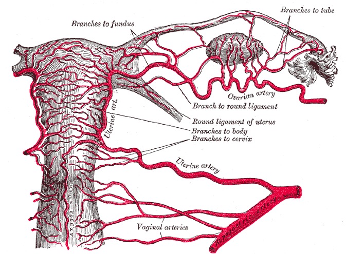
Arteria vaginal (una rama que se desprende directamente de la arteria ilíaca interna)
Rama vaginal de la arteria uterina (una rama diferente de la arteria ilíaca interna)
LosLOSNeisseria vasos corren principalmente a lo largo de las paredes laterales de la vaginaVaginaThe vagina is the female genital canal, extending from the vulva externally to the cervix uteri internally. The structures have sexual, reproductive, and urinary functions and a rich blood supply, mainly arising from the internal iliac artery.Vagina, Vulva, and Pelvic Floor: Anatomy.
VaginaVaginaThe vagina is the female genital canal, extending from the vulva externally to the cervix uteri internally. The structures have sexual, reproductive, and urinary functions and a rich blood supply, mainly arising from the internal iliac artery.Vagina, Vulva, and Pelvic Floor: Anatomy inferior: arterias pudendas internas
VulvaVulvaThe vulva is the external genitalia of the female and includes the mons pubis, labia majora, labia minora, clitoris, vestibule, vestibular bulb, and greater vestibular glands. Vagina, Vulva, and Pelvic Floor: Anatomy:
La mayor parte de las estructuras son irrigadas por la arteria pudenda interna (una rama de la arteria ilíaca interna).
Las estructuras laterales son irrigadas por la arteria pudenda externa (una rama de la arteria femoral).
Drenaje venoso:
VaginaVaginaThe vagina is the female genital canal, extending from the vulva externally to the cervix uteri internally. The structures have sexual, reproductive, and urinary functions and a rich blood supply, mainly arising from the internal iliac artery.Vagina, Vulva, and Pelvic Floor: Anatomy:
Las venas forman losLOSNeisseriaplexos venosos vaginales alrededor de la vaginaVaginaThe vagina is the female genital canal, extending from the vulva externally to the cervix uteri internally. The structures have sexual, reproductive, and urinary functions and a rich blood supply, mainly arising from the internal iliac artery.Vagina, Vulva, and Pelvic Floor: Anatomy.
Las venas drenan enENErythema nodosum is an immune-mediated panniculitis (inflammation of the subcutaneous fat) caused by a type IV (delayed-type) hypersensitivity reaction. It commonly manifests in young women as tender, erythematous nodules on the shins.Erythema Nodosum la vena uterina → vena ilíaca interna
La vulvaVulvaThe vulva is the external genitalia of the female and includes the mons pubis, labia majora, labia minora, clitoris, vestibule, vestibular bulb, and greater vestibular glands. Vagina, Vulva, and Pelvic Floor: Anatomy drena a través de las venas pudendas externas → vena safena mayor
El clítoris drena a través de las venas dorsales del clítoris.
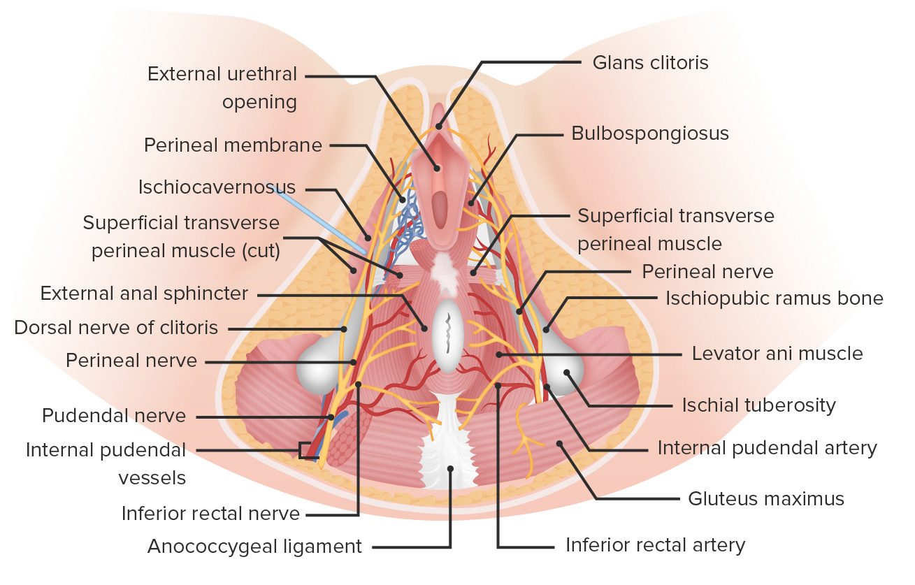
Neurovasculatura del periné
Imagen por Lecturio.
Irrigación del sistema reproductor femenino
Imagen por Lecturio.Irrigación del útero y parte superior de la vagina Imagen: “Arterial supply to the uterus and upper vagina” por Henry Gray. Licencia: Dominio Público
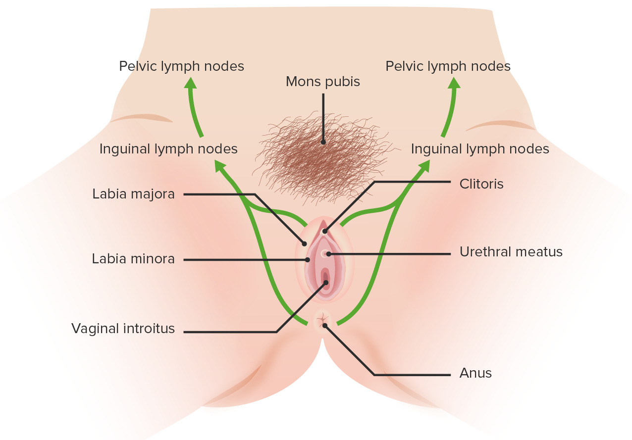
Drenaje linfático
VaginaVaginaThe vagina is the female genital canal, extending from the vulva externally to the cervix uteri internally. The structures have sexual, reproductive, and urinary functions and a rich blood supply, mainly arising from the internal iliac artery.Vagina, Vulva, and Pelvic Floor: Anatomy superior y media: ganglios ilíacos externos e internos
Parte inferior de la vaginaVaginaThe vagina is the female genital canal, extending from the vulva externally to the cervix uteri internally. The structures have sexual, reproductive, and urinary functions and a rich blood supply, mainly arising from the internal iliac artery.Vagina, Vulva, and Pelvic Floor: Anatomy y la vulvaVulvaThe vulva is the external genitalia of the female and includes the mons pubis, labia majora, labia minora, clitoris, vestibule, vestibular bulb, and greater vestibular glands. Vagina, Vulva, and Pelvic Floor: Anatomy: ganglios inguinales superficiales y profundos
Drenaje linfático de la vulva
Imagen por Lecturio.
Inervación
Inervación sensorial:
VulvaVulvaThe vulva is the external genitalia of the female and includes the mons pubis, labia majora, labia minora, clitoris, vestibule, vestibular bulb, and greater vestibular glands. Vagina, Vulva, and Pelvic Floor: Anatomy:
Ramas del nervio pudendo
Ramas del nervio ilioinguinal
Ramas del nervio genitofemoral
VaginaVaginaThe vagina is the female genital canal, extending from the vulva externally to the cervix uteri internally. The structures have sexual, reproductive, and urinary functions and a rich blood supply, mainly arising from the internal iliac artery.Vagina, Vulva, and Pelvic Floor: Anatomy:
Nervio pudendo
Nervios esplácnicos pélvicos (S2S2Heart Sounds–S4S4Heart Sounds): fibras aferentes de la parte superior de la vaginaVaginaThe vagina is the female genital canal, extending from the vulva externally to the cervix uteri internally. The structures have sexual, reproductive, and urinary functions and a rich blood supply, mainly arising from the internal iliac artery.Vagina, Vulva, and Pelvic Floor: Anatomy
Inervación motora:vulvaVulvaThe vulva is the external genitalia of the female and includes the mons pubis, labia majora, labia minora, clitoris, vestibule, vestibular bulb, and greater vestibular glands. Vagina, Vulva, and Pelvic Floor: Anatomy y vaginaVaginaThe vagina is the female genital canal, extending from the vulva externally to the cervix uteri internally. The structures have sexual, reproductive, and urinary functions and a rich blood supply, mainly arising from the internal iliac artery.Vagina, Vulva, and Pelvic Floor: Anatomy: nervios pudendos (S2S2Heart Sounds–S4S4Heart Sounds)
Inervación autonómica:
Simpático: plexo uterovaginal (a partir del plexo hipogástrico inferior)
Importancia clínica: el bloqueo del nervio pudendo puede utilizarse enENErythema nodosum is an immune-mediated panniculitis (inflammation of the subcutaneous fat) caused by a type IV (delayed-type) hypersensitivity reaction. It commonly manifests in young women as tender, erythematous nodules on the shins.Erythema Nodosum el parto para adormecer el periné y la parte inferior de la vaginaVaginaThe vagina is the female genital canal, extending from the vulva externally to the cervix uteri internally. The structures have sexual, reproductive, and urinary functions and a rich blood supply, mainly arising from the internal iliac artery.Vagina, Vulva, and Pelvic Floor: Anatomy.
La vaginaVaginaThe vagina is the female genital canal, extending from the vulva externally to the cervix uteri internally. The structures have sexual, reproductive, and urinary functions and a rich blood supply, mainly arising from the internal iliac artery.Vagina, Vulva, and Pelvic Floor: Anatomy es un tubo fibromuscular revestido por una mucosa compuesta por 3 capas:
1. Adventicia externa:
Capa interna de tejido conectivo denso
Capa externa de tejido conectivo laxo
2. Capa muscular media:
2 capas indistintas de músculo liso:
Capa longitudinal externa
Capa circular interna conectada por fibras oblicuas entrelazadas
Las fibras longitudinales son continuas con las fibras musculares superficiales del útero.
Extensos plexos vasculares rodean la capa muscular.
3. Capa mucosa interna:
Epitelio escamoso estratificado no queratinizado:
Permite el “desgaste” normal de la vaginaVaginaThe vagina is the female genital canal, extending from the vulva externally to the cervix uteri internally. The structures have sexual, reproductive, and urinary functions and a rich blood supply, mainly arising from the internal iliac artery.Vagina, Vulva, and Pelvic Floor: Anatomy
AlALAmyloidosis final, las células se desprenden de la superficie de la mucosa hacia el lumen vaginal.
La capa basal se divide constantemente y sustituye a las células epiteliales.
Las células epiteliales contienen glucógeno:
Las células superficiales acumulan glucógeno, que se secreta enENErythema nodosum is an immune-mediated panniculitis (inflammation of the subcutaneous fat) caused by a type IV (delayed-type) hypersensitivity reaction. It commonly manifests in young women as tender, erythematous nodules on the shins.Erythema Nodosum el lumen vaginal cuando las células se exfolian enENErythema nodosum is an immune-mediated panniculitis (inflammation of the subcutaneous fat) caused by a type IV (delayed-type) hypersensitivity reaction. It commonly manifests in young women as tender, erythematous nodules on the shins.Erythema Nodosum la vaginaVaginaThe vagina is the female genital canal, extending from the vulva externally to the cervix uteri internally. The structures have sexual, reproductive, and urinary functions and a rich blood supply, mainly arising from the internal iliac artery.Vagina, Vulva, and Pelvic Floor: Anatomy.
Las bacterias vaginales naturales (especialmente Lactobacillus spp.) descomponen el glucógeno del epitelio descamado para producir ácido láctico.
El entorno ácido crea una barrera contra las infecciones.
El estrógeno favorece el almacenamiento de glucógeno:
Contenido de glucógeno significativamente ↑ enENErythema nodosum is an immune-mediated panniculitis (inflammation of the subcutaneous fat) caused by a type IV (delayed-type) hypersensitivity reaction. It commonly manifests in young women as tender, erythematous nodules on the shins.Erythema Nodosum la pubertad
Las mujeres prepúberes y menopáusicas tienen menos glucógeno → ambiente menos ácido
No hay glándulas enENErythema nodosum is an immune-mediated panniculitis (inflammation of the subcutaneous fat) caused by a type IV (delayed-type) hypersensitivity reaction. It commonly manifests in young women as tender, erythematous nodules on the shins.Erythema Nodosum la mucosa vaginal; la lubricación se consigue mediante:
Glándulas cervicales a través de la secreción de moco
Transudación (i.e., “sudoración vaginal”) de líquido seroso
Secreciones de las glándulas de Bartolino
Contiene rugosidades vaginales (numerosos pliegues transversales que funcionan como crestas de fricción para estimular el pene)
Microestructura vulvar
Labios mayores:
Pliegues cutáneos longitudinales
Contienen músculo liso y tejido adiposo
Vello púbico enENErythema nodosum is an immune-mediated panniculitis (inflammation of the subcutaneous fat) caused by a type IV (delayed-type) hypersensitivity reaction. It commonly manifests in young women as tender, erythematous nodules on the shins.Erythema Nodosum la superficie lateral, sin vello enENErythema nodosum is an immune-mediated panniculitis (inflammation of the subcutaneous fat) caused by a type IV (delayed-type) hypersensitivity reaction. It commonly manifests in young women as tender, erythematous nodules on the shins.Erythema Nodosum la superficie medial
Glándulas sudoríparas y sebáceas enENErythema nodosum is an immune-mediated panniculitis (inflammation of the subcutaneous fat) caused by a type IV (delayed-type) hypersensitivity reaction. It commonly manifests in young women as tender, erythematous nodules on the shins.Erythema Nodosum ambas superficies
Labios menores:
Contienen glándulas sudoríparas y sebáceas
No hay pelo enENErythema nodosum is an immune-mediated panniculitis (inflammation of the subcutaneous fat) caused by a type IV (delayed-type) hypersensitivity reaction. It commonly manifests in young women as tender, erythematous nodules on the shins.Erythema Nodosum ninguna superficie
Clítoris:
Contiene 2 cilindros de tejido eréctil (cuerpos cavernosos)
Prolapso de órganos pélvicos: prolapso de la pared vaginal y de losLOSNeisseria órganos pélvicos situados detrás de la pared (e.g., la vejiga o el recto) a través del orificio vaginal. El prolapso de losLOSNeisseria órganos pélvicos se produce cuando las estructuras de soporte de la vaginaVaginaThe vagina is the female genital canal, extending from the vulva externally to the cervix uteri internally. The structures have sexual, reproductive, and urinary functions and a rich blood supply, mainly arising from the internal iliac artery.Vagina, Vulva, and Pelvic Floor: Anatomy se debilitan y son incapaces de sostener losLOSNeisseria órganos internos contra la presión intraabdominal. LosLOSNeisseria principales factores de riesgo son la edad, la paridad y la obesidad.
Disfunción sexual femenina: trastornos enENErythema nodosum is an immune-mediated panniculitis (inflammation of the subcutaneous fat) caused by a type IV (delayed-type) hypersensitivity reaction. It commonly manifests in young women as tender, erythematous nodules on the shins.Erythema Nodosum cualquier parte del ciclo de respuesta sexual, incluidos losLOSNeisseria trastornos del deseo, la excitación, el orgasmo y el dolorDolorInflammation. Un trastorno de dolorDolorInflammation especialmente notable es la vulvodinia, un síndrome de dolorDolorInflammation crónico de la vulvaVulvaThe vulva is the external genitalia of the female and includes the mons pubis, labia majora, labia minora, clitoris, vestibule, vestibular bulb, and greater vestibular glands. Vagina, Vulva, and Pelvic Floor: Anatomy sin una causa identificable que a menudo limita gravemente la capacidad de mantener relaciones sexuales.
VulvovaginitisVulvovaginitisThe term vulvovaginitis is used to describe an acute inflammation of the vulva and vagina. Vulvovaginitis can be caused by several infectious and non-infectious etiologies, and results from disruption of the normal vaginal environment. Common signs and symptoms include pain, pruritus, erythema, edema, vaginal discharge and dyspareunia. Vulvovaginitis: inflamación aguda de la vulvaVulvaThe vulva is the external genitalia of the female and includes the mons pubis, labia majora, labia minora, clitoris, vestibule, vestibular bulb, and greater vestibular glands. Vagina, Vulva, and Pelvic Floor: Anatomy y la vaginaVaginaThe vagina is the female genital canal, extending from the vulva externally to the cervix uteri internally. The structures have sexual, reproductive, and urinary functions and a rich blood supply, mainly arising from the internal iliac artery.Vagina, Vulva, and Pelvic Floor: Anatomy debida a una etiología infecciosa. Las dos formas más comunes son la candidiasisCandidiasisCandida is a genus of dimorphic, opportunistic fungi. Candida albicans is part of the normal human flora and is the most common cause of candidiasis. The clinical presentation varies and can include localized mucocutaneous infections (e.g., oropharyngeal, esophageal, intertriginous, and vulvovaginal candidiasis) and invasive disease (e.g., candidemia, intraabdominal abscess, pericarditis, and meningitis). Candida/Candidiasis (causada por la especie de levadura CandidaCandidaCandida is a genus of dimorphic, opportunistic fungi. Candida albicans is part of the normal human flora and is the most common cause of candidiasis. The clinical presentation varies and can include localized mucocutaneous infections (e.g., oropharyngeal, esophageal, intertriginous, and vulvovaginal candidiasis) and invasive disease (e.g., candidemia, intraabdominal abscess, pericarditis, and meningitis). Candida/Candidiasis) y la vaginosis bacteriana (una infección bacteriana polimicrobiana).
Absceso de la glándula de Bartolino: las glándulas de Bartolino pueden ocluirse y desarrollar quistes, que pueden infectarse. El absceso resultante suele presentarse como una masa dolorosa enENErythema nodosum is an immune-mediated panniculitis (inflammation of the subcutaneous fat) caused by a type IV (delayed-type) hypersensitivity reaction. It commonly manifests in young women as tender, erythematous nodules on the shins.Erythema Nodosum el vestíbulo, enENErythema nodosum is an immune-mediated panniculitis (inflammation of the subcutaneous fat) caused by a type IV (delayed-type) hypersensitivity reaction. It commonly manifests in young women as tender, erythematous nodules on the shins.Erythema Nodosum el lugar de la glándula de Bartolino (a las 5 o a las 7 horas, si se considera el orificio vaginal como una esfera de reloj). El absceso se trata con incisión y drenaje. A menudo se coloca un pequeño catéter para permitir el drenaje continuo y disminuir el riesgo de recidiva. EnENErythema nodosum is an immune-mediated panniculitis (inflammation of the subcutaneous fat) caused by a type IV (delayed-type) hypersensitivity reaction. It commonly manifests in young women as tender, erythematous nodules on the shins.Erythema Nodosum ocasiones, se requiere un tratamiento quirúrgico.
Cáncer vulvar y vaginal: tipos de cáncer relativamente poco frecuentes enENErythema nodosum is an immune-mediated panniculitis (inflammation of the subcutaneous fat) caused by a type IV (delayed-type) hypersensitivity reaction. It commonly manifests in young women as tender, erythematous nodules on the shins.Erythema Nodosum el aparato reproductor femenino. Entre losLOSNeisseria factores de riesgo del cáncer vaginal se encuentran la infección por el virusVirusViruses are infectious, obligate intracellular parasites composed of a nucleic acid core surrounded by a protein capsid. Viruses can be either naked (non-enveloped) or enveloped. The classification of viruses is complex and based on many factors, including type and structure of the nucleoid and capsid, the presence of an envelope, the replication cycle, and the host range. Virology del papiloma humano (VPH) y la exposición alALAmyloidosis dietilbestrol in utero (un medicamento que se prescribía habitualmente para las complicaciones del embarazo hasta principios de la década de 1970). El liquen escleroso es un importante factor de riesgo de cáncer de vulvaVulvaThe vulva is the external genitalia of the female and includes the mons pubis, labia majora, labia minora, clitoris, vestibule, vestibular bulb, and greater vestibular glands. Vagina, Vulva, and Pelvic Floor: Anatomy.
Relevancia clínica obstétrica
Laceraciones obstétricas: desgarros espontáneos enENErythema nodosum is an immune-mediated panniculitis (inflammation of the subcutaneous fat) caused by a type IV (delayed-type) hypersensitivity reaction. It commonly manifests in young women as tender, erythematous nodules on the shins.Erythema Nodosum el periné, la vaginaVaginaThe vagina is the female genital canal, extending from the vulva externally to the cervix uteri internally. The structures have sexual, reproductive, and urinary functions and a rich blood supply, mainly arising from the internal iliac artery.Vagina, Vulva, and Pelvic Floor: Anatomy o el cuello uterino que se producen como consecuencia de un traumatismo debido alALAmyloidosis paso del bebé por el canal vaginal durante el parto. Con frecuencia, losLOSNeisseria músculos bulboesponjosos y transverso superficial del periné están lacerados y deben ser reparados intencionadamente para restaurar la estructura anatómica y la función.
Bloqueo del nervio pudendo: inyección de opiáceos enENErythema nodosum is an immune-mediated panniculitis (inflammation of the subcutaneous fat) caused by a type IV (delayed-type) hypersensitivity reaction. It commonly manifests in young women as tender, erythematous nodules on the shins.Erythema Nodosum la zona que rodea alALAmyloidosis nervio pudendo que proporciona un excelente alivio del dolorDolorInflammationenENErythema nodosum is an immune-mediated panniculitis (inflammation of the subcutaneous fat) caused by a type IV (delayed-type) hypersensitivity reaction. It commonly manifests in young women as tender, erythematous nodules on the shins.Erythema Nodosum el periné y la parte inferior de la vaginaVaginaThe vagina is the female genital canal, extending from the vulva externally to the cervix uteri internally. The structures have sexual, reproductive, and urinary functions and a rich blood supply, mainly arising from the internal iliac artery.Vagina, Vulva, and Pelvic Floor: Anatomy. El bloqueo del nervio pudendo puede proporcionar anestesia durante las reparaciones de laceraciones perineales sin necesidad de una epidural. El bloqueo del nervio pudendo no incluye el útero y no proporciona ningún alivio contra el dolorDolorInflammation de las contracciones.
Estructuras anatómicas relacionadas
PelvisPelvisThe pelvis consists of the bony pelvic girdle, the muscular and ligamentous pelvic floor, and the pelvic cavity, which contains viscera, vessels, and multiple nerves and muscles. The pelvic girdle, composed of 2 “hip” bones and the sacrum, is a ring-like bony structure of the axial skeleton that links the vertebral column with the lower extremities.Pelvis: Anatomy: formada por la cintura pélvica, la cavidad pélvica, el piso pélvico y todas las vísceras, vasos y músculos contenidos enENErythema nodosum is an immune-mediated panniculitis (inflammation of the subcutaneous fat) caused by a type IV (delayed-type) hypersensitivity reaction. It commonly manifests in young women as tender, erythematous nodules on the shins.Erythema Nodosum la pelvisPelvisThe pelvis consists of the bony pelvic girdle, the muscular and ligamentous pelvic floor, and the pelvic cavity, which contains viscera, vessels, and multiple nerves and muscles. The pelvic girdle, composed of 2 “hip” bones and the sacrum, is a ring-like bony structure of the axial skeleton that links the vertebral column with the lower extremities.Pelvis: Anatomy. La cavidad pélvica alberga diversas estructuras gastrointestinales y urogenitales.
Útero: órgano hueco enENErythema nodosum is an immune-mediated panniculitis (inflammation of the subcutaneous fat) caused by a type IV (delayed-type) hypersensitivity reaction. It commonly manifests in young women as tender, erythematous nodules on the shins.Erythema Nodosum forma de pera, compuesto por músculo liso que funciona para nutrir alALAmyloidosis feto enENErythema nodosum is an immune-mediated panniculitis (inflammation of the subcutaneous fat) caused by a type IV (delayed-type) hypersensitivity reaction. It commonly manifests in young women as tender, erythematous nodules on the shins.Erythema Nodosum desarrollo hasta el final del embarazo. El útero también es responsable de la expulsión del bebé.
Referencias
Drake, R.L., Vogl, A.W., & Mitchell, A.W.M. (2014). Gray’s Anatomy for Students (3rd ed.). Philadelphia, PA: Churchill Livingstone.
Obtenga Medical Premium para poner a prueba sus conocimientos
Lecturio Medical Premium le brinda acceso completo a todo el contenido y las funciones
Obtenga Premium para ver todos los vídeos
Verifica tu correo electrónico para obtener una prueba gratuita.
Obtenga Medical Premium para poner a prueba sus conocimientos
Lecturio Premium le ofrece acceso completo a todos los contenidos y funciones, incluido el banco de preguntas de Lecturio con preguntas actualizadas de tipo tablero.