Las lesiones traumáticas del tracto genitourinario incluyen lesiones en EN Erythema nodosum is an immune-mediated panniculitis (inflammation of the subcutaneous fat) caused by a type IV (delayed-type) hypersensitivity reaction. It commonly manifests in young women as tender, erythematous nodules on the shins. Erythema Nodosum los LOS Neisseria riñones, el uréter, la vejiga, la uretra o los LOS Neisseria genitales. Por lo general, las lesiones del tracto genitourinario por sí solas no son mortales, pero pueden estar asociadas con otras lesiones potencialmente más significativas. El sistema genitourinario se divide en EN Erythema nodosum is an immune-mediated panniculitis (inflammation of the subcutaneous fat) caused by a type IV (delayed-type) hypersensitivity reaction. It commonly manifests in young women as tender, erythematous nodules on the shins. Erythema Nodosum el tracto genitourinario superior (riñones y uréteres) e inferior (vejiga, uretra y genitales externos). Los LOS Neisseria mecanismos incluyen lesiones contusas y penetrantes. El diagnóstico se basa en EN Erythema nodosum is an immune-mediated panniculitis (inflammation of the subcutaneous fat) caused by a type IV (delayed-type) hypersensitivity reaction. It commonly manifests in young women as tender, erythematous nodules on the shins. Erythema Nodosum un examen físico completo y en EN Erythema nodosum is an immune-mediated panniculitis (inflammation of the subcutaneous fat) caused by a type IV (delayed-type) hypersensitivity reaction. It commonly manifests in young women as tender, erythematous nodules on the shins. Erythema Nodosum imagenología. El tratamiento depende de la gravedad de la lesión y va VA Ventilation: Mechanics of Breathing desde la simple observación y medidas de soporte hasta intervenciones quirúrgicas mayores. El diagnóstico y la intervención oportuna son cruciales para prevenir complicaciones y garantizar resultados óptimos.
Last updated: Jan 29, 2026
Tracto genitourinario superior:
Tracto genitourinario inferior:
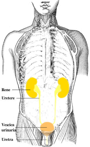
Órganos del tracto urinario
Imagen: “Urinary tract it” por Lennert B. Licencia: CC BY 2.5Lesiones contusas:
Lesiones penetrantes:
Fisiopatología:
Presentación clínica:
Diagnóstico:
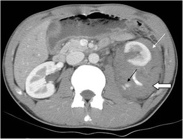
Lesión renal izquierda grado IV por accidente automovilístico
La TC con contraste en fase arterial en corte axial mostró extravasación del medio de contraste (flecha negra), hematoma perirrenal (flecha ancha) y hematoma pararrenal (flecha blanca).
Tratamiento:
Mecanismos:
Síntomas:
Diagnóstico:
Tratamiento:
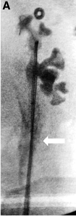
Traumatismo ureteral iatrogénico sostenido durante la instrumentación del uréter
La pielografía intraoperatoria muestra extravasación de contraste (flecha) del uréter izquierdo.
Extraperitoneales:
Intraperitoneales:
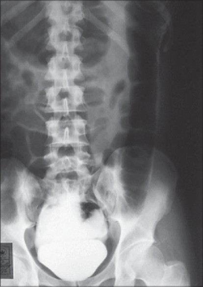
Ruptura vesical intraperitoneal: cistografía que muestra fuga de contraste en la cavidad peritoneal
Imagen: “Intraperitoneal bladder rupture mimicking acute renal failure” por Arun KG. Licencia: CC BY 2.0Síntomas:
Lesiones de la uretra anterior:
Lesiones de la uretra posterior:
Diagnóstico: uretrografía retrógrada
Tratamiento:
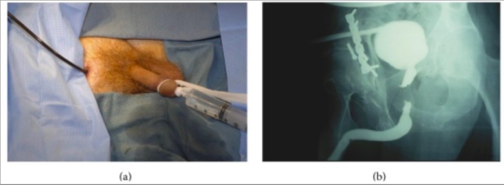
Individuo con antecedentes de traumatismo pélvico:
(a): Se realiza una uretrografía retrógrada mientras se inyecta simultáneamente contraste en la uretra posterior a través del cistoscopio flexible con la punta en la uretra prostática distal.
(b): Las imágenes demuestran con precisión la longitud y la localización del defecto.

Fractura de pene: fotografía clínica que muestra un pene edematizado con deformidad en “berenjena” y desviación hacia el lado derecho
Imagen: “Synergism of clinical evaluation and penile sonographic imaging in diagnosis of penile fracture: a case report” por Bello JO. Licencia: CC BY 2.0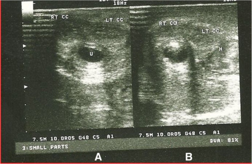
Sonouretrograma de fractura de pene
A: Sonouretrografía en la cara distal del cuerpo del pene, que muestra un cuerpo cavernoso derecho (RT CC, por sus siglas en inglés) e izquierdo normal (LT CC, por sus siglas en inglés) y una uretra normal (U)
B: Sonouretrografía en el 1/3 proximal del cuerpo del pene que muestra un cuerpo cavernoso derecho normal y una rotura del cuerpo cavernoso izquierdo con hematoma asociado (H)