Las porfirias son un grupo de trastornos metabólicos causados por una alteración en EN Erythema nodosum is an immune-mediated panniculitis (inflammation of the subcutaneous fat) caused by a type IV (delayed-type) hypersensitivity reaction. It commonly manifests in young women as tender, erythematous nodules on the shins. Erythema Nodosum la síntesis del hemo. En EN Erythema nodosum is an immune-mediated panniculitis (inflammation of the subcutaneous fat) caused by a type IV (delayed-type) hypersensitivity reaction. It commonly manifests in young women as tender, erythematous nodules on the shins. Erythema Nodosum la mayoría de los LOS Neisseria casos, la porfiria es causada por un defecto enzimático hereditario. Los LOS Neisseria patrones de la enfermedad difieren según la enzima afectada, y las variantes de porfiria pueden diferenciarse clínicamente entre formas agudas y no agudas. Los LOS Neisseria pacientes con porfiria presentan erupciones cutáneas por fotosensibilidad y, a veces, síntomas sistémicos como dolor Dolor Inflammation abdominal y neuropatía. Las porfirias se tratan evitando los LOS Neisseria desencadenantes, como la exposición al AL Amyloidosis sol y el consumo de alcohol. Cuando ocurren brotes, la terapia se dirige hacia el alivio sintomático.
Last updated: Apr 24, 2025
Las porfirias son trastornos metabólicos raros causados por deficiencias en EN Erythema nodosum is an immune-mediated panniculitis (inflammation of the subcutaneous fat) caused by a type IV (delayed-type) hypersensitivity reaction. It commonly manifests in young women as tender, erythematous nodules on the shins. Erythema Nodosum la síntesis de hemo.
Las porfirias se deben a defectos enzimáticos en EN Erythema nodosum is an immune-mediated panniculitis (inflammation of the subcutaneous fat) caused by a type IV (delayed-type) hypersensitivity reaction. It commonly manifests in young women as tender, erythematous nodules on the shins. Erythema Nodosum la biosíntesis del hemo:
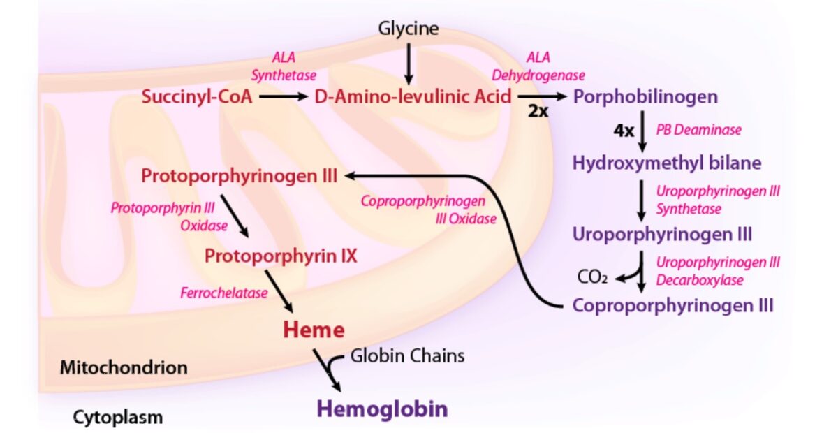
Vía enzimática de la síntesis de hemo:
ALA: ácido aminolevulínico
CoA: coenzima A
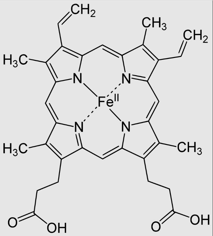
Hemo B (tipo más común de hemo)
Imagen: “Heme B” por Yikrazuul. Licencia: Dominio Público| Tipo de porfiria | Enzima defectuosa |
|---|---|
| Porfiria cutánea tardía | Uroporfirinógeno III descarboxilasa |
| Porfiria aguda intermitente | Porfobilinógeno desaminasa |
| Protoporfiria ligada al AL Amyloidosis cromosoma X | Ácido δ-aminolevulínico sintasa 2 |
| Porfiria eritropoyética congénita | Uroporfirinógeno III sintasa |
| Coproporfiria hereditaria | Coproporfirinógeno oxidasa |
| Porfiria variegata | Protoporfirinógeno oxidasa |
| Protoporfiria eritropoyética | Ferroquelatasa |
Más comúnmente, las porfirias se deben a un defecto enzimático heredado dentro de la ruta de biosíntesis del hemo. Rara vez se pueden adquirir más adelante en EN Erythema nodosum is an immune-mediated panniculitis (inflammation of the subcutaneous fat) caused by a type IV (delayed-type) hypersensitivity reaction. It commonly manifests in young women as tender, erythematous nodules on the shins. Erythema Nodosum la vida:
La presentación clínica depende del patrón de afectación de órganos.
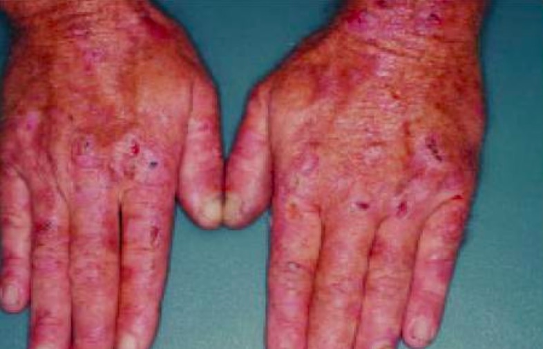
Erosiones, costras y ampollas son evidentes en las manos de este paciente con porfiria cutánea tardía.
Imagen: “Erosions, crust, and blisters are evident on the hands of this patient with porphyria cutanea tarda” por Department of Internal Medicine, Minia University, Minia, Egypt. Licencia: CC BY 2.5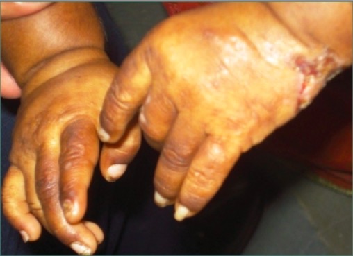
Fotografía que muestra la presencia de lesiones y cicatrices en ambas manos de un paciente con porfiria eritropoyética congénita
Imagen: “Erythrodontia in congenital erythropoietic porphyria” por Department of Oral Pathology and Microbiology, Kamineni Institute of Dental Sciences, Narketpalli, Andhra Pradesh, India. Licencia: CC BY 2.0El diagnóstico de porfirias generalmente se realiza mediante análisis de sangre especializados.
No existe una cura conocida para la porfiria.