Domina Conceptos Médicos

Estudia para la escuela de medicina y tus examenes con Lecturio.

Osteocondritis Disecante

La osteocondritis disecante es un trastorno ortopédico caracterizado por el desprendimiento de un segmento focal de hueso y cartílago subcondral como resultado de una necrosis Necrosis The death of cells in an organ or tissue due to disease, injury or failure of the blood supply. Ischemic Cell Damage aséptica focal. Esto puede ocurrir a cualquier edad, pero se observa con mayor frecuencia en EN Erythema nodosum is an immune-mediated panniculitis (inflammation of the subcutaneous fat) caused by a type IV (delayed-type) hypersensitivity reaction. It commonly manifests in young women as tender, erythematous nodules on the shins. Erythema Nodosum adolescentes que participan en EN Erythema nodosum is an immune-mediated panniculitis (inflammation of the subcutaneous fat) caused by a type IV (delayed-type) hypersensitivity reaction. It commonly manifests in young women as tender, erythematous nodules on the shins. Erythema Nodosum deportes competitivos. Los LOS Neisseria pacientes pueden estar asintomáticos o pueden presentar dolor Dolor Inflammation en EN Erythema nodosum is an immune-mediated panniculitis (inflammation of the subcutaneous fat) caused by a type IV (delayed-type) hypersensitivity reaction. It commonly manifests in young women as tender, erythematous nodules on the shins. Erythema Nodosum las articulaciones, rigidez e inflamación que empeora con la actividad. El diagnóstico se puede hacer con imágenes. El tratamiento depende de la gravedad, pero puede incluir actividad restringida con carga de peso, fisioterapia o cirugía.

Last updated: Dec 15, 2025

Editorial responsibility: Stanley Oiseth, Lindsay Jones, Evelin Maza

Descripción General

Definición

La osteocondritis disecante es un trastorno caracterizado por necrosis Necrosis The death of cells in an organ or tissue due to disease, injury or failure of the blood supply. Ischemic Cell Damage aséptica del hueso subcondral que provoca un desprendimiento focal y el desplazamiento del hueso y el cartílago hacia el espacio articular. El término osteocondritis es un nombre inapropiado, ya que el análisis histológico revela una falta de células inflamatorias.

Epidemiología

  • Prevalencia: 15–29 por cada 100 000 pacientes
  • Edad máxima: 12–19 años
  • Hombres > mujeres
  • Mayor incidencia en EN Erythema nodosum is an immune-mediated panniculitis (inflammation of the subcutaneous fat) caused by a type IV (delayed-type) hypersensitivity reaction. It commonly manifests in young women as tender, erythematous nodules on the shins. Erythema Nodosum deportistas jóvenes

Etiología y Fisiopatología

Etiología

Se desconoce la etiología exacta. Se han propuesto muchas teorías y es probable que la osteocondritis disecante sea multifactorial.

  • Trauma:
    • Microtrauma Microtrauma Small injuries caused by external force applied to the body including bones, muscles, nerves and tendons. Osgood-Schlatter Disease repetitivo (estrés mecánico de deportes competitivos)
    • Trauma directo (lesión aislada)
  • Isquemia
  • Predisposición genética

Fisiopatología

Patogenia de la osteocondritis disecante:

  • Microtrauma Microtrauma Small injuries caused by external force applied to the body including bones, muscles, nerves and tendons. Osgood-Schlatter Disease repetitivo → lesión del hueso subcondral → fragmentación
  • Se desarrolla un defecto entre la lesión osteocondral y el hueso subyacente.
  • Resulta en EN Erythema nodosum is an immune-mediated panniculitis (inflammation of the subcutaneous fat) caused by a type IV (delayed-type) hypersensitivity reaction. It commonly manifests in young women as tender, erythematous nodules on the shins. Erythema Nodosum hipovascularidad → osteonecrosis de la lesión
  • Afectación del cartílago suprayacente → inestabilidad del fragmento
  • El hueso subcondral y el cartílago pueden separarse parcial o totalmente.

Etapas de progresión en EN Erythema nodosum is an immune-mediated panniculitis (inflammation of the subcutaneous fat) caused by a type IV (delayed-type) hypersensitivity reaction. It commonly manifests in young women as tender, erythematous nodules on the shins. Erythema Nodosum la osteocondritis disecante:

  • Etapa I: pequeña área de compresión del hueso subcondral
  • Etapa II: desprendimiento parcial de un fragmento osteocondral
  • Etapa III:
    • Desprendimiento completo de un fragmento
    • Permanece dentro del lecho del cráter subyacente
  • Etapa IV:
    • Desprendimiento completo de un fragmento
    • Desplazado del lecho del cráter (cuerpo suelto)
Osteocondritis disecante 4 etapas

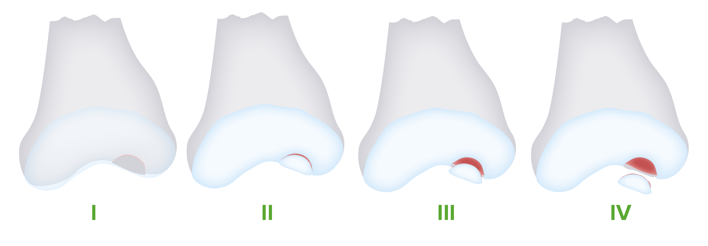
La osteocondritis disecante progresa a través de 4 etapas. Con la lesión continua, la lesión se fragmenta y se separa del hueso subyacente, y eventualmente se desplaza en el espacio articular (cuerpo suelto).

Imagen por Lecturio.

Presentación Clínica

Síntomas

Algunos pacientes pueden estar asintomáticos y la osteocondritis disecante se encuentra incidentalmente en EN Erythema nodosum is an immune-mediated panniculitis (inflammation of the subcutaneous fat) caused by a type IV (delayed-type) hypersensitivity reaction. It commonly manifests in young women as tender, erythematous nodules on the shins. Erythema Nodosum la imagenología. Sin embargo, las personas sintomáticas pueden experimentar:

  • Dolor Dolor Inflammation crónico en EN Erythema nodosum is an immune-mediated panniculitis (inflammation of the subcutaneous fat) caused by a type IV (delayed-type) hypersensitivity reaction. It commonly manifests in young women as tender, erythematous nodules on the shins. Erythema Nodosum la articulación afectada:
    • Aumenta con la actividad
    • Se alivia en EN Erythema nodosum is an immune-mediated panniculitis (inflammation of the subcutaneous fat) caused by a type IV (delayed-type) hypersensitivity reaction. It commonly manifests in young women as tender, erythematous nodules on the shins. Erythema Nodosum reposo
  • Síntomas progresivos, como:
    • Rigidez articular
    • Inflamación intermitente
    • Sensación de bloqueo de la articulación (puede indicar un cuerpo suelto)
    • Inestabilidad articular
  • Articulaciones más comúnmente afectadas:

Examen físico

  • Dolor Dolor Inflammation localizado a la palpación en EN Erythema nodosum is an immune-mediated panniculitis (inflammation of the subcutaneous fat) caused by a type IV (delayed-type) hypersensitivity reaction. It commonly manifests in young women as tender, erythematous nodules on the shins. Erythema Nodosum el sitio de la lesión, particularmente:
    • Cóndilo femoral medial de la rodilla
    • Articulación radiocapitelar del codo
    • Articulación tibioastragalina del tobillo
  • Rango de movimiento doloroso
  • Rango de movimiento:
    • Generalmente normal si la rodilla es la articulación afectada
    • A menudo disminuido si el codo es la articulación afectada
  • Derrame articular
  • Crepitación
  • Prueba de Wilson:
    • Dolor Dolor Inflammation con extensión de rodilla y rotación interna.
    • Se ha HA Hemolytic anemia (HA) is the term given to a large group of anemias that are caused by the premature destruction/hemolysis of circulating red blood cells (RBCs). Hemolysis can occur within (intravascular hemolysis) or outside the blood vessels (extravascular hemolysis). Hemolytic Anemia descrito con la osteocondritis disecante, pero carece de sensibilidad.
  • Trastornos de la marcha:
    • Puede verse en EN Erythema nodosum is an immune-mediated panniculitis (inflammation of the subcutaneous fat) caused by a type IV (delayed-type) hypersensitivity reaction. It commonly manifests in young women as tender, erythematous nodules on the shins. Erythema Nodosum algunos pacientes con afectación de la rodilla.
    • La pierna afectada rota externamente con la deambulación.
prueba de wilson

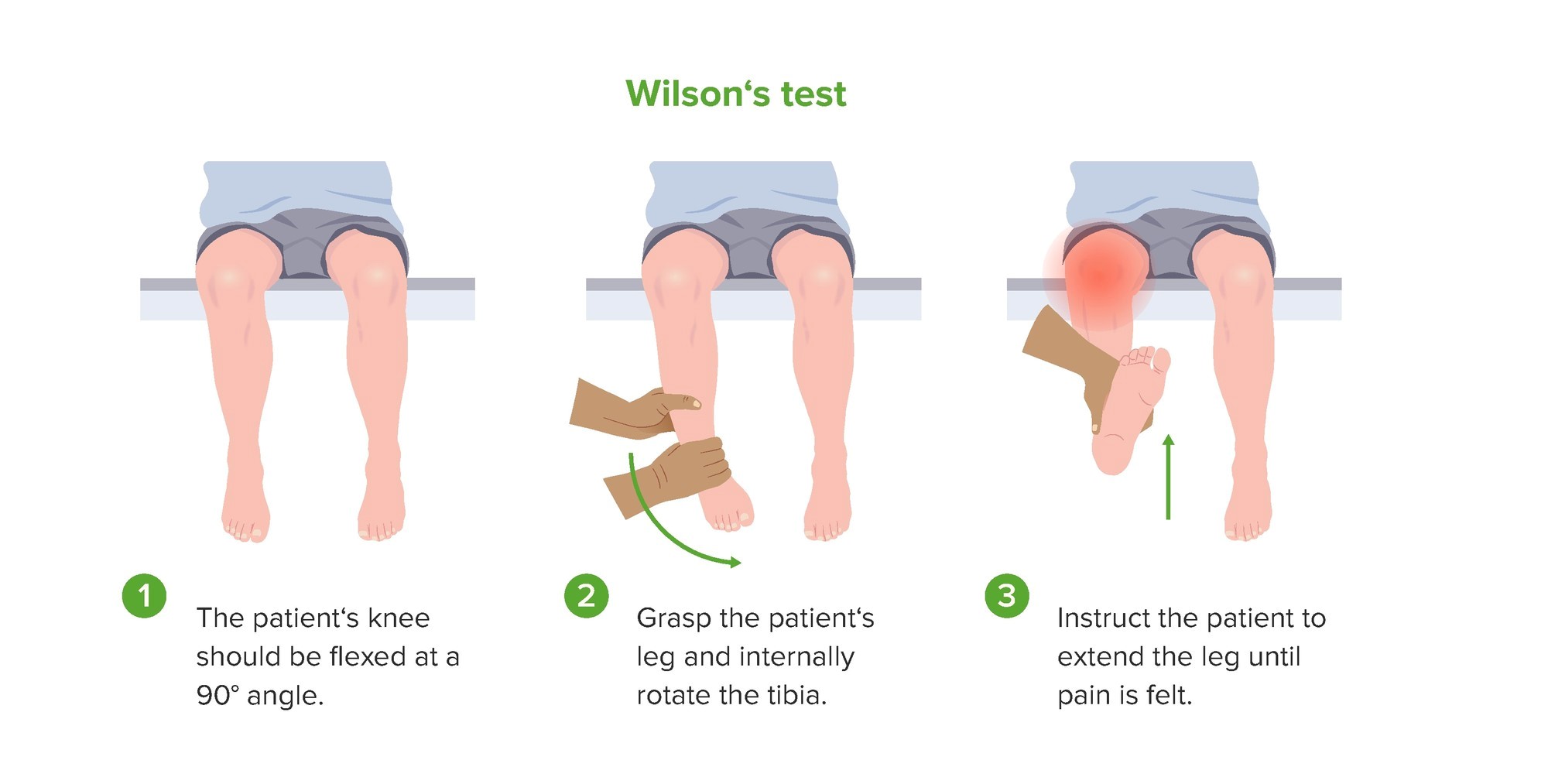
Demostración de la prueba de Wilson, que puede ser positiva en pacientes con osteocondritis disecante de rodilla.

Imagen por Lecturio.

Diagnóstico

Se debe sospechar un diagnóstico de osteocondritis disecante en EN Erythema nodosum is an immune-mediated panniculitis (inflammation of the subcutaneous fat) caused by a type IV (delayed-type) hypersensitivity reaction. It commonly manifests in young women as tender, erythematous nodules on the shins. Erythema Nodosum adolescentes con una presentación característica de dolor Dolor Inflammation articular y participación en EN Erythema nodosum is an immune-mediated panniculitis (inflammation of the subcutaneous fat) caused by a type IV (delayed-type) hypersensitivity reaction. It commonly manifests in young women as tender, erythematous nodules on the shins. Erythema Nodosum actividades repetitivas y deportivas.

Radiografía

Las radiografías de la articulación afectada suelen ser diagnósticas y son el método de prueba inicial de elección. Sin embargo, estas imágenes pueden ser normales en EN Erythema nodosum is an immune-mediated panniculitis (inflammation of the subcutaneous fat) caused by a type IV (delayed-type) hypersensitivity reaction. It commonly manifests in young women as tender, erythematous nodules on the shins. Erythema Nodosum las primeras etapas de la enfermedad.

Rodilla:

  • Hallazgos:
    • A menudo se detecta por primera vez una translucidez subcortical en EN Erythema nodosum is an immune-mediated panniculitis (inflammation of the subcutaneous fat) caused by a type IV (delayed-type) hypersensitivity reaction. It commonly manifests in young women as tender, erythematous nodules on the shins. Erythema Nodosum forma de media luna.
    • Fragmento óseo subcondral
  • Ubicaciones frecuentes de las lesiones:
    • Cóndilo medial (77%)
    • Cóndilo lateral (17%)
    • Rótula (7%)
Osteocondritis disecante radiografía de rodilla

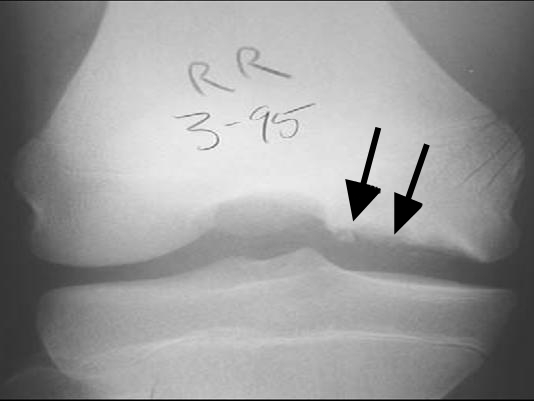
Radiografía de rodilla de un paciente con osteocondritis disecante:
Obsérvese la fragmentación del hueso y la radiotransparencia debajo (flechas).

Imagen: ““Osteochondritis dissecans diagram” por Kristin M Houghton. Licencia: CC BY 2.0

Codo:

  • Aplanamiento lateral del capitellum
  • Esclerosis subcondral
  • Fragmentación del capitellum
Osteocondritis disecante codo radiografía

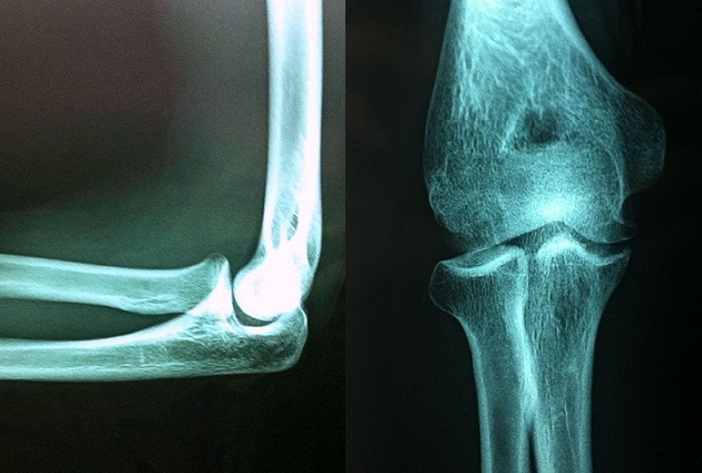
Imágenes de rayos X de un atleta de 16 años con osteocondritis disecante del codo.
Se muestra un foco radiolúcido del capitellum sobre la cabeza radial.

Imagen : “Osteochondritis dissecans of the capitellum in adolescents: report of a case and review of the literature” por Erraji M, Kharraji A, Abbassi N, Najib A, Yacoubi H. Licencia: CC BY 2.0

Tobillo:

  • Normal en EN Erythema nodosum is an immune-mediated panniculitis (inflammation of the subcutaneous fat) caused by a type IV (delayed-type) hypersensitivity reaction. It commonly manifests in young women as tender, erythematous nodules on the shins. Erythema Nodosum ⅓ de los LOS Neisseria casos
  • Lesión en EN Erythema nodosum is an immune-mediated panniculitis (inflammation of the subcutaneous fat) caused by a type IV (delayed-type) hypersensitivity reaction. It commonly manifests in young women as tender, erythematous nodules on the shins. Erythema Nodosum forma de copa en EN Erythema nodosum is an immune-mediated panniculitis (inflammation of the subcutaneous fat) caused by a type IV (delayed-type) hypersensitivity reaction. It commonly manifests in young women as tender, erythematous nodules on the shins. Erythema Nodosum el astrágalo
Osteocondritis disecante radiografía de tobillo

Imágenes de rayos X de un tobillo que muestran osteocondritis disecante del astrágalo superomedial.
Obsérvese la radiotransparencia y la fragmentación (flechas).

Imagen: “CT and projectional radiography of osteochondritis dissecans – annotated” por Mikael Häggström. Licencia: CC0 1.0, recortada por Lecturio.

RM

  • Debe considerarse en EN Erythema nodosum is an immune-mediated panniculitis (inflammation of the subcutaneous fat) caused by a type IV (delayed-type) hypersensitivity reaction. It commonly manifests in young women as tender, erythematous nodules on the shins. Erythema Nodosum pacientes sintomáticos con radiografías normales
  • Más sensible para detectar cambios subcondrales y se puede utilizar en EN Erythema nodosum is an immune-mediated panniculitis (inflammation of the subcutaneous fat) caused by a type IV (delayed-type) hypersensitivity reaction. It commonly manifests in young women as tender, erythematous nodules on the shins. Erythema Nodosum la estadificación
  • Se puede usar gadolinio para evaluar el suministro de sangre al AL Amyloidosis área.
  • Utilizado con frecuencia en EN Erythema nodosum is an immune-mediated panniculitis (inflammation of the subcutaneous fat) caused by a type IV (delayed-type) hypersensitivity reaction. It commonly manifests in young women as tender, erythematous nodules on the shins. Erythema Nodosum la planificación preoperatoria
Resonancia magnética de rodilla con osteocondritis disecante

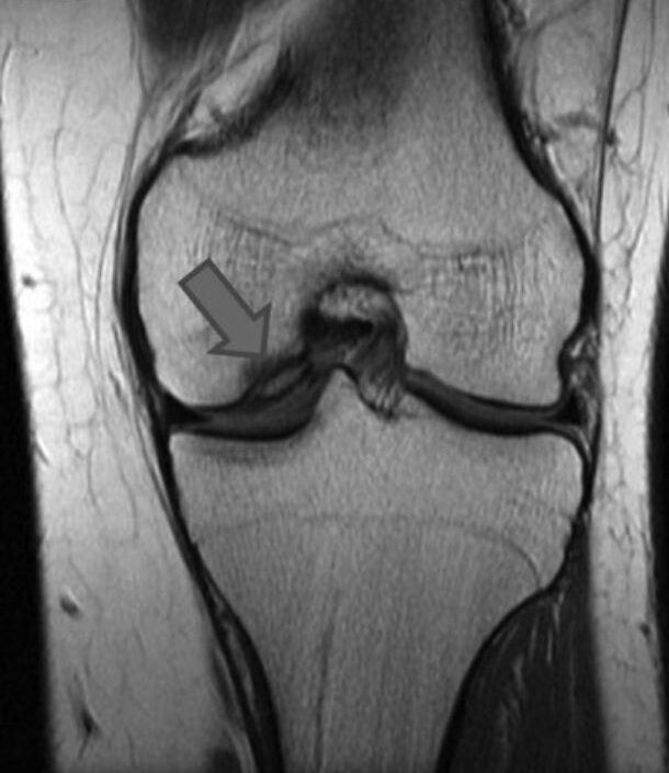
RM de una rodilla que muestra un área de osteocondritis disecante que afecta el cóndilo femoral medial

Imagen: “The present state of treatments for articular cartilage defects in the knee” por Perera JR, Gikas PD, Bentley G.  Licencia: CC BY 3.0 , recortada por Lecturio.

Tratamiento

El objetivo principal del tratamiento es lograr la curación de la lesión osteocondral y devolver la función completa a la articulación afectada. Es posible que se necesiten referir para las especialidades de medicina deportiva o cirugía ortopédica.

Tratamiento conservador

  • Limitar la carga de peso y la actividad:
    • De importancia crítica para prevenir la progresión.
    • El regreso a la actividad puede ocurrir cuando:
      • Desista el dolor Dolor Inflammation
      • Mejore el rango de movimiento activo
      • Evidencia de curación
  • Inmovilización de la articulación
  • Fisioterapia
  • Indicaciones para el tratamiento conservador:
    • Pacientes esqueléticamente inmaduros con fisis abiertas y sin un cuerpo extraño intraarticular
    • Pacientes adultos que tienen un fragmento pequeño, estable y asintomático.
  • Pacientes asintomáticos con hallazgos radiográficos incidentales de lesiones osteocondrales:
    • Repetir radiografías cada 6 meses.
    • Dar seguimiento al AL Amyloidosis paciente hasta que llegue a la madurez esquelética o hasta que la lesión haya cicatrizado.

Tratamiento quirúrgico

  • Indicaciones:
    • Signos de inestabilidad articular
    • Lesión en EN Erythema nodosum is an immune-mediated panniculitis (inflammation of the subcutaneous fat) caused by a type IV (delayed-type) hypersensitivity reaction. It commonly manifests in young women as tender, erythematous nodules on the shins. Erythema Nodosum expansión en EN Erythema nodosum is an immune-mediated panniculitis (inflammation of the subcutaneous fat) caused by a type IV (delayed-type) hypersensitivity reaction. It commonly manifests in young women as tender, erythematous nodules on the shins. Erythema Nodosum las radiografías
    • Tratamiento no quirúrgico fallido
    • Cuerpo suelto intraarticular
  • Técnicas:
    • Perforación subcondral artroscópica
    • Fijación de lesión inestable
    • Escisión artroscópica, curetaje y perforación
    • Trasplante osteocondral autólogo
  • La elección depende de la madurez esquelética, el estadio de la enfermedad y el tamaño de la lesión.

Pronóstico

  • La mayoría de los LOS Neisseria pacientes con lesiones estables tendrán curación espontánea con medidas conservadoras.
  • Las tasas de éxito para lesiones inestables varían según la gravedad y la técnica quirúrgica: 30%–100%
  • Los LOS Neisseria pacientes tienen un mayor riesgo de desarrollar osteoartritis de la articulación.
  • Factores relacionados con un mejor pronóstico:
    • Edad más joven
    • Cartílago articular intacto
    • Lesiones osteocondrales más pequeñas
    • Lesiones estables

Diagnóstico Diferencial

  • Desgarro de menisco: una lesión aguda que a menudo se observa en EN Erythema nodosum is an immune-mediated panniculitis (inflammation of the subcutaneous fat) caused by a type IV (delayed-type) hypersensitivity reaction. It commonly manifests in young women as tender, erythematous nodules on the shins. Erythema Nodosum pacientes más jóvenes. La presentación puede incluir una sensación de desgarro o estallido durante la lesión, dolor Dolor Inflammation de rodilla, disminución del rango de movimiento, bloqueo y derrame articular. El diagnóstico se basa en EN Erythema nodosum is an immune-mediated panniculitis (inflammation of the subcutaneous fat) caused by a type IV (delayed-type) hypersensitivity reaction. It commonly manifests in young women as tender, erythematous nodules on the shins. Erythema Nodosum el examen físico y la imagenología. El tratamiento incluye medidas conservadoras y cirugía para las lesiones graves.
  • Síndrome de plica: irritación e inflamación del componente sinovial medial de la rodilla, que puede ocurrir con traumatismos repetitivos. Los LOS Neisseria pacientes pueden experimentar dolor Dolor Inflammation en EN Erythema nodosum is an immune-mediated panniculitis (inflammation of the subcutaneous fat) caused by a type IV (delayed-type) hypersensitivity reaction. It commonly manifests in young women as tender, erythematous nodules on the shins. Erythema Nodosum la rodilla, a menudo en EN Erythema nodosum is an immune-mediated panniculitis (inflammation of the subcutaneous fat) caused by a type IV (delayed-type) hypersensitivity reaction. It commonly manifests in young women as tender, erythematous nodules on the shins. Erythema Nodosum la cara medial de la articulación, con una sensación de atrapamiento o estallido. El diagnóstico definitivo lo proporciona la artroscopia. El tratamiento consiste en EN Erythema nodosum is an immune-mediated panniculitis (inflammation of the subcutaneous fat) caused by a type IV (delayed-type) hypersensitivity reaction. It commonly manifests in young women as tender, erythematous nodules on the shins. Erythema Nodosum medidas conservadoras, fisioterapia, inyecciones intraarticulares de esteroides y cirugía artroscópica para casos refractarios.
  • Síndrome patelofemoral: causa más común de dolor Dolor Inflammation en EN Erythema nodosum is an immune-mediated panniculitis (inflammation of the subcutaneous fat) caused by a type IV (delayed-type) hypersensitivity reaction. It commonly manifests in young women as tender, erythematous nodules on the shins. Erythema Nodosum la parte anterior de la rodilla, que puede deberse a una mala alineación de la pierna, desequilibrio muscular, hiperactividad o traumatismo. El síndrome patelofemoral produce un dolor Dolor Inflammation sordo y generalizado en EN Erythema nodosum is an immune-mediated panniculitis (inflammation of the subcutaneous fat) caused by a type IV (delayed-type) hypersensitivity reaction. It commonly manifests in young women as tender, erythematous nodules on the shins. Erythema Nodosum la parte anterior de la rodilla que se exacerba con la actividad o la sedestación prolongada. El síndrome femororrotuliano es un diagnóstico de exclusión, debiéndose descartar otras etiologías intraarticulares. En EN Erythema nodosum is an immune-mediated panniculitis (inflammation of the subcutaneous fat) caused by a type IV (delayed-type) hypersensitivity reaction. It commonly manifests in young women as tender, erythematous nodules on the shins. Erythema Nodosum el tratamiento se utilizan medidas conservadoras y fisioterapia.
  • Codo de “ligas menores”: lesión por uso excesivo que produce apofisitis del epicóndilo medial del codo. Esta afección a menudo se asocia con lanzamientos repetitivos, comúnmente en EN Erythema nodosum is an immune-mediated panniculitis (inflammation of the subcutaneous fat) caused by a type IV (delayed-type) hypersensitivity reaction. It commonly manifests in young women as tender, erythematous nodules on the shins. Erythema Nodosum lanzadores de béisbol preadolescentes y adolescentes. Los LOS Neisseria pacientes desarrollan dolor Dolor Inflammation en EN Erythema nodosum is an immune-mediated panniculitis (inflammation of the subcutaneous fat) caused by a type IV (delayed-type) hypersensitivity reaction. It commonly manifests in young women as tender, erythematous nodules on the shins. Erythema Nodosum el codo, particularmente en EN Erythema nodosum is an immune-mediated panniculitis (inflammation of the subcutaneous fat) caused by a type IV (delayed-type) hypersensitivity reaction. It commonly manifests in young women as tender, erythematous nodules on the shins. Erythema Nodosum el epicóndilo medial. El diagnóstico es clínico y el tratamiento incluye medidas conservadoras y fisioterapia.
  • Enfermedad de Panner: osteocondrosis de la epífisis del capitellum humeral que consiste en EN Erythema nodosum is an immune-mediated panniculitis (inflammation of the subcutaneous fat) caused by a type IV (delayed-type) hypersensitivity reaction. It commonly manifests in young women as tender, erythematous nodules on the shins. Erythema Nodosum osificación, necrosis Necrosis The death of cells in an organ or tissue due to disease, injury or failure of the blood supply. Ischemic Cell Damage y regeneración atípicas. A diferencia de la osteocondritis disecante, este trastorno se observa a edades más tempranas y no progresa a un cuerpo suelto. Los LOS Neisseria pacientes presentan dolor Dolor Inflammation en EN Erythema nodosum is an immune-mediated panniculitis (inflammation of the subcutaneous fat) caused by a type IV (delayed-type) hypersensitivity reaction. It commonly manifests in young women as tender, erythematous nodules on the shins. Erythema Nodosum el codo, rigidez y rango de movimiento limitado. El diagnóstico se realiza mediante imagenología y el tratamiento incluye medidas conservadoras.
  • Epicondilitis medial: también se conoce como “codo de golfista”. Este trastorno es causado por una fuerza en EN Erythema nodosum is an immune-mediated panniculitis (inflammation of the subcutaneous fat) caused by a type IV (delayed-type) hypersensitivity reaction. It commonly manifests in young women as tender, erythematous nodules on the shins. Erythema Nodosum valgo en EN Erythema nodosum is an immune-mediated panniculitis (inflammation of the subcutaneous fat) caused by a type IV (delayed-type) hypersensitivity reaction. It commonly manifests in young women as tender, erythematous nodules on the shins. Erythema Nodosum el codo que provoca la inflamación del tendón del grupo flexor de la muñeca en EN Erythema nodosum is an immune-mediated panniculitis (inflammation of the subcutaneous fat) caused by a type IV (delayed-type) hypersensitivity reaction. It commonly manifests in young women as tender, erythematous nodules on the shins. Erythema Nodosum su inserción en EN Erythema nodosum is an immune-mediated panniculitis (inflammation of the subcutaneous fat) caused by a type IV (delayed-type) hypersensitivity reaction. It commonly manifests in young women as tender, erythematous nodules on the shins. Erythema Nodosum el epicóndilo medial. Los LOS Neisseria pacientes tienen dolor Dolor Inflammation y sensibilidad en EN Erythema nodosum is an immune-mediated panniculitis (inflammation of the subcutaneous fat) caused by a type IV (delayed-type) hypersensitivity reaction. It commonly manifests in young women as tender, erythematous nodules on the shins. Erythema Nodosum la parte media del codo. El dolor Dolor Inflammation es provocado por la flexión de la muñeca y la pronación contra resistencia. El diagnóstico es clínico y el tratamiento implica medidas conservadoras y fisioterapia.
  • Epicondilitis lateral: también conocida como “codo de tenista”: Esta condición resulta de la inflamación de los LOS Neisseria tendones extensores del antebrazo en EN Erythema nodosum is an immune-mediated panniculitis (inflammation of the subcutaneous fat) caused by a type IV (delayed-type) hypersensitivity reaction. It commonly manifests in young women as tender, erythematous nodules on the shins. Erythema Nodosum su inserción en EN Erythema nodosum is an immune-mediated panniculitis (inflammation of the subcutaneous fat) caused by a type IV (delayed-type) hypersensitivity reaction. It commonly manifests in young women as tender, erythematous nodules on the shins. Erythema Nodosum el epicóndilo lateral. Los LOS Neisseria pacientes tienen dolor Dolor Inflammation lateral en EN Erythema nodosum is an immune-mediated panniculitis (inflammation of the subcutaneous fat) caused by a type IV (delayed-type) hypersensitivity reaction. It commonly manifests in young women as tender, erythematous nodules on the shins. Erythema Nodosum el codo que puede irradiarse al AL Amyloidosis antebrazo. El dolor Dolor Inflammation será provocado por la extensión de la muñeca con resistencia. El diagnóstico es clínico y el tratamiento implica medidas conservadoras y fisioterapia.
  • Esguince de tobillo: lesión del tobillo, con mayor frecuencia por inversión del pie. Los LOS Neisseria pacientes tienen dolor Dolor Inflammation, sensibilidad e inflamación en EN Erythema nodosum is an immune-mediated panniculitis (inflammation of the subcutaneous fat) caused by a type IV (delayed-type) hypersensitivity reaction. It commonly manifests in young women as tender, erythematous nodules on the shins. Erythema Nodosum la parte anterolateral del tobillo (ubicación más común). El diagnóstico es clínico. Se puede realizar imagenología para excluir otras causas de dolor Dolor Inflammation en EN Erythema nodosum is an immune-mediated panniculitis (inflammation of the subcutaneous fat) caused by a type IV (delayed-type) hypersensitivity reaction. It commonly manifests in young women as tender, erythematous nodules on the shins. Erythema Nodosum el tobillo. El tratamiento incluye medidas conservadoras y fisioterapia.

Referencias

  1. Pallin, D. J. (2018). Knee and lower leg. In Walls, R. M., et al. (Eds.). Rosen’s emergency medicine: Concepts and clinical practice. pp. 614‒633.e2. https://www.clinicalkey.es/#!/content/3-s2.0-B9780323354790000507
  2. O’Keefe, K. P. (2020). Hip and knee pain. In Tintinalli, J. E., et al. (Eds.). Tintinalli’s emergency medicine: A comprehensive study guide. New York, NY: McGraw-Hill Education. https://accessmedicine.mhmedical.com/content.aspx?aid=1167030636
  3. Ferri, F., Ferri, F. (2020). Ferri’s clinical advisor 2020: 5 books in 1. Philadelphia: Elsevier.
  4. Park, N.H., Kim, H.S., Yi, S.Y., Min, B.C. (2013). Multiple osteochondritis dissecans of knee joint in a patient with Wilson disease, focusing on magnetic resonance findings. Knee Surg Relat Res 25(4):225‒229. https://doi.org/10.5792/ksrr.2013.25.4.225
  5. Cooper, G., Warren, R. (2023). Osteochondritis dissecans. In DeBerardino, T.M. (Ed.). Medscape. Retrieved March 27, 2025, from https://emedicine.medscape.com/article/1253074-overview
  6. Wood, D., Davis, D.D., Carter, K.R. (2023). Osteochondritis dissecans. StatPearls. Retrieved March 27, 2025, from https://www.ncbi.nlm.nih.gov/books/NBK526091/
  7. Hergenroeder, A.C., Harvey, B.S. (2025). Osteochondritis dissecans (OCD): Clinical manifestations and diagnosis. In Wiley, J.F. (Ed.). UpToDate. Retrieved March 27, 2025, from https://www.uptodate.com/contents/osteochondritis-dissecans-ocd-clinical-manifestations-and-diagnosis
  8. Hergenroeder, A.C., Harvey, B.S. (2025). Management of osteochondritis dissecans (OCD). In Wiley, J.F. (Ed.). UpToDate. Retrieved March 27, 2025, from https://www.uptodate.com/contents/management-of-osteochondritis-dissecans-ocd
  9. Orazi, S., Andriolo, L., Franceschini, M., Di Martino, A., Zaffagnini, S., & Filardo, G. (2024). Single-plug osteochondral autograft transplantation for knee osteochondritis dissecans: Clinical improvement and long-term survivorship at a minimum ten year follow-up. International Orthopaedics, 48(10), 2625–2632. https://doi.org/10.1007/s00264-024-06267-5

¡Crea tu cuenta gratis o inicia una sesión para seguir leyendo!

Regístrate ahora y obtén acceso gratuito a Lecturio con páginas de concepto, videos médicos y cuestionarios para tu educación médica.

User Reviews

Que tengas una sesión de estudio alegre y navideña 🎁 Ahorra 50% en todos los planes >>

Details