Una oclusión de un vaso retiniano es un bloqueo en EN Erythema nodosum is an immune-mediated panniculitis (inflammation of the subcutaneous fat) caused by a type IV (delayed-type) hypersensitivity reaction. It commonly manifests in young women as tender, erythematous nodules on the shins. Erythema Nodosum una arteria o vena principal de la retina Retina The ten-layered nervous tissue membrane of the eye. It is continuous with the optic nerve and receives images of external objects and transmits visual impulses to the brain. Its outer surface is in contact with the choroid and the inner surface with the vitreous body. The outermost layer is pigmented, whereas the inner nine layers are transparent. Eye: Anatomy. Según la ubicación, la oclusión se puede clasificar como oclusión de la arteria central de la retina Retina The ten-layered nervous tissue membrane of the eye. It is continuous with the optic nerve and receives images of external objects and transmits visual impulses to the brain. Its outer surface is in contact with the choroid and the inner surface with the vitreous body. The outermost layer is pigmented, whereas the inner nine layers are transparent. Eye: Anatomy, oclusión de la rama de la arteria retiniana, oclusión de la vena central de la retina Retina The ten-layered nervous tissue membrane of the eye. It is continuous with the optic nerve and receives images of external objects and transmits visual impulses to the brain. Its outer surface is in contact with the choroid and the inner surface with the vitreous body. The outermost layer is pigmented, whereas the inner nine layers are transparent. Eye: Anatomy u oclusión de la rama de la vena retiniana. Típicamente, la oclusión de un vaso retiniano es un evento tromboembólico. Los LOS Neisseria factores de riesgo incluyen hipertensión, diabetes Diabetes Diabetes mellitus (DM) is a metabolic disease characterized by hyperglycemia and dysfunction of the regulation of glucose metabolism by insulin. Type 1 DM is diagnosed mostly in children and young adults as the result of autoimmune destruction of β cells in the pancreas and the resulting lack of insulin. Type 2 DM has a significant association with obesity and is characterized by insulin resistance. Diabetes Mellitus mellitus y enfermedad valvular cardíaca. La oclusión del vaso central de la retina Retina The ten-layered nervous tissue membrane of the eye. It is continuous with the optic nerve and receives images of external objects and transmits visual impulses to the brain. Its outer surface is in contact with the choroid and the inner surface with the vitreous body. The outermost layer is pigmented, whereas the inner nine layers are transparent. Eye: Anatomy se caracteriza por una pérdida súbita de la visión, unilateral e indolora y/o una pérdida de visión transitoria ( amaurosis fugax Amaurosis fugax Transient complete or partial monocular blindness due to retinal ischemia. This may be caused by emboli from the carotid artery (usually in association with carotid stenosis) and other locations that enter the central retinal artery. Carotid Artery Stenosis). Las opciones de tratamiento son limitadas en EN Erythema nodosum is an immune-mediated panniculitis (inflammation of the subcutaneous fat) caused by a type IV (delayed-type) hypersensitivity reaction. It commonly manifests in young women as tender, erythematous nodules on the shins. Erythema Nodosum todos los LOS Neisseria casos y generalmente inefectivas. Cuando la mácula está involucrada, el pronóstico es especialmente malo y conduce a la pérdida permanente de la visión.
Last updated: Dec 15, 2025
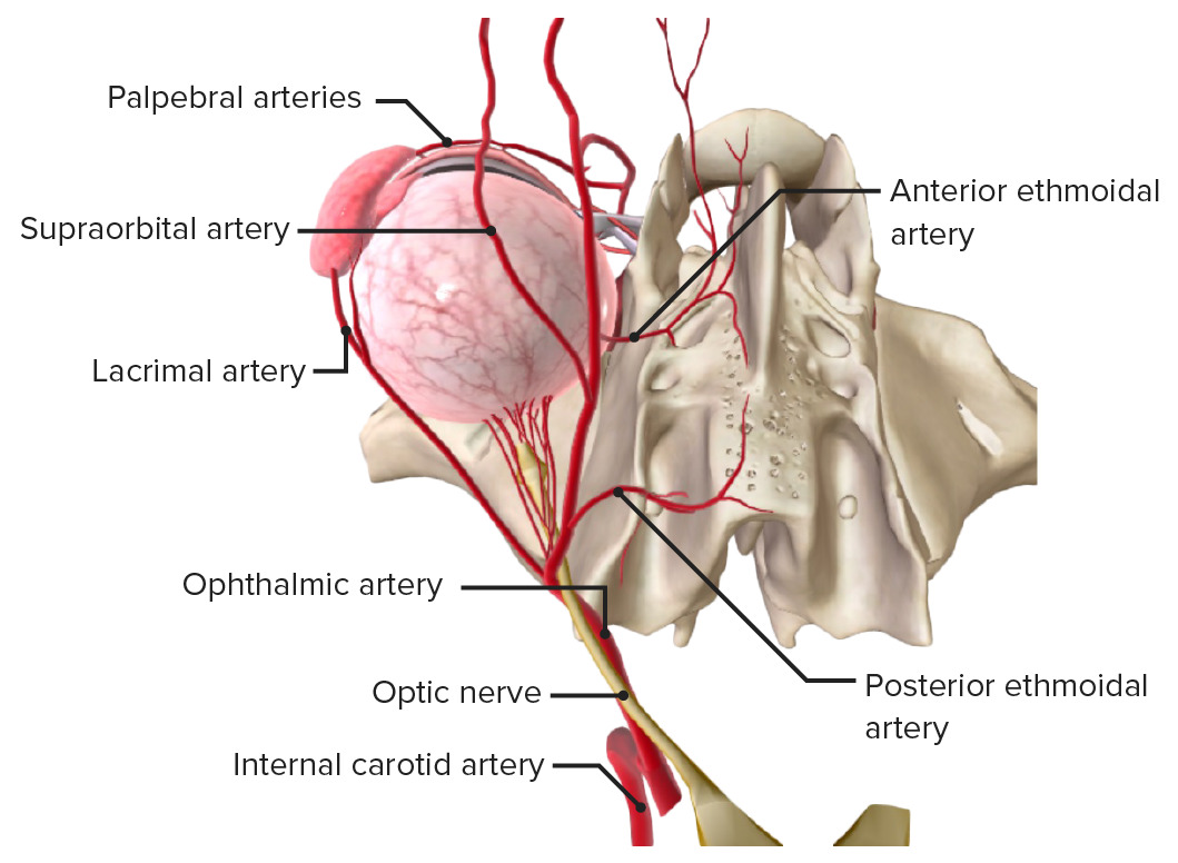
La arteria carótida con la arteria oftálmica y ramas. Tenga en cuenta la arteria central de la retina y las arterias coroideas posteriores, que entran en el ojo alrededor del nervio óptico.
Imagen de BioDigital, editada por Lecturio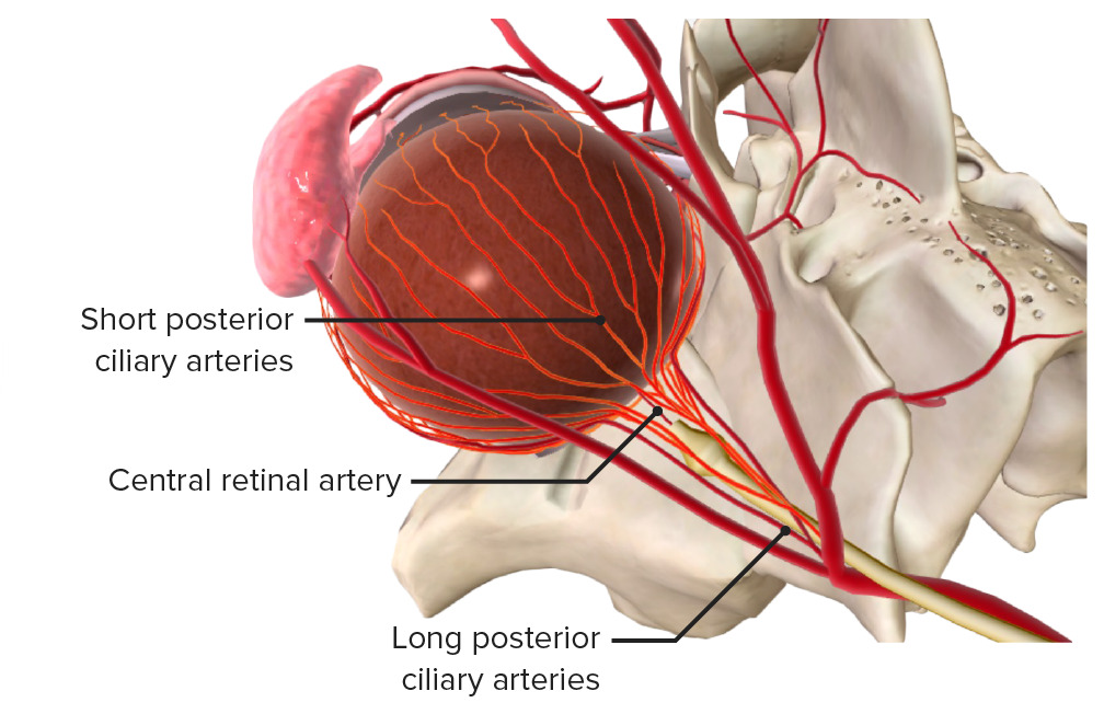
Vista superior: la arteria carótida interna se ramifica en la arteria oftálmica, que luego se divide en la arteria central de la retina y las arterias ciliares posteriores.
Imagen de BioDigital, editada por Lecturio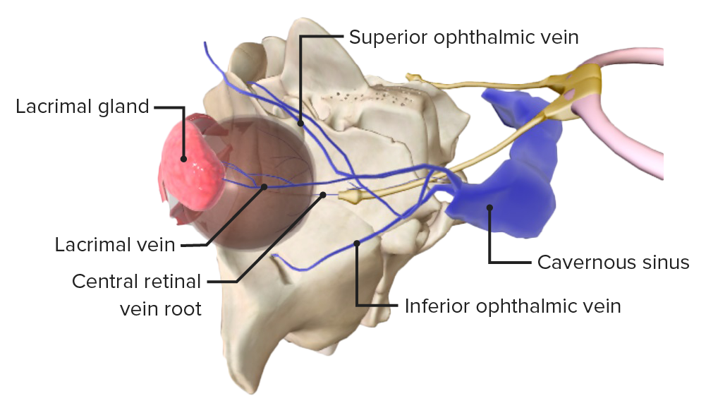
La vena oftálmica superior y el seno cavernoso, en el que drena la vena central de la retina.
Imagen de BioDigital, editada por LecturioLas causas exactas no se conocen, aunque existe una asociación con las siguientes condiciones:
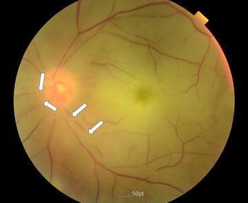
Imagen que muestra una fotografía de fondo de ojo en color del ojo izquierdo con oclusión de la arteria central de la retina; atenuación de la arteria retiniana (flechas) y palidez del disco óptico
Imagen: “Color fundus photography of the left eye” por Division of Cardiology, Department of Internal Medicine, Pohang St. Mary’s Hospital, Pohang, Republic of Korea.. Licencia: CC BY 4.0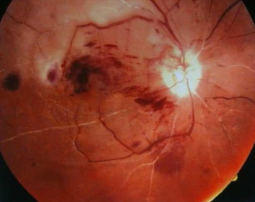
Fotografía de fondo de ojo derecho que indica oclusión de la arteria central de la retina/oclusión de la vena central de la retina: en esta imagen se ven una apariencia de “sangre y trueno” y una mancha “rojo cereza” (un paciente con lupus eritematoso sistémico)
Imagen: “Chorea and retinal vessel occlusion” por Division of Cardiology, Department of Internal Medicine, Pohang St. Mary’s Hospital, Pohang, Republic of Korea. Licencia: CC BY 2.0Oclusión de la arteria central de la retina Retina The ten-layered nervous tissue membrane of the eye. It is continuous with the optic nerve and receives images of external objects and transmits visual impulses to the brain. Its outer surface is in contact with the choroid and the inner surface with the vitreous body. The outermost layer is pigmented, whereas the inner nine layers are transparent. Eye: Anatomy
Oclusión de la vena central de la retina Retina The ten-layered nervous tissue membrane of the eye. It is continuous with the optic nerve and receives images of external objects and transmits visual impulses to the brain. Its outer surface is in contact with the choroid and the inner surface with the vitreous body. The outermost layer is pigmented, whereas the inner nine layers are transparent. Eye: Anatomy
El diagnóstico de ambas condiciones suele ser clínico, pero se pueden ordenar investigaciones adicionales.
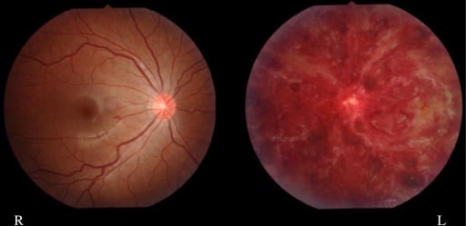
Imagen de una niña de 15 años con hemorragia intrarretiniana izquierda (oclusión de la vena central de la retina) (paciente con antecedentes de lupus sistémico y anticuerpos antifosfolipídicos)
Imagen: “Finding of eye fundus” por Department of Pediatrics and Child Neurology, Oita University Faculty of Medicine, Hasama, Yufu, Oita 879-5593, Japan. Licencia: CC BY 2.0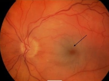
Ojo izquierdo que muestra una mancha “rojo cereza” con palidez retiniana típica de la oclusión de la arteria central de la retina (flecha)
Imagen: “Acute central retinal artery occlusion” por James Paget University Hospital NHS Foundation Trust, Lowestoft Road, Gorleston, Great Yarmouth NR31 6LA, Norfolk, UK. Licencia: CC BY 2.0