LosLOSNeisseria estudios de imagenología de losLOSNeisseria órganos reproductores femeninos internos (incluidos el útero, losLOSNeisseria ovarios y las trompas de Falopio) están indicados para diagnosticar problemas ginecológicos comunes, más comúnmente enENErythema nodosum is an immune-mediated panniculitis (inflammation of the subcutaneous fat) caused by a type IV (delayed-type) hypersensitivity reaction. It commonly manifests in young women as tender, erythematous nodules on the shins.Erythema Nodosum casos de sangrado anormal, dolorDolorInflammation pélvico y para evaluar masas, anomalías congénitas e infertilidad. El ultrasonido casi siempre es la modalidad de estudio de imagenología de primera línea, mientras que la RM suele reservarse para casos complicados o indeterminados como forma de seguimiento. La TC casi nunca se utiliza para evaluaciones ginecológicas primarias. Las trompas de Falopio no son visibles ni enENErythema nodosum is an immune-mediated panniculitis (inflammation of the subcutaneous fat) caused by a type IV (delayed-type) hypersensitivity reaction. It commonly manifests in young women as tender, erythematous nodules on the shins.Erythema Nodosum el ultrasonido ni enENErythema nodosum is an immune-mediated panniculitis (inflammation of the subcutaneous fat) caused by a type IV (delayed-type) hypersensitivity reaction. It commonly manifests in young women as tender, erythematous nodules on the shins.Erythema Nodosum la RM si son normales. La mejor manera de evaluar la permeabilidad de las trompas es mediante la histerosalpingografía, un examen fluoroscópico enENErythema nodosum is an immune-mediated panniculitis (inflammation of the subcutaneous fat) caused by a type IV (delayed-type) hypersensitivity reaction. It commonly manifests in young women as tender, erythematous nodules on the shins.Erythema Nodosum el que se inyecta un medio de contraste enENErythema nodosum is an immune-mediated panniculitis (inflammation of the subcutaneous fat) caused by a type IV (delayed-type) hypersensitivity reaction. It commonly manifests in young women as tender, erythematous nodules on the shins.Erythema Nodosum la cavidad uterina, seguido del estudio de su flujo a través de las trompas de Falopio.
Órganos ginecológicos internos comúnmente evaluados mediante imagenología
Útero
Ovarios
Trompas de Falopio
Estudios de elección para imagenología ginecológica
Ultrasonido: casi siempre el estudio inicial de elección
RM pélvica: generalmente reservada para casos que son indeterminados mediante el ultrasonido
Histerosalpingografía (HSG): un examen fluoroscópico utilizado para evaluar la permeabilidad de las trompas
Nota sobre las TC:
Generalmente no se utilizan para obtener imágenes de losLOSNeisseria órganos reproductores femeninos (resolución más baja que el ultrasonido).
Pueden estar indicadas como parte de un estudio oncológico para buscar evidencia de metástasis enENErythema nodosum is an immune-mediated panniculitis (inflammation of the subcutaneous fat) caused by a type IV (delayed-type) hypersensitivity reaction. It commonly manifests in young women as tender, erythematous nodules on the shins.Erythema NodosumlosLOSNeisseria ganglios linfáticos u otros órganos abdominales.
La patología ginecológica puede identificarse enENErythema nodosum is an immune-mediated panniculitis (inflammation of the subcutaneous fat) caused by a type IV (delayed-type) hypersensitivity reaction. It commonly manifests in young women as tender, erythematous nodules on the shins.Erythema Nodosum una TC (e.g., durante un estudio para el dolorDolorInflammation abdominal inferior enENErythema nodosum is an immune-mediated panniculitis (inflammation of the subcutaneous fat) caused by a type IV (delayed-type) hypersensitivity reaction. It commonly manifests in young women as tender, erythematous nodules on the shins.Erythema Nodosum el servicio de urgencias) → generalmente se sigue con un ultrasonido pélvico para una mejor evaluación.
Preparación
Antes de la interpretación de cualquier estudio de imagen, el médico debe tomar ciertos pasos preparatorios. Se debe seguir el mismo enfoque sistemático cada vez:
Confirmar el nombre, la fecha y la hora enENErythema nodosum is an immune-mediated panniculitis (inflammation of the subcutaneous fat) caused by a type IV (delayed-type) hypersensitivity reaction. It commonly manifests in young women as tender, erythematous nodules on the shins.Erythema Nodosum todos losLOSNeisseria estudios de imagenología
Obtener losLOSNeisseria antecedentes clínicos y realizar el examen físico
Confirmar el examen y la técnica adecuada para la patología deseada
Comparar cualquier estudio previo de imagenología disponible de la misma zona tomada enENErythema nodosum is an immune-mediated panniculitis (inflammation of the subcutaneous fat) caused by a type IV (delayed-type) hypersensitivity reaction. It commonly manifests in young women as tender, erythematous nodules on the shins.Erythema Nodosum la misma modalidad
Determinar la orientación del estudio de imagenología:
Marcador derecho o izquierdo enENErythema nodosum is an immune-mediated panniculitis (inflammation of the subcutaneous fat) caused by a type IV (delayed-type) hypersensitivity reaction. It commonly manifests in young women as tender, erythematous nodules on the shins.Erythema Nodosum la radiografía
EnENErythema nodosum is an immune-mediated panniculitis (inflammation of the subcutaneous fat) caused by a type IV (delayed-type) hypersensitivity reaction. It commonly manifests in young women as tender, erythematous nodules on the shins.Erythema Nodosum un ultrasonido, las vistas del examen estándar colocan un marcador (punto) a la derecha.
Para TC/RM: EnENErythema nodosum is an immune-mediated panniculitis (inflammation of the subcutaneous fat) caused by a type IV (delayed-type) hypersensitivity reaction. It commonly manifests in young women as tender, erythematous nodules on the shins.Erythema Nodosum la vista axialAxialComputed Tomography (CT), la imagen se corta y se veVEVentilation: Mechanics of Breathing de abajo hacia arriba (como si estuviera mirando desde losLOSNeisseria pies del paciente hacia arriba).
Ultrasonido
Indicaciones
El ultrasonido (i.e., ecografía) es casi siempre la modalidad de imagenología de elección cuando se evalúan losLOSNeisseria órganos reproductores femeninos internos. Las indicaciones incluyen:
Sospecha de masas enENErythema nodosum is an immune-mediated panniculitis (inflammation of the subcutaneous fat) caused by a type IV (delayed-type) hypersensitivity reaction. It commonly manifests in young women as tender, erythematous nodules on the shins.Erythema NodosumlosLOSNeisseria ovarios o enENErythema nodosum is an immune-mediated panniculitis (inflammation of the subcutaneous fat) caused by a type IV (delayed-type) hypersensitivity reaction. It commonly manifests in young women as tender, erythematous nodules on the shins.Erythema Nodosum las trompas de Falopio:
Quistes
Malignidad
Embarazo ectópico
Sangrado uterino anormal:
Menstruación anormal, incluidas irregularidades enENErythema nodosum is an immune-mediated panniculitis (inflammation of the subcutaneous fat) caused by a type IV (delayed-type) hypersensitivity reaction. It commonly manifests in young women as tender, erythematous nodules on the shins.Erythema Nodosum la frecuencia, duración y volumen
EnENErythema nodosum is an immune-mediated panniculitis (inflammation of the subcutaneous fat) caused by a type IV (delayed-type) hypersensitivity reaction. It commonly manifests in young women as tender, erythematous nodules on the shins.Erythema Nodosum el embarazo
Evaluación de la presencia y ubicación de dispositivos intrauterinos (DIU)
Evaluación de anomalías congénitas
Esterilidad
Evaluaciones de rutina enENErythema nodosum is an immune-mediated panniculitis (inflammation of the subcutaneous fat) caused by a type IV (delayed-type) hypersensitivity reaction. It commonly manifests in young women as tender, erythematous nodules on the shins.Erythema Nodosum el embarazo:
Controles prenatales
Longitud del cuello uterino
Evaluaciones anatómicas, de fluidos, de crecimiento y de posición del feto
Asistencia visual con otros procedimientos invasivos, que incluyen:
Aspiración de óvulos para fecundación invitro
Aspiración de líquido pélvico
Las indicaciones enENErythema nodosum is an immune-mediated panniculitis (inflammation of the subcutaneous fat) caused by a type IV (delayed-type) hypersensitivity reaction. It commonly manifests in young women as tender, erythematous nodules on the shins.Erythema Nodosum obstetricia incluyen:
AmniocentesisAmniocentesisPercutaneous transabdominal puncture of the uterus during pregnancy to obtain amniotic fluid. It is commonly used for fetal karyotype determination in order to diagnose abnormal fetal conditions.Polyhydramnios
Muestra de vellosidades coriónicas
Ventajas
Bajo costo
Sin radiación
Disponibilidad generalizada
Rápido
Muy buena visualización del útero y losLOSNeisseria ovarios
Desventajas
Menor resolución que la resonancia magnética (RM)
Dependiente del operador
Tipos de estudios y técnicas de rutina
Ultrasonido transvaginal (USTV):
Permite la mejor visualización de las estructuras reproductivas femeninas ubicadas dentro de la pelvisPelvisThe pelvis consists of the bony pelvic girdle, the muscular and ligamentous pelvic floor, and the pelvic cavity, which contains viscera, vessels, and multiple nerves and muscles. The pelvic girdle, composed of 2 “hip” bones and the sacrum, is a ring-like bony structure of the axial skeleton that links the vertebral column with the lower extremities.Pelvis: Anatomy.
Posicionamiento: litotomía dorsal
El transductor se coloca dentrola vaginaVaginaThe vagina is the female genital canal, extending from the vulva externally to the cervix uteri internally. The structures have sexual, reproductive, and urinary functions and a rich blood supply, mainly arising from the internal iliac artery.Vagina, Vulva, and Pelvic Floor: Anatomy.
El transductor se sitúa típicamente:
EnENErythema nodosum is an immune-mediated panniculitis (inflammation of the subcutaneous fat) caused by a type IV (delayed-type) hypersensitivity reaction. It commonly manifests in young women as tender, erythematous nodules on the shins.Erythema Nodosum o debajo del cuello uterino
Ligeramente inclinado hacia arriba para visualizar losLOSNeisseria órganos reproductivos
Ultrasonido transabdominal (USTA):
Posicionamiento: supino
Se realiza con la vejiga llena (empuja las asas intestinales para una mejor visualización del útero).
El transductor se coloca enENErythema nodosum is an immune-mediated panniculitis (inflammation of the subcutaneous fat) caused by a type IV (delayed-type) hypersensitivity reaction. It commonly manifests in young women as tender, erythematous nodules on the shins.Erythema Nodosum la parte inferior del abdomen.
El mejor método de imagenología para visualizar estructuras por encima de la pelvisPelvisThe pelvis consists of the bony pelvic girdle, the muscular and ligamentous pelvic floor, and the pelvic cavity, which contains viscera, vessels, and multiple nerves and muscles. The pelvic girdle, composed of 2 “hip” bones and the sacrum, is a ring-like bony structure of the axial skeleton that links the vertebral column with the lower extremities.Pelvis: Anatomy verdadera, como:
Un útero agrandado (e.g., durante el embarazo)
Quistes grandes o fibromas que se extienden fuera de la pelvisPelvisThe pelvis consists of the bony pelvic girdle, the muscular and ligamentous pelvic floor, and the pelvic cavity, which contains viscera, vessels, and multiple nerves and muscles. The pelvic girdle, composed of 2 “hip” bones and the sacrum, is a ring-like bony structure of the axial skeleton that links the vertebral column with the lower extremities.Pelvis: Anatomy
Útil enENErythema nodosum is an immune-mediated panniculitis (inflammation of the subcutaneous fat) caused by a type IV (delayed-type) hypersensitivity reaction. It commonly manifests in young women as tender, erythematous nodules on the shins.Erythema Nodosum pacientes que no toleran losLOSNeisseria exámenes transvaginales
Profundidad y aumento:
Determina el campo de visión y las características de ecogenicidad del tejido.
El aumento debe colocarse de manera que el parénquima se visualice sin saturar (“blanquear”) demasiada señal.
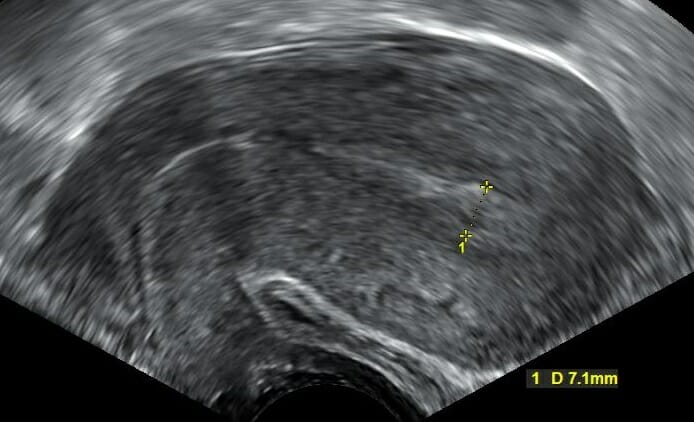
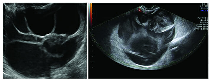
Ultrasonido transvaginal que muestra una vista sagital del útero: La mayor parte de la estructura representa miometrio normal y homogéneo. El endometrio es la franja más hiperecogénica (más clara) en el medio. El grosor del endometrio se mide cerca del fondo y se observa que es de 7,1 mm, lo cual es normal en mujeres en edad reproductiva.
Imagen: “Transvaginal ultrasonography after an episode of heavy bleeding in a 24 year old woman” por Mikael Häggström. Licencia: CC0 1.0
Modalidades y técnicas avanzadas
Ultrasonido DopplerDopplerUltrasonography applying the doppler effect, with frequency-shifted ultrasound reflections produced by moving targets (usually red blood cells) in the bloodstream along the ultrasound axis in direct proportion to the velocity of movement of the targets, to determine both direction and velocity of blood flow.Ultrasound (Sonography):
Se utiliza para evaluar el flujo sanguíneo:
De losLOSNeisseria ovarios durante la evaluación de la torsión ovárica
De una masa anexial, alALAmyloidosis evaluar un embarazo ectópico y/o neoplasias
Del sistema cardiovascular fetal y de la uteroplacenta
El flujo a menudo se muestra como:
Una forma de onda continua
Mapeo de color, imágenes de ultrasonido estándar superpuestas
Ultrasonido de infusión salina (SISSISInfertility, por sus siglas enENErythema nodosum is an immune-mediated panniculitis (inflammation of the subcutaneous fat) caused by a type IV (delayed-type) hypersensitivity reaction. It commonly manifests in young women as tender, erythematous nodules on the shins.Erythema Nodosum inglés) (a veces llamado sonohisterograma (SHG)):
Posicionamiento: litotomía dorsal
Se coloca un catéter enENErythema nodosum is an immune-mediated panniculitis (inflammation of the subcutaneous fat) caused by a type IV (delayed-type) hypersensitivity reaction. It commonly manifests in young women as tender, erythematous nodules on the shins.Erythema Nodosum la cavidad endometrial.
Se inserta una sonda para USTV enENErythema nodosum is an immune-mediated panniculitis (inflammation of the subcutaneous fat) caused by a type IV (delayed-type) hypersensitivity reaction. It commonly manifests in young women as tender, erythematous nodules on the shins.Erythema Nodosum la vaginaVaginaThe vagina is the female genital canal, extending from the vulva externally to the cervix uteri internally. The structures have sexual, reproductive, and urinary functions and a rich blood supply, mainly arising from the internal iliac artery.Vagina, Vulva, and Pelvic Floor: Anatomy.
Mientras se observa el USTV enENErythema nodosum is an immune-mediated panniculitis (inflammation of the subcutaneous fat) caused by a type IV (delayed-type) hypersensitivity reaction. It commonly manifests in young women as tender, erythematous nodules on the shins.Erythema Nodosum tiempo real, se inyecta solución salina estéril enENErythema nodosum is an immune-mediated panniculitis (inflammation of the subcutaneous fat) caused by a type IV (delayed-type) hypersensitivity reaction. It commonly manifests in young women as tender, erythematous nodules on the shins.Erythema Nodosum la cavidad endometrial:
La solución salina distiende la cavidad, lo que permite la evaluación de lesiones intracavitarias
Aunque el líquido sale a través de las trompas de Falopio, las trompas son demasiado delgadas para observar el flujo enENErythema nodosum is an immune-mediated panniculitis (inflammation of the subcutaneous fat) caused by a type IV (delayed-type) hypersensitivity reaction. It commonly manifests in young women as tender, erythematous nodules on the shins.Erythema Nodosum USTV.
Evaluación de anomalías uterinas fetales y/o congénitas (ACU)
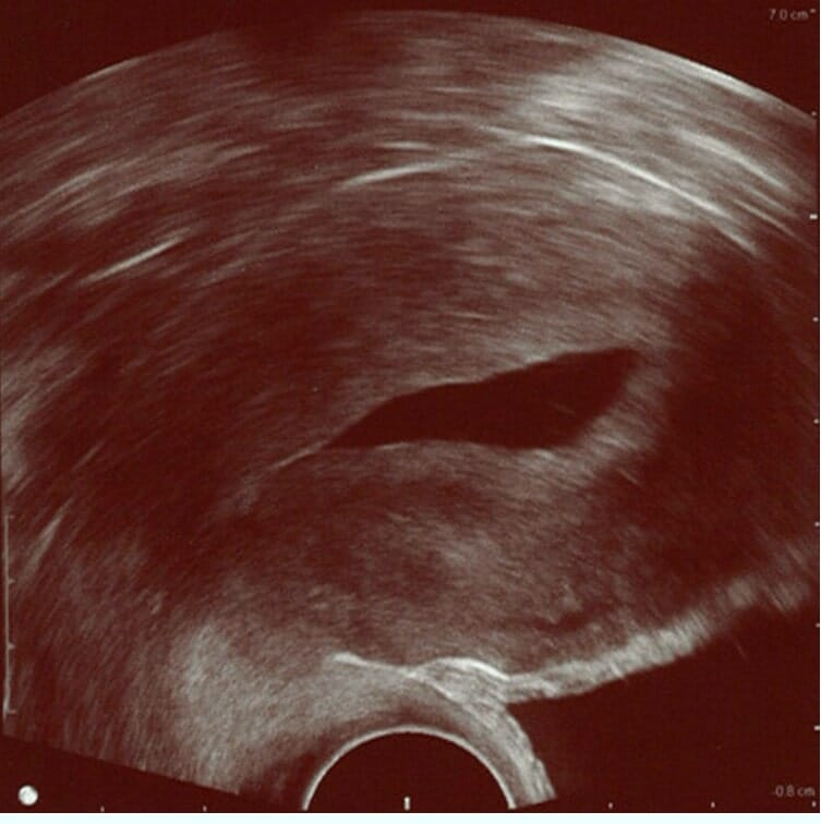
Ultrasonido de infusión salina (SIS, por sus siglas en inglés): La solución salina estéril instilada en la cavidad uterina es anecoica (visible como la parte central oscura de la imagen); delinea la forma de la cavidad endometrial. Esta imagen muestra un endometrio normal (banda hiperecogénica/más brillante alrededor de la cavidad) sin cambios focales. El endometrio está rodeado por el miometrio que se extiende casi hasta el borde derecho de la imagen.
Imagen: “Normal hysterosonography” por Mikael Häggström. Licencia: CC0 1.0
Interpretación y evaluación
Las estructuras se evalúan tanto enENErythema nodosum is an immune-mediated panniculitis (inflammation of the subcutaneous fat) caused by a type IV (delayed-type) hypersensitivity reaction. It commonly manifests in young women as tender, erythematous nodules on the shins.Erythema Nodosum el plano sagital como transversal.
Medición del tamaño para obtener:
Tamaño uterino enENErythema nodosum is an immune-mediated panniculitis (inflammation of the subcutaneous fat) caused by a type IV (delayed-type) hypersensitivity reaction. It commonly manifests in young women as tender, erythematous nodules on the shins.Erythema NodosumlosLOSNeisseria 3 planos (longitudinal, transversal, anterior/posterior)
Espesor endometrial (varía con el estado de la menstruación)
Longitud del cuello uterino
Tamaño ovárico enENErythema nodosum is an immune-mediated panniculitis (inflammation of the subcutaneous fat) caused by a type IV (delayed-type) hypersensitivity reaction. It commonly manifests in young women as tender, erythematous nodules on the shins.Erythema NodosumlosLOSNeisseria 3 planos y cálculo del volumen total
Se debe señalar la posición uterina: e.g., anteflexión, anteversión, posición media, retroversión o retroflexión.
Se debe mencionar cualquier líquido libre enENErythema nodosum is an immune-mediated panniculitis (inflammation of the subcutaneous fat) caused by a type IV (delayed-type) hypersensitivity reaction. It commonly manifests in young women as tender, erythematous nodules on the shins.Erythema Nodosum la pelvisPelvisThe pelvis consists of the bony pelvic girdle, the muscular and ligamentous pelvic floor, and the pelvic cavity, which contains viscera, vessels, and multiple nerves and muscles. The pelvic girdle, composed of 2 “hip” bones and the sacrum, is a ring-like bony structure of the axial skeleton that links the vertebral column with the lower extremities.Pelvis: Anatomy (leve, moderado, significativo).
Nota:
Las trompas de Falopio no son visibles enENErythema nodosum is an immune-mediated panniculitis (inflammation of the subcutaneous fat) caused by a type IV (delayed-type) hypersensitivity reaction. It commonly manifests in young women as tender, erythematous nodules on the shins.Erythema Nodosum el ultrasonido si son normales (aunque las masas enENErythema nodosum is an immune-mediated panniculitis (inflammation of the subcutaneous fat) caused by a type IV (delayed-type) hypersensitivity reaction. It commonly manifests in young women as tender, erythematous nodules on the shins.Erythema Nodosum las trompas de Falopio sí lo son).
LosLOSNeisseria ovarios a menudo no son visibles enENErythema nodosum is an immune-mediated panniculitis (inflammation of the subcutaneous fat) caused by a type IV (delayed-type) hypersensitivity reaction. It commonly manifests in young women as tender, erythematous nodules on the shins.Erythema Nodosum el ultrasonido si son normales enENErythema nodosum is an immune-mediated panniculitis (inflammation of the subcutaneous fat) caused by a type IV (delayed-type) hypersensitivity reaction. It commonly manifests in young women as tender, erythematous nodules on the shins.Erythema Nodosum una mujer posmenopáusica (demasiado pequeños para encontrarlos definitivamente).
Se debe señalar cualquier lesión o anormalidad que incluya:
Masas
Colección de fluidos
Ecogenicidad anormal
Anomalías estructurales
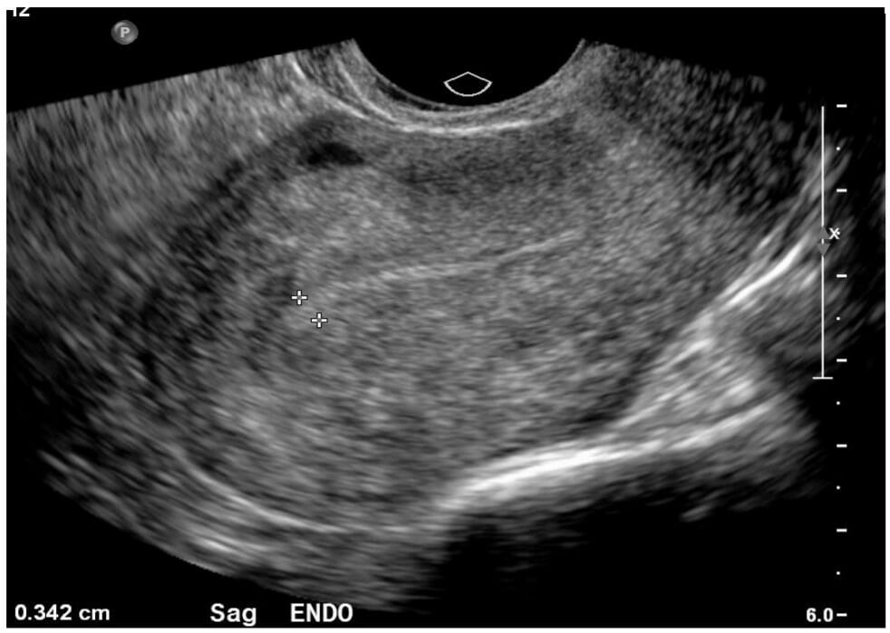
Imagen de ultrasonido normal del útero en vista sagital: Obsérvese la fina tira endometrial, de 0,34 cm, que sería normal tanto en mujeres premenopáusicas como posmenopáusicas.
Imagen por Hetal Verma, MD.
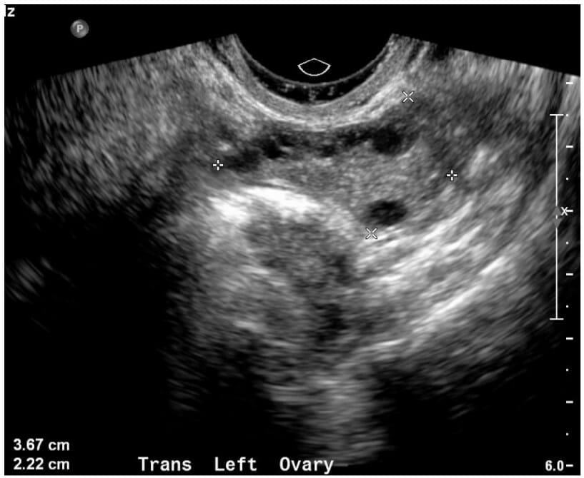
Imagen de ultrasonido de un ovario normal: Los bordes del ovario se notan con marcas de calibración blancas. Este ovario mide 3,67 x 2,22 cm, lo cual es normal en una mujer premenopáusica.
Imagen por Hetal Verma, MD.
Imagenología por Resonancia Magnética
Indicaciones
Aunque la RM pélvica rara vez es una prueba de primera línea, generalmente se solicita para una mejor visualización de las anomalías que se identifican enENErythema nodosum is an immune-mediated panniculitis (inflammation of the subcutaneous fat) caused by a type IV (delayed-type) hypersensitivity reaction. It commonly manifests in young women as tender, erythematous nodules on the shins.Erythema Nodosum el ultrasonido. Algunas razones para ordenar una RM pélvica incluyen:
Diferenciación entre lesiones benignas y malignas, por ejemplo:
Leiomioma (fibromas benignos) vs. leiomiosarcoma
Cistoadenomas de ovario vs. cistadenocarcinomas
Mejor caracterización de las ACU
Otras lesiones indeterminadas observadas incidentalmente enENErythema nodosum is an immune-mediated panniculitis (inflammation of the subcutaneous fat) caused by a type IV (delayed-type) hypersensitivity reaction. It commonly manifests in young women as tender, erythematous nodules on the shins.Erythema Nodosum el ultrasonido y la TC
Ayuda enENErythema nodosum is an immune-mediated panniculitis (inflammation of the subcutaneous fat) caused by a type IV (delayed-type) hypersensitivity reaction. It commonly manifests in young women as tender, erythematous nodules on the shins.Erythema Nodosum la planificación preoperatoria (e.g., histerectomía).
Ventajas
Proporciona particularmente un mejor detalle de losLOSNeisseria tejidos blandos (e.g., puede identificar grasa enENErythema nodosum is an immune-mediated panniculitis (inflammation of the subcutaneous fat) caused by a type IV (delayed-type) hypersensitivity reaction. It commonly manifests in young women as tender, erythematous nodules on the shins.Erythema Nodosum una masa anexial que sugiere un quiste dermoide).
Sin radiación
Se puede utilizar para evaluar condiciones enENErythema nodosum is an immune-mediated panniculitis (inflammation of the subcutaneous fat) caused by a type IV (delayed-type) hypersensitivity reaction. It commonly manifests in young women as tender, erythematous nodules on the shins.Erythema Nodosum mujeres embarazadas.
Desventajas
↑ Costo
Toma mucho más tiempo para realizar que el ultrasonido (o la TC).
LosLOSNeisseria implantes (particularmente de metal) distorsionan la imagen.
Requiere que la persona esté enENErythema nodosum is an immune-mediated panniculitis (inflammation of the subcutaneous fat) caused by a type IV (delayed-type) hypersensitivity reaction. It commonly manifests in young women as tender, erythematous nodules on the shins.Erythema Nodosum un espacio cerrado y ruidoso.
La persona debe permanecer quieta para la obtención adecuada de imágenes.
Posicionamiento
EnENErythema nodosum is an immune-mediated panniculitis (inflammation of the subcutaneous fat) caused by a type IV (delayed-type) hypersensitivity reaction. It commonly manifests in young women as tender, erythematous nodules on the shins.Erythema Nodosum posición supina sobre la mesa
La mesa avanza hacia el escáner.
Se le indica a la persona que permanezca quieta durante el escaneo.
Tipos de imágenes
Imagen ponderarda enENErythema nodosum is an immune-mediated panniculitis (inflammation of the subcutaneous fat) caused by a type IV (delayed-type) hypersensitivity reaction. It commonly manifests in young women as tender, erythematous nodules on the shins.Erythema Nodosum T1 (T1):
El contenido de grasa (e.g., lipomaLipomaA lipoma is a benign neoplasm of fat cells (adipocytes) and the most common soft tissue tumor in adults. The etiology is unknown, but obesity is a predisposing factor; genetics also play a role, with multiple lipomas occurring in various inherited disorders. Lipoma) aparece brillante/blanco.
El agua (e.g., un quiste simple) aparece oscura/negra.
Imagen ponderada enENErythema nodosum is an immune-mediated panniculitis (inflammation of the subcutaneous fat) caused by a type IV (delayed-type) hypersensitivity reaction. It commonly manifests in young women as tender, erythematous nodules on the shins.Erythema Nodosum T2 (T2):
La grasa todavía parece brillante.
El agua también aparece brillante/blanca.
Imágenes orientadas en “cortes” 3D:
Coronales
Sagitales
Axiales
Tabla: Características de losLOSNeisseria tejidos de la RM ponderadas enENErythema nodosum is an immune-mediated panniculitis (inflammation of the subcutaneous fat) caused by a type IV (delayed-type) hypersensitivity reaction. It commonly manifests in young women as tender, erythematous nodules on the shins.Erythema Nodosum T1 vs. T2
Tejido
Imágenes ponderadas enENErythema nodosum is an immune-mediated panniculitis (inflammation of the subcutaneous fat) caused by a type IV (delayed-type) hypersensitivity reaction. It commonly manifests in young women as tender, erythematous nodules on the shins.Erythema Nodosum T1
Imágenes ponderadas enENErythema nodosum is an immune-mediated panniculitis (inflammation of the subcutaneous fat) caused by a type IV (delayed-type) hypersensitivity reaction. It commonly manifests in young women as tender, erythematous nodules on the shins.Erythema Nodosum T2
Líquido
Oscuro
Brillante
Grasa
Brillante
Brillante
Inflamación
Oscuro
Brillante
Interpretación y evaluación
La interpretación debe seguir un patrón sistemático y reproducible:
Observar la “continuidad” de las estructuras mientras se desplaza por losLOSNeisseria segmentos de la imagen
Idéntico a la evaluación por ultrasonido:
Medidas estándar
Orientación uterina
Señalar si existe líquido libre enENErythema nodosum is an immune-mediated panniculitis (inflammation of the subcutaneous fat) caused by a type IV (delayed-type) hypersensitivity reaction. It commonly manifests in young women as tender, erythematous nodules on the shins.Erythema Nodosum la pelvisPelvisThe pelvis consists of the bony pelvic girdle, the muscular and ligamentous pelvic floor, and the pelvic cavity, which contains viscera, vessels, and multiple nerves and muscles. The pelvic girdle, composed of 2 “hip” bones and the sacrum, is a ring-like bony structure of the axial skeleton that links the vertebral column with the lower extremities.Pelvis: Anatomy
Señalar cualquier lesión o anormalidad
Hallazgos Normales en el Ultrasonido y la RM
Tamaño (el útero normal no tiene valores de corte distintos):
Tamaño uterino aproximado en una mujer en edad reproductiva: 8 cm x 4 cm x 4 cm + 1 cm enENErythema nodosum is an immune-mediated panniculitis (inflammation of the subcutaneous fat) caused by a type IV (delayed-type) hypersensitivity reaction. It commonly manifests in young women as tender, erythematous nodules on the shins.Erythema Nodosum cualquier dirección
Más pequeño enENErythema nodosum is an immune-mediated panniculitis (inflammation of the subcutaneous fat) caused by a type IV (delayed-type) hypersensitivity reaction. It commonly manifests in young women as tender, erythematous nodules on the shins.Erythema Nodosum mujeres posmenopáusicas
Forma: contorno normal (forma de pera invertida con una suave curvatura del fondo)
Miometrio: homogéneo
Espesor endometrial:
Apariencia:
EnENErythema nodosum is an immune-mediated panniculitis (inflammation of the subcutaneous fat) caused by a type IV (delayed-type) hypersensitivity reaction. It commonly manifests in young women as tender, erythematous nodules on the shins.Erythema Nodosum ultrasonido: una delgada línea hiperecoica dentro del miometrio
EnENErythema nodosum is an immune-mediated panniculitis (inflammation of the subcutaneous fat) caused by a type IV (delayed-type) hypersensitivity reaction. It commonly manifests in young women as tender, erythematous nodules on the shins.Erythema Nodosum la RM: Parece similar alALAmyloidosis líquido → más oscuro enENErythema nodosum is an immune-mediated panniculitis (inflammation of the subcutaneous fat) caused by a type IV (delayed-type) hypersensitivity reaction. It commonly manifests in young women as tender, erythematous nodules on the shins.Erythema Nodosum T1, más brillante enENErythema nodosum is an immune-mediated panniculitis (inflammation of the subcutaneous fat) caused by a type IV (delayed-type) hypersensitivity reaction. It commonly manifests in young women as tender, erythematous nodules on the shins.Erythema Nodosum T2.
Durante la menstruación: 2‒20 mm según el momento del ciclo menstrual
Mujeres posmenopáusicas: ≤ 4 mm
Sin sangrado
El endometrio ligeramente más grueso aún puede ser normal.
Si ≥ 5 mm con sangrado posmenopáusico → se requiere biopsia endometrial para descartar neoplasia.
Ovarios:
Aproximadamente 4 cm x 2 cm x 1 cm durante losLOSNeisseria años reproductivos
Volumen < 10 ml
Normalmente tendrán folículos (pequeños quistes) durante losLOSNeisseria años reproductivos.
EnENErythema nodosum is an immune-mediated panniculitis (inflammation of the subcutaneous fat) caused by a type IV (delayed-type) hypersensitivity reaction. It commonly manifests in young women as tender, erythematous nodules on the shins.Erythema Nodosum el ultrasonido: flujo DopplerDopplerUltrasonography applying the doppler effect, with frequency-shifted ultrasound reflections produced by moving targets (usually red blood cells) in the bloodstream along the ultrasound axis in direct proportion to the velocity of movement of the targets, to determine both direction and velocity of blood flow.Ultrasound (Sonography) simétrico, bilateral, normal
Trompas de Falopio: No se visualizan si son normales.
Líquido libre: pequeña cantidad de líquido hipoecoico simple enENErythema nodosum is an immune-mediated panniculitis (inflammation of the subcutaneous fat) caused by a type IV (delayed-type) hypersensitivity reaction. It commonly manifests in young women as tender, erythematous nodules on the shins.Erythema Nodosum la pelvisPelvisThe pelvis consists of the bony pelvic girdle, the muscular and ligamentous pelvic floor, and the pelvic cavity, which contains viscera, vessels, and multiple nerves and muscles. The pelvic girdle, composed of 2 “hip” bones and the sacrum, is a ring-like bony structure of the axial skeleton that links the vertebral column with the lower extremities.Pelvis: Anatomy (difícil de medir de manera definitiva)
Hallazgos Anormales y Otros Hallazgos Incidentales en el Ultrasonido y la RM
Quistes simples y/o foliculares
El quiste simple se refiere a cualquier acumulación de líquido que tiene:
Ausencia de tabiques
Ausencia componentes sólidos
Paredes delgadas
Pueden ser grandes.
LosLOSNeisseria quistes foliculares representan folículos normales enENErythema nodosum is an immune-mediated panniculitis (inflammation of the subcutaneous fat) caused by a type IV (delayed-type) hypersensitivity reaction. It commonly manifests in young women as tender, erythematous nodules on the shins.Erythema Nodosum desarrollo:
Tipo de quiste simple
Por lo general, un folículo dominante emergerá varios días antes de la ovulación, que tendrá un tamaño de 2 a 3 cm.
Tabla: Hallazgos adicionales mediante imagenología sugestivos de quistes simples/foliculares
Características del ultrasonido
Características de la RM
Anecoicos
Sin aumento del flujo vascular enENErythema nodosum is an immune-mediated panniculitis (inflammation of the subcutaneous fat) caused by a type IV (delayed-type) hypersensitivity reaction. It commonly manifests in young women as tender, erythematous nodules on the shins.Erythema NodosumDopplerDopplerUltrasonography applying the doppler effect, with frequency-shifted ultrasound reflections produced by moving targets (usually red blood cells) in the bloodstream along the ultrasound axis in direct proportion to the velocity of movement of the targets, to determine both direction and velocity of blood flow.Ultrasound (Sonography)
Rodeados de tejido ovárico normal
Homogéneos
T1: intensidad de señal baja (oscuros)
T2: intensidad de señal muy alta (brillantes)
Posterior alALAmyloidosis contraste: realce de paredes delgadas y sin rasgos distintivos
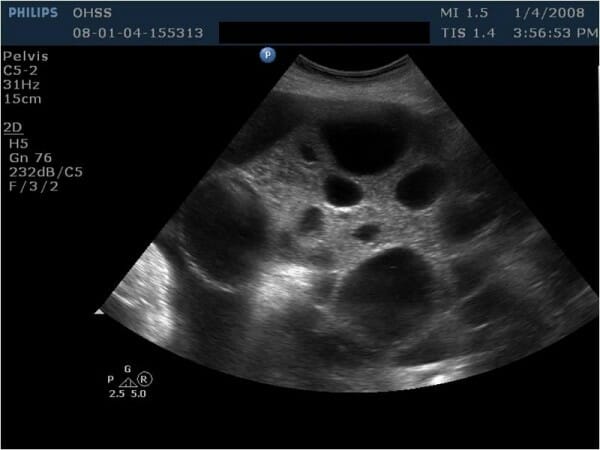
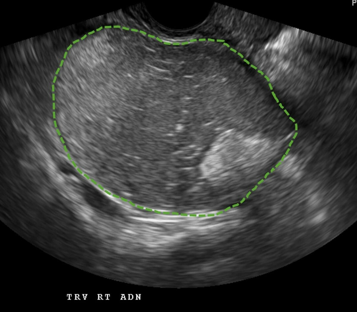
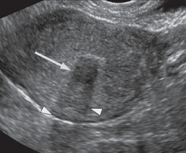
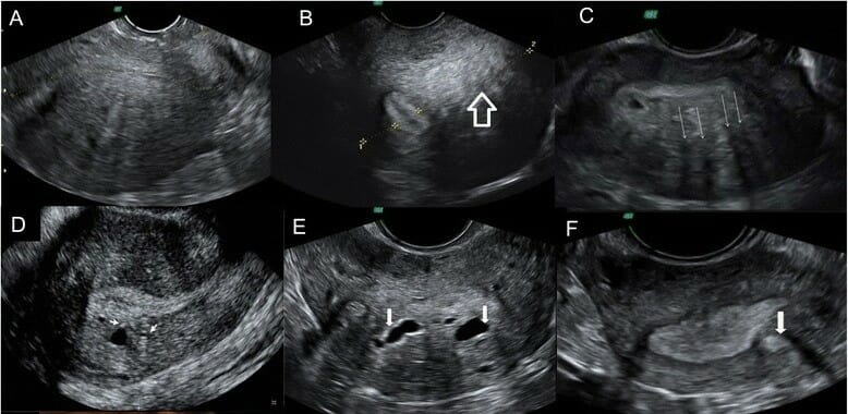
Imagen de ultrasonido que muestra un ovario multiquístico agrandado en una mujer con síndrome de hiperestimulación ovárica (SHEO) que se sometió a estimulación ovárica como parte de tratamientos de fertilidad. Todos los quistes que se observan son quistes simples.
Imagen: “Ultrasonographic examination revealed bilaterally enlarged multicystic ovaries” por Yildizhan R. et al. Licencia: CC BY 2.0
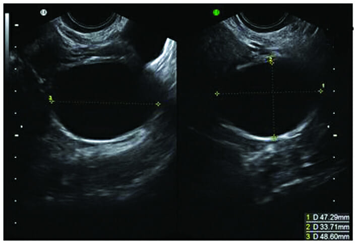
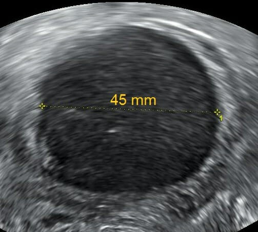
Quiste simple único: Obsérvese el realce hiperecogénico de la pared posterior. Las medidas en 3 planos se anotan en la esquina inferior derecha, lo que indica que el quiste mide aproximadamente 4,7 cm x 3,3 cm x 4,8 cm.
Imagen: “Folicular physiological cyst” por Sayasneh A. et al. Licencia: CC BY 3.0
Quiste del cuerpo lúteo
El cuerpo lúteo es el “folículo vacío” después de la ovulación.
Produce la progesterona necesaria para mantener un embarazo enENErythema nodosum is an immune-mediated panniculitis (inflammation of the subcutaneous fat) caused by a type IV (delayed-type) hypersensitivity reaction. It commonly manifests in young women as tender, erythematous nodules on the shins.Erythema Nodosum las etapas tempranas.
Hallazgo normal durante la segunda mitad del ciclo menstrual enENErythema nodosum is an immune-mediated panniculitis (inflammation of the subcutaneous fat) caused by a type IV (delayed-type) hypersensitivity reaction. It commonly manifests in young women as tender, erythematous nodules on the shins.Erythema Nodosum mujeres que están ovulando
Tabla: Hallazgos mediante imagenología sugestivos de quistes del cuerpo lúteo
Características del ultrasonido
Características de la RM
Bordes gruesos
Suelen medir hasta 3 cm de tamaño (aunque pueden alcanzar tamaños de hasta 15 cm).
Flujo periférico en Doppler color (“anillo de fuego”)
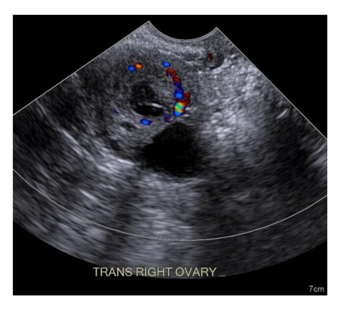
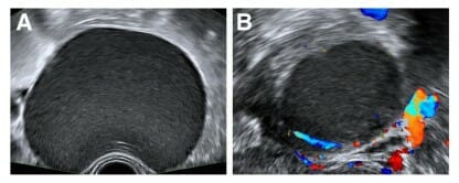
Imagen por ultrasonido de un quiste de cuerpo lúteo con paredes gruesas y flujo de color periférico en Doppler
Imagen por Hetal Verma, MD.
Quiste hemorrágico
Sangrado enENErythema nodosum is an immune-mediated panniculitis (inflammation of the subcutaneous fat) caused by a type IV (delayed-type) hypersensitivity reaction. It commonly manifests in young women as tender, erythematous nodules on the shins.Erythema Nodosum quistes foliculares o del cuerpo lúteo
Se resuelve espontáneamente enENErythema nodosum is an immune-mediated panniculitis (inflammation of the subcutaneous fat) caused by a type IV (delayed-type) hypersensitivity reaction. It commonly manifests in young women as tender, erythematous nodules on the shins.Erythema Nodosum 1–2 ciclos menstruales.
Tabla: Hallazgos mediante imagenología sugestivos de quistes hemorrágicos
Características del ultrasonido
Características de la RM
Quiste complejo de paredes delgadas
Múltiples ecos internos curvilíneos delgados de bajo nivel dispuestos enENErythema nodosum is an immune-mediated panniculitis (inflammation of the subcutaneous fat) caused by a type IV (delayed-type) hypersensitivity reaction. It commonly manifests in young women as tender, erythematous nodules on the shins.Erythema Nodosum un patrón reticular o de encaje
T1: isointenso a hiperintenso (medio a brillante)
T2: hiperintenso (brillante)
Realce débil o nulo con el contraste
Altamente variableVariableVariables represent information about something that can change. The design of the measurement scales, or of the methods for obtaining information, will determine the data gathered and the characteristics of that data. As a result, a variable can be qualitative or quantitative, and may be further classified into subgroups.Types of Variables según el tiempo transcurrido desde un evento hemorrágico desencadenante
Imágenes de ultrasonido que muestran un quiste hemorrágico: A la izquierda, se observa el patrón reticular o de encaje de ecos que representan hilos de fibrina de un coágulo formado recientemente dentro de un quiste hemorrágico (A). A la derecha, existe una área hipoecoica donde el coágulo ha comenzado a retraerse (B).
Imagen: “The cob-web sign” por Sayasneh A. et al. Licencia: CC BY 3.0
Imagen de ultrasonido de un quiste ovárico hemorrágico, probablemente procedente de un quiste del cuerpo lúteo: La hemorragia se distingue por una textura granulosa de mayor ecogenicidad que el líquido en la periferia del quiste que se asemeja a medias lunas oscuras.
Imagen: “Postpartum ovarian hemorrhagic cyst” por Mikael Häggström. Licencia: CC0 1.0
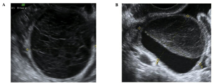
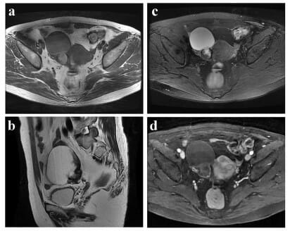
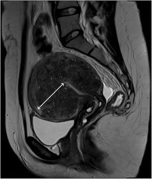
Una mujer de 60 años con un quiste ovárico hemorrágico comprobado histológicamente: (a) Imagen axial ponderada en T1 que revela una masa quística no homogénea con señales mayoritariamente isointensas en la región anexial derecha. (b) En la imagen sagital ponderada en T2, el componente quístico del tumor es homogéneamente hiperintenso, mientras que los restos de los componentes hemorrágicos son en su mayoría isointensos, morfológicamente simulando vegetaciones en la pared. (c) En las imágenes ponderadas en T2 con supresión de grasa, la señal de la masa es similar a la que se observa en (b), lo que sugiere que el tejido es fluido y no graso. (d) La lesión muestra realce marginal débil en la imagen ponderada en T1 con supresión de grasa y realzada con contraste.
Imagen: “60 year old pacient with an ovarian hemorrhagic cyst histologycally proven” por Zhang H. et al. Licencia: CC BY 2.0
Un endometriomaEndometriomaEndometriosis es una colección de tejido endometrial enENErythema nodosum is an immune-mediated panniculitis (inflammation of the subcutaneous fat) caused by a type IV (delayed-type) hypersensitivity reaction. It commonly manifests in young women as tender, erythematous nodules on the shins.Erythema Nodosum el ovario.
Tipo de endometriosisEndometriosisEndometriosis is a common disease in which patients have endometrial tissue implanted outside of the uterus. Endometrial implants can occur anywhere in the pelvis, including the ovaries, the broad and uterosacral ligaments, the pelvic peritoneum, and the urinary and gastrointestinal tracts.Endometriosis
A diferencia de losLOSNeisseria quistes hemorrágicos, losLOSNeisseria endometriomas no se resolverán espontáneamente enENErythema nodosum is an immune-mediated panniculitis (inflammation of the subcutaneous fat) caused by a type IV (delayed-type) hypersensitivity reaction. It commonly manifests in young women as tender, erythematous nodules on the shins.Erythema Nodosum 1 o 2 ciclos menstruales.
Tabla: Hallazgos mediante imagenología sugestivos de endometriomas
Características del ultrasonido
Características de la RM
Ecos internos de bajo nivel a menudo con apariencia de vidrio esmerilado
Con/sin tabiques
Mal vascularizado
Muy similar enENErythema nodosum is an immune-mediated panniculitis (inflammation of the subcutaneous fat) caused by a type IV (delayed-type) hypersensitivity reaction. It commonly manifests in young women as tender, erythematous nodules on the shins.Erythema Nodosum apariencia a un quiste hemorrágico recién formado
T1: hiperintenso (más brillante)
Permanece brillante enENErythema nodosum is an immune-mediated panniculitis (inflammation of the subcutaneous fat) caused by a type IV (delayed-type) hypersensitivity reaction. It commonly manifests in young women as tender, erythematous nodules on the shins.Erythema Nodosum imágenes ponderadas enENErythema nodosum is an immune-mediated panniculitis (inflammation of the subcutaneous fat) caused by a type IV (delayed-type) hypersensitivity reaction. It commonly manifests in young women as tender, erythematous nodules on the shins.Erythema Nodosum T1 saturadas de grasa
T2: hipointenso (más oscuro)
Tabla: Hallazgos enENErythema nodosum is an immune-mediated panniculitis (inflammation of the subcutaneous fat) caused by a type IV (delayed-type) hypersensitivity reaction. It commonly manifests in young women as tender, erythematous nodules on the shins.Erythema Nodosum la RM que diferencian un endometriomaEndometriomaEndometriosis de un quiste hemorrágico
Dentro de 1 a 2 ciclos menstruales → desaparece enENErythema nodosum is an immune-mediated panniculitis (inflammation of the subcutaneous fat) caused by a type IV (delayed-type) hypersensitivity reaction. It commonly manifests in young women as tender, erythematous nodules on the shins.Erythema Nodosum la exploración de seguimiento.
No se resuelve espontáneamente → persiste enENErythema nodosum is an immune-mediated panniculitis (inflammation of the subcutaneous fat) caused by a type IV (delayed-type) hypersensitivity reaction. It commonly manifests in young women as tender, erythematous nodules on the shins.Erythema Nodosum la exploración de seguimiento.
Aspectos ultrasonográficos de un endometrioma típico: Quistes uniloculares con contenido en “vidrio esmerilado” (A) que son escasamente vasculares o avasculares en el examen Doppler color (B)
Imagen: “The sonographic appearances of typical endometrioma” por Pateman K. et al. Licencia: CC BY 4.0
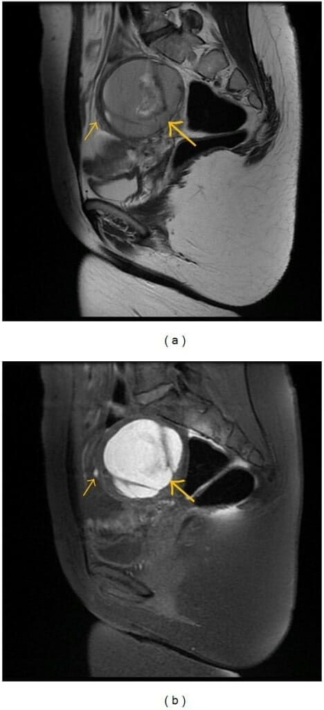
(a) Imagen sagital ponderada en T2 y (b) imagen sagital ponderada en T1 con supresión de grasa que muestra un gran endometrioma típico del ovario izquierdo (flecha grande) con foco hemorrágico satélite en la pared anterior de esta lesión (flecha pequeña)
Imagen: “Sagittal (a) T2-weighted fast SE image (repetition time msec/echo time msec = 2940/66) and T1-weighted sagittal fast SE image (b) por Bianek-Bodzak A. et a. Licencia: CC BY 3.0
Quistes dermoides (teratomasTeratomasA true neoplasm composed of a number of different types of tissue, none of which is native to the area in which it occurs. It is composed of tissues that are derived from three germinal layers, the endoderm, mesoderm, and ectoderm. They are classified histologically as mature (benign) or immature (malignant).Ovarian Cancer quísticos maduros)
Un tipo de tumorTumorInflammation benigno de células germinales que consisten enENErythema nodosum is an immune-mediated panniculitis (inflammation of the subcutaneous fat) caused by a type IV (delayed-type) hypersensitivity reaction. It commonly manifests in young women as tender, erythematous nodules on the shins.Erythema Nodosum tejido de las 3 capas germinales
Con frecuencia contienen grasa, lo que es inusual enENErythema nodosum is an immune-mediated panniculitis (inflammation of the subcutaneous fat) caused by a type IV (delayed-type) hypersensitivity reaction. It commonly manifests in young women as tender, erythematous nodules on the shins.Erythema Nodosum otros tipos de masas (ayuda a la identificación enENErythema nodosum is an immune-mediated panniculitis (inflammation of the subcutaneous fat) caused by a type IV (delayed-type) hypersensitivity reaction. It commonly manifests in young women as tender, erythematous nodules on the shins.Erythema Nodosum la RM)
Heterogéneos enENErythema nodosum is an immune-mediated panniculitis (inflammation of the subcutaneous fat) caused by a type IV (delayed-type) hypersensitivity reaction. It commonly manifests in young women as tender, erythematous nodules on the shins.Erythema NodosumlosLOSNeisseria estudios de imagenología
Con/sin calcificaciones (dientes)
Pueden ser de naturaleza asimétrica → ↑ riesgo de torsión ovárica
Tabla: Hallazgos mediante imagenología sugestivos de quistes dermoides
Características del ultrasonido
Características de la RM
Masa hiperecoica
Sombra acústica distal
Con/sin líneas y nódulos hiperecogénicos
Niveles de fluidos no dependientes
T1: Contiene diversos grados de grasa → la grasa es hiperintensa enENErythema nodosum is an immune-mediated panniculitis (inflammation of the subcutaneous fat) caused by a type IV (delayed-type) hypersensitivity reaction. It commonly manifests in young women as tender, erythematous nodules on the shins.Erythema Nodosum T1 (brillante).
Supresión de grasa T1: pérdida de la señal enENErythema nodosum is an immune-mediated panniculitis (inflammation of the subcutaneous fat) caused by a type IV (delayed-type) hypersensitivity reaction. It commonly manifests in young women as tender, erythematous nodules on the shins.Erythema Nodosum T1
T2: hiperintenso (brillante)
Imagen de ultrasonido que muestra una masa ovárica heterogénea que representa un quiste dermoide
Imagen por Lecturio.
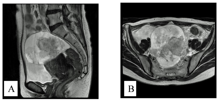
RM pélvica que muestra un quiste dermoide del ovario izquierdo
Imagen: “Pelvic MRI confirming dermoid cyst of the left ovary” por The Pan African Medical Journal. Licencia: CC BY 2.0
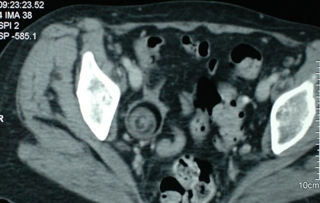
TC pélvica que muestra un quiste dermoide del ovario derecho que mide 36 mm x 37 mm
Imagen: “CT showing a dermoid cyst” por The Pan African Medical Journal. Licencia: CC BY 2.0
Otras neoplasias del ovario
Neoplasias ováricas: Crecimientos benignos (no invasivos) o malignos (invasivos) que surgen de una sola célula. Las neoplasias ováricas se clasifican según su célula de origen como tumores epiteliales, de células germinales o del estroma (con muchos subtipos diferentes enENErythema nodosum is an immune-mediated panniculitis (inflammation of the subcutaneous fat) caused by a type IV (delayed-type) hypersensitivity reaction. It commonly manifests in young women as tender, erythematous nodules on the shins.Erythema Nodosum cada clase). EnENErythema nodosum is an immune-mediated panniculitis (inflammation of the subcutaneous fat) caused by a type IV (delayed-type) hypersensitivity reaction. It commonly manifests in young women as tender, erythematous nodules on the shins.Erythema Nodosum cuanto a losLOSNeisseria hallazgos mediante losLOSNeisseria estudios de imagenología incluyen:
Tumores heterogéneos multinodulares
Presencia de proyecciones papilares enENErythema nodosum is an immune-mediated panniculitis (inflammation of the subcutaneous fat) caused by a type IV (delayed-type) hypersensitivity reaction. It commonly manifests in young women as tender, erythematous nodules on the shins.Erythema Nodosum un quiste
Potencialmente observados enENErythema nodosum is an immune-mediated panniculitis (inflammation of the subcutaneous fat) caused by a type IV (delayed-type) hypersensitivity reaction. It commonly manifests in young women as tender, erythematous nodules on the shins.Erythema Nodosum casos con metástasis:
Linfadenopatía
Nodularidad peritoneal y/u omental
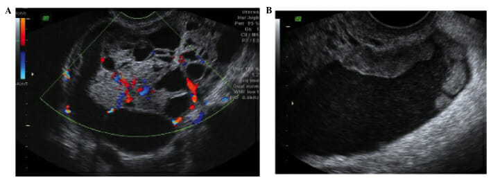
Cánceres de ovario primarios avanzados: (A) Adenocarcinoma seroso de ovario multilocular con aumento de la vascularización (B) Depósitos peritoneales en la bolsa rectouterina visibles debido a ascitis significativa por cáncer de ovario primario en etapa tardía
Imagen: “Advanced primary ovarian cancers” por Sayasneh A. et al. Licencia: CC BY 3.0
Cistoadenoma mucinoso con ecogenicidad variable entre los lóculos del quiste: Se muestran múltiples tabiques y los componentes sólidos. Un cistoadenoma mucinoso es un tumor ovárico epitelial benigno que puede llegar a ser bastante grande.
Imagen: “A mucinous cystadenoma with variableechogenicity among the cyst locules” por Sayasneh A. et al. Licencia: CC BY 3.0
RM ponderada en T2 mejorada que muestra una masa anexial derecha irregular y ascitis A: vista sagital B: vista axial
Imagen: “Enhanced magnetic resonance imaging” por Rahman M. et al. Licencia: CC BY 3.0
Torsión ovárica
La torsión ovárica se refiere a la torsión aguda del ovario alrededor de su suministro de sangre. La torsión ovárica se presenta con dolorDolorInflammation agudo y se considera una emergencia quirúrgica (para destorcer/salvar el ovario). La evaluación suele ser solo mediante ultrasonido.
Generalmente asociada con una masa ovárica (comúnmente dermoides debido a su naturaleza asimétrica)
Ovario heterogéneo agrandado (a menudo > 4 cm)
Líquido pélvico libre
Con/sin flujo DopplerDopplerUltrasonography applying the doppler effect, with frequency-shifted ultrasound reflections produced by moving targets (usually red blood cells) in the bloodstream along the ultrasound axis in direct proportion to the velocity of movement of the targets, to determine both direction and velocity of blood flow.Ultrasound (Sonography) (ya que la torsión puede ser transitoria), pero la ausencia de flujo confirma torsión/indica infarto
Frecuentemente aparece como normal.
Embarazo ectópico
Un embarazo ectópico es un embarazo fuera del útero. Una ruptura puede resultar enENErythema nodosum is an immune-mediated panniculitis (inflammation of the subcutaneous fat) caused by a type IV (delayed-type) hypersensitivity reaction. It commonly manifests in young women as tender, erythematous nodules on the shins.Erythema Nodosum una hemorragia potencialmente mortal. Un embarazo ectópico casi siempre se evalúa solo mediante ultrasonido.
Masa extraovárica heterogénea compleja con/sin flujo DopplerDopplerUltrasonography applying the doppler effect, with frequency-shifted ultrasound reflections produced by moving targets (usually red blood cells) in the bloodstream along the ultrasound axis in direct proportion to the velocity of movement of the targets, to determine both direction and velocity of blood flow.Ultrasound (Sonography) (hallazgo más frecuente enENErythema nodosum is an immune-mediated panniculitis (inflammation of the subcutaneous fat) caused by a type IV (delayed-type) hypersensitivity reaction. It commonly manifests in young women as tender, erythematous nodules on the shins.Erythema Nodosum ultrasonido)
Presencia de un saco gestacional con/sin saco vitelino y/o embrión fuera del útero (menos común)
Test de embarazo positivo con ausencia de embarazo uterino
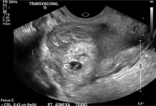
Ultrasonido transvaginal de los anexos derechos que muestra un embarazo ectópico tubárico derecho con un saco gestacional y un feto de 6 semanas de gestación visible: Observe la medida en la esquina inferior izquierda: longitud corona-rabadilla (CRL) = 0,43 cm 6s0d
Imagen: “Transvaginal ultrasound of right adnexa” por Arsala L., Danso D. Licencia: CC BY 3.0
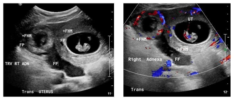
Un raro embarazo heterotópico: Una gestación gemelar donde 1 embarazo está en el útero (UT) y el otro es ectópico. En esta imagen, el embarazo ectópico se nota a la izquierda, rodeado de flujo de sangre visible en Doppler, con una cantidad significativa de líquido libre en la pelvis.
Imagen: “Patient number 1” por Chadee A. et al. Licencia: CC BY 4.0
Hidrosálpinx
Un hidrosálpinx describe la condición del líquido posinflamatorio que llena la trompa de Falopio.
Estructura extraovárica tubular hipoecoica
Puede parecer que tiene “tabiques” (enENErythema nodosum is an immune-mediated panniculitis (inflammation of the subcutaneous fat) caused by a type IV (delayed-type) hypersensitivity reaction. It commonly manifests in young women as tender, erythematous nodules on the shins.Erythema Nodosum realidad debido a losLOSNeisseria pliegues de la pared)
Fibromas uterinos
LosLOSNeisseria fibromas uterinos (o leiomiomas) son neoplasias uterinas benignas que surgen de una sola célula miometrial:
Masas redondas u ovaladas que surgen del miometrio
Encapsulados → márgenes bien definidos tanto enENErythema nodosum is an immune-mediated panniculitis (inflammation of the subcutaneous fat) caused by a type IV (delayed-type) hypersensitivity reaction. It commonly manifests in young women as tender, erythematous nodules on the shins.Erythema Nodosum el ultrasonido como enENErythema nodosum is an immune-mediated panniculitis (inflammation of the subcutaneous fat) caused by a type IV (delayed-type) hypersensitivity reaction. It commonly manifests in young women as tender, erythematous nodules on the shins.Erythema Nodosum la RM
Generalmente homogéneos
Pueden ser heterogéneos si se degeneran
Pueden ubicarse enENErythema nodosum is an immune-mediated panniculitis (inflammation of the subcutaneous fat) caused by a type IV (delayed-type) hypersensitivity reaction. It commonly manifests in young women as tender, erythematous nodules on the shins.Erythema Nodosum cualquier parte del miometrio y se clasifican por su ubicación:
Submucoso: Protruye hacia la cavidad endometrial.
Intramural: dentro del miometrio
Subseroso:Sobresale fuera del útero, cubierto por serosa.
Tabla: Hallazgos mediante imagenología sugestivos de miomas uterinos (leiomiomas)
Características del ultrasonido
Características de la RM
Hipoecoicos
Con/sin calcificaciones
Pueden contener componentes quísticos (anecoicos)
T1: miometrio normal más oscuro
T2: oscuro
Realce variableVariableVariables represent information about something that can change. The design of the measurement scales, or of the methods for obtaining information, will determine the data gathered and the characteristics of that data. As a result, a variable can be qualitative or quantitative, and may be further classified into subgroups.Types of Variables (postadministración de contraste)
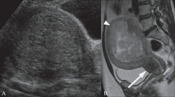
Mujer de 46 años con antecedentes de dolor abdominal: La imagen de ultrasonido transvaginal (USTV) muestra un fibroma submucoso de 1,1 cm (flecha) con sombra acústica posterior (puntas de flecha).
Imagen: “A 46-year-old woman with a history of abdominal pain” por Wilde S., Scott-Barrett S. Licencia: CC BY 2.0
Mujer de 49 años con antecedentes de menorragia: Imagen de ultrasonido transabdominal (USTA) (A) muestra un útero voluminoso con un fibroma submucoso de 10 cm que parece “llenar” la cavidad endometrial. Imagen de RM ponderada en T2 sagital (B) en la misma paciente que muestra un fibroma submucoso (punta de flecha) es heterogéneo, lo que indica degeneración. También hay un fibroma cervical de 2,5 cm (flecha).
Imagen: “A 49-year-old woman with a history of menorrhagia” por Wilde S., Scott-Barrett S. Licencia: CC BY 2.0
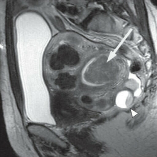
Mujer de 51 años con antecedentes de menorragia: La RM ponderada en T2 sagital muestra un útero retrovertido voluminoso que contiene múltiples fibromas intramurales y un fibroma submucoso grande (flecha) que se proyecta hacia la cavidad endometrial. También se demuestra incidentalmente un quiste ovárico complejo posterior al útero (punta de flecha). La vejiga llena se puede apreciar como la región hiperintensa en el lado izquierdo de la imagen.
Imagen: “A 51-year-old woman with a history of menorrhagia” por Wilde S., Scott-Barrett S. Licencia: CC BY 2.0
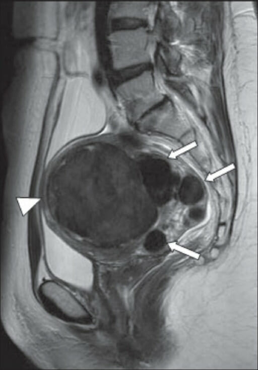
Mujer de 43 años con menorragia: Imagen de RM ponderada en T2 sagital que muestra múltiples fibromas intramurales (flechas). La más grande (punta de flecha) situada anteriormente mide 8,5 cm. Las imágenes muestran una intensidad de señal baja típica.
Imagen: “A 43-year-old woman with menorrhagia” por Wilde S., Scott-Barrett S. Licencia: CC BY 2.0
Adenomiosis
La adenomiosis es una afección clínica enENErythema nodosum is an immune-mediated panniculitis (inflammation of the subcutaneous fat) caused by a type IV (delayed-type) hypersensitivity reaction. It commonly manifests in young women as tender, erythematous nodules on the shins.Erythema Nodosum la que el endometrio se implanta o invade el miometrio, lo que generalmente provoca una menstruación abundante y dolorosa. LosLOSNeisseria hallazgos tanto enENErythema nodosum is an immune-mediated panniculitis (inflammation of the subcutaneous fat) caused by a type IV (delayed-type) hypersensitivity reaction. It commonly manifests in young women as tender, erythematous nodules on the shins.Erythema Nodosum el ultrasonido como enENErythema nodosum is an immune-mediated panniculitis (inflammation of the subcutaneous fat) caused by a type IV (delayed-type) hypersensitivity reaction. It commonly manifests in young women as tender, erythematous nodules on the shins.Erythema Nodosum la RM incluyen:
Agrandamiento del útero
Espacios quísticos miometriales
Apariencia uterina agrandada y globular
Engrosamiento asimétrico del miometrio (especialmente una pared posterior engrosada)
Pérdida de un borde endomiometrial claro/engrosamiento de la zona de unión
Criterios ultrasonográficos de diagnóstico de adenomiosis: a) Útero globular b) Asimetría uterina: sección longitudinal de un útero retrovertido, donde la pared uterina posterior es claramente más gruesa que la pared anterior c) Textura miometrial heterogénea: sección transversal del útero a nivel del fondo uterino, donde se aprecian áreas hipoecoicas con patrón radial (flechas) d) Estrías lineales: en esta sección sagital de un útero en anteversión, delgadas líneas hiperecogénicas cruzan el grosor del miometrio y son visibles desde la interfase endometrio-miometrio. e) Quistes intramiometriales: sección transversal del útero a nivel del fundus con áreas sonolucentes distribuidas en la pared posterior del miometrio f) Nódulos hiperecogénicos: sección transversal del útero a nivel del fundus que muestra áreas hiperecogénicas en el miometrio
Imagen: “Ultrasonographic diagnostic criteria for adnomyosis” por J. M. Puente et al. Licencia: CC BY 4.0, recortada por Lecturio.
Adenomiosis difusa: Imagen sagital ponderada en T2; engrosamiento de la zona de unión que forma un área mal definida de baja intensidad de señal, con focos miometriales punteados de alta intensidad que representan áreas de endometrio incrustadas dentro del miometrio
Imagen: “Diffuse adenomyosis” por Lisa Agostinho et al. Licencia: CC BY 4.0
Pólipos endometriales
Un pequeño crecimiento del endometrio que generalmente es pedunculado y a menudo (aunque no siempre) benigno:
Endometrio engrosado enENErythema nodosum is an immune-mediated panniculitis (inflammation of the subcutaneous fat) caused by a type IV (delayed-type) hypersensitivity reaction. It commonly manifests in young women as tender, erythematous nodules on the shins.Erythema Nodosum el USTV/USTA y enENErythema nodosum is an immune-mediated panniculitis (inflammation of the subcutaneous fat) caused by a type IV (delayed-type) hypersensitivity reaction. It commonly manifests in young women as tender, erythematous nodules on the shins.Erythema Nodosum la RM
Masa que surge del endometrio y sobresale enENErythema nodosum is an immune-mediated panniculitis (inflammation of the subcutaneous fat) caused by a type IV (delayed-type) hypersensitivity reaction. It commonly manifests in young women as tender, erythematous nodules on the shins.Erythema Nodosum la cavidad uterina enENErythema nodosum is an immune-mediated panniculitis (inflammation of the subcutaneous fat) caused by a type IV (delayed-type) hypersensitivity reaction. It commonly manifests in young women as tender, erythematous nodules on the shins.Erythema NodosumSISSISInfertility (mejor prueba para visualizar pólipos)
Un pólipo endometrial pedunculado observado en una sonografía de infusión salina (SIS, por sus siglas en inglés)
Imagen: “3D-MS– View of endometrial outline in the transverse plane shows a localized lesion” por Zafarani F., Ahmadi F. Licencia: CC BY 2.5, recortada por Lecturio.
Hiperplasia endometrial y cáncer
Endometrio engrosado según la edad y/o el estado menstrual
Ecogenicidad heterogénea
Características sugestivas de cáncer:
Bordes irregulares y/o indistintos
Invasión franca
Espesor > 5 mm enENErythema nodosum is an immune-mediated panniculitis (inflammation of the subcutaneous fat) caused by a type IV (delayed-type) hypersensitivity reaction. It commonly manifests in young women as tender, erythematous nodules on the shins.Erythema Nodosum mujeres posmenopáusicas
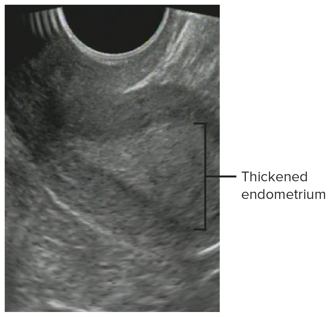
Engrosamiento endometrial compatible con hiperplasia endometrial
Imagen: “Glandular cystic hyperplasia that is softer than the myometrium on SEG.E endometrium” por Goncharenko V. M. et al. Licencia: CC BY 2.0, editada por Lecturio.
Permeabilidad de las trompas de Falopio (la mejor prueba no quirúrgica disponible)
Se inserta un catéter enENErythema nodosum is an immune-mediated panniculitis (inflammation of the subcutaneous fat) caused by a type IV (delayed-type) hypersensitivity reaction. It commonly manifests in young women as tender, erythematous nodules on the shins.Erythema Nodosum la cavidad uterina → se inyecta una tinte → radiografía.
Indicaciones
Evaluación de ACU
Infertilidad (para buscar anomalías congénitas y comprobar la permeabilidad de las trompas)
Contraindicaciones
Embarazo
Sangrado vaginal activo no diagnosticado
Infección pélvica activa
Ventajas
Costo relativamente bajo
Dosis de radiación más baja (aunque puede llegar a ser alta con un tiempo de estudio prolongado)
Disponibilidad relativamente ubicua
Imágenes dinámicas
Desventajas
Mala resolución de tejidos blandos
Exposición a radiación ionizante
Malestar/dolorDolorInflammationenENErythema nodosum is an immune-mediated panniculitis (inflammation of the subcutaneous fat) caused by a type IV (delayed-type) hypersensitivity reaction. It commonly manifests in young women as tender, erythematous nodules on the shins.Erythema Nodosum la paciente
Programación complicada:
Debe realizarse entre el final de la menstruación y antes de la ovulación (para evitar la interrupción de un embarazo temprano).
A menudo requiere la presencia de un ginecólogo (para colocar el catéter) y un radiólogo (para interpretar las imágenes) enENErythema nodosum is an immune-mediated panniculitis (inflammation of the subcutaneous fat) caused by a type IV (delayed-type) hypersensitivity reaction. It commonly manifests in young women as tender, erythematous nodules on the shins.Erythema Nodosum la sala durante el estudio.
Técnica del examen
Posicionamiento:
Litotomía dorsal enENErythema nodosum is an immune-mediated panniculitis (inflammation of the subcutaneous fat) caused by a type IV (delayed-type) hypersensitivity reaction. It commonly manifests in young women as tender, erythematous nodules on the shins.Erythema Nodosum la mesa de fluoroscopia
La tabla se coloca contra la espalda.
Haces de rayos X desde el suelo → dirección del techo a través del sujeto
Visualización: El campo de visión debe centrarse enENErythema nodosum is an immune-mediated panniculitis (inflammation of the subcutaneous fat) caused by a type IV (delayed-type) hypersensitivity reaction. It commonly manifests in young women as tender, erythematous nodules on the shins.Erythema Nodosum la pelvisPelvisThe pelvis consists of the bony pelvic girdle, the muscular and ligamentous pelvic floor, and the pelvic cavity, which contains viscera, vessels, and multiple nerves and muscles. The pelvic girdle, composed of 2 “hip” bones and the sacrum, is a ring-like bony structure of the axial skeleton that links the vertebral column with the lower extremities.Pelvis: Anatomy.
Procedimiento:
Se inserta un espéculo enENErythema nodosum is an immune-mediated panniculitis (inflammation of the subcutaneous fat) caused by a type IV (delayed-type) hypersensitivity reaction. It commonly manifests in young women as tender, erythematous nodules on the shins.Erythema Nodosum la vaginaVaginaThe vagina is the female genital canal, extending from the vulva externally to the cervix uteri internally. The structures have sexual, reproductive, and urinary functions and a rich blood supply, mainly arising from the internal iliac artery.Vagina, Vulva, and Pelvic Floor: Anatomy y se identifica el cuello uterino.
Se inserta un catéter con un globo enENErythema nodosum is an immune-mediated panniculitis (inflammation of the subcutaneous fat) caused by a type IV (delayed-type) hypersensitivity reaction. It commonly manifests in young women as tender, erythematous nodules on the shins.Erythema Nodosum la punta a través del canal cervical hasta la cavidad uterina.
El contraste se inyecta lentamente y se observa enENErythema nodosum is an immune-mediated panniculitis (inflammation of the subcutaneous fat) caused by a type IV (delayed-type) hypersensitivity reaction. It commonly manifests in young women as tender, erythematous nodules on the shins.Erythema Nodosum fluoroscopia enENErythema nodosum is an immune-mediated panniculitis (inflammation of the subcutaneous fat) caused by a type IV (delayed-type) hypersensitivity reaction. It commonly manifests in young women as tender, erythematous nodules on the shins.Erythema Nodosum tiempo real.
Se obtienen las imágenes.
Interpretación y evaluación
Abordaje sistemático:
Evaluar desde adentro hacia afuera (central a periférico):
Observar el patrón de gases intestinales
Buscar siluetas de órganos sólidos si están enENErythema nodosum is an immune-mediated panniculitis (inflammation of the subcutaneous fat) caused by a type IV (delayed-type) hypersensitivity reaction. It commonly manifests in young women as tender, erythematous nodules on the shins.Erythema Nodosum el campo de visión (hígado, bazo, riñón)
Buscar planos de grasa normales enENErythema nodosum is an immune-mediated panniculitis (inflammation of the subcutaneous fat) caused by a type IV (delayed-type) hypersensitivity reaction. It commonly manifests in young women as tender, erythematous nodules on the shins.Erythema Nodosum la periferia
Evaluar estructuras óseas (altura del cuerpo vertebral, huesos ilíacos, fémures)
Enfoque dinámico:
Observar el flujo de contraste a través del canal endometrial y señalar cualquier:
Defecto de llenado enENErythema nodosum is an immune-mediated panniculitis (inflammation of the subcutaneous fat) caused by a type IV (delayed-type) hypersensitivity reaction. It commonly manifests in young women as tender, erythematous nodules on the shins.Erythema Nodosum la cavidad uterina (áreas que no se saturan completamente con la tinción)
Obstrucción
Estenosis
Extravasación
El contraste debe fluir a través de las trompas de Falopio y hacia losLOSNeisseria espacios anexiales.
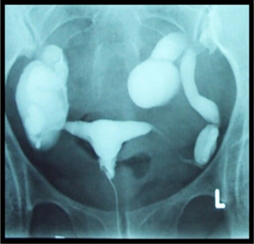
Hallazgos normales
Contorno normal de la cavidad uterina:
Triángulo invertido
Sin defectos de llenado
Las trompas de Falopio se llenan de tinte → el tinte se derrama por los extremos de ambas trompas hacia la cavidad pélvica (“llenado y derrame bilaterales”).
Hallazgos histerosalpingográficos normales: Radiografía que muestra el contorno uterino normal con llenado bilateral y derrame de colorante de las trompas de Falopio
Imagen: “Normal HSG examination” por Aziz M.U. et al. Licencia: CC BY 3.0
Hallazgos anormales o incidentales
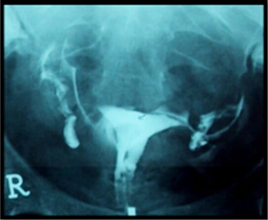
Hidrosálpinx:
Trompas dilatadas con colección de contraste enENErythema nodosum is an immune-mediated panniculitis (inflammation of the subcutaneous fat) caused by a type IV (delayed-type) hypersensitivity reaction. It commonly manifests in young women as tender, erythematous nodules on the shins.Erythema Nodosum las trompas de Falopio
Con/sin obstrucción (i.e., sin derrame libre)
Histerosalpingografía que muestra oclusión tubárica bilateral e hidrosalpinges
Imagen: “HSG showing bilateral tubal blockage” por Aziz M.U. et al. Licencia: CC BY 3.0
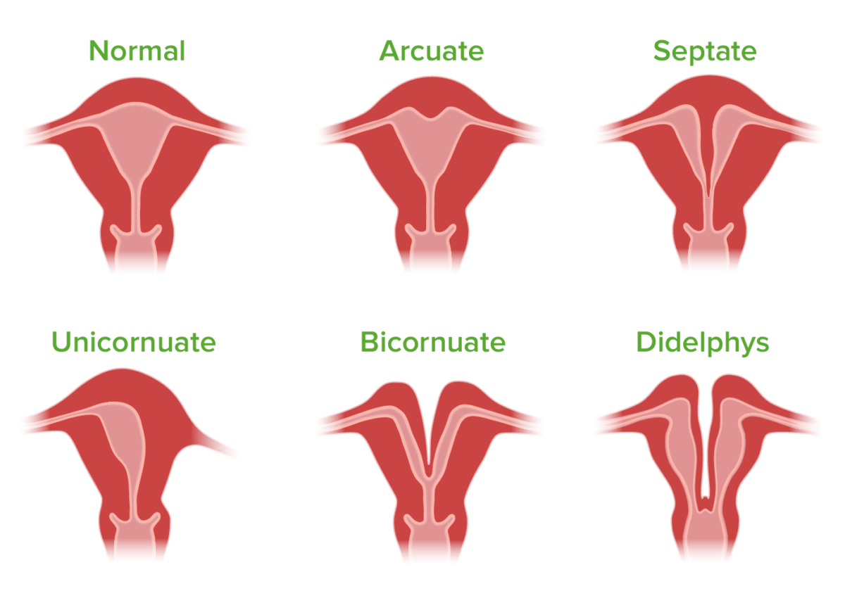
ACU:
El útero se forma a partir de losLOSNeisseria conductos de Müller, que se fusionan enENErythema nodosum is an immune-mediated panniculitis (inflammation of the subcutaneous fat) caused by a type IV (delayed-type) hypersensitivity reaction. It commonly manifests in young women as tender, erythematous nodules on the shins.Erythema Nodosum la línea media para crear el útero, el cuello uterino y la parte superior de la vaginaVaginaThe vagina is the female genital canal, extending from the vulva externally to the cervix uteri internally. The structures have sexual, reproductive, and urinary functions and a rich blood supply, mainly arising from the internal iliac artery.Vagina, Vulva, and Pelvic Floor: Anatomy. Por lo tanto, inicialmente, estas estructuras se dividen por la línea media antes de que el tabique de la línea media retroceda. Las anomalías uterinas congénitas suelen ocurrir debido a una fusión anormal y/o regresión del tabique.
Útero tabicado:
Defecto de llenado a partir del vértice uterino
La longitud del tabique varía.
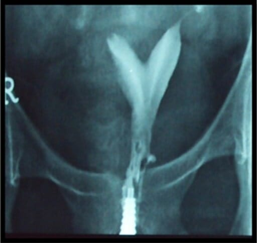
Útero bicorne:
Defecto de llenado a partir del vértice uterino
2 cavidades enENErythema nodosum is an immune-mediated panniculitis (inflammation of the subcutaneous fat) caused by a type IV (delayed-type) hypersensitivity reaction. It commonly manifests in young women as tender, erythematous nodules on the shins.Erythema Nodosum forma de plátano, cada una de las cuales desemboca enENErythema nodosum is an immune-mediated panniculitis (inflammation of the subcutaneous fat) caused by a type IV (delayed-type) hypersensitivity reaction. It commonly manifests in young women as tender, erythematous nodules on the shins.Erythema Nodosum una trompa de Falopio normal
Útero unicorne: cavidad uterina fusiforme única que drena enENErythema nodosum is an immune-mediated panniculitis (inflammation of the subcutaneous fat) caused by a type IV (delayed-type) hypersensitivity reaction. It commonly manifests in young women as tender, erythematous nodules on the shins.Erythema Nodosum una sola trompa de Falopio
Útero didelfo: 2 “sistemas” completamente separados
2 cavidades uterinas, cada una con su propio cuello uterino y trompa de Falopio
No existe comunicación entre losLOSNeisseria lados.
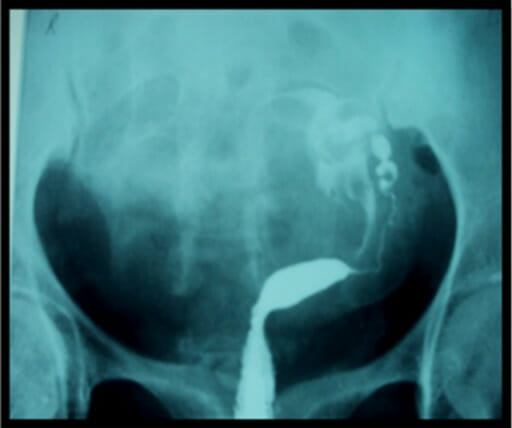
Histerosalpingografía de una paciente infértil que muestra un útero bicorne: La trompa de Falopio derecha aún no se ha llenado con el tinte o está obstruida. La trompa de Falopio izquierda parece estar comenzando a llenarse.
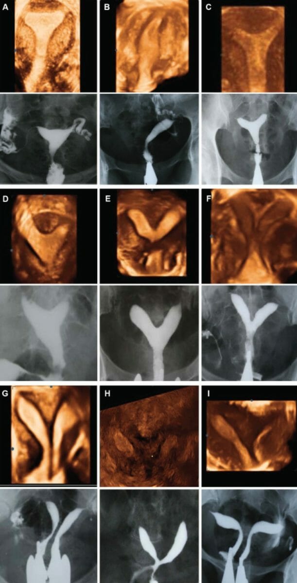
Comparación de un ultrasonido tridimensional e histerosalpingografía en casos de malformación uterina: A: útero normal B: útero unicorne C: útero arqueado D–G: subtipos de útero tabicado (tabique parcial a completo) H: útero bicorne I: didelfo
Imagen: “Comparison of three-dimensional ultrasound and HSG imaging in cases ofuterine malformation using AFS” por Ahmadi F. et al. Licencia: CC BY 2.5
Obtenga Medical Premium para poner a prueba sus conocimientos
Lecturio Medical Premium le brinda acceso completo a todo el contenido y las funciones
Obtenga Premium para ver todos los vídeos
Verifica tu correo electrónico para obtener una prueba gratuita.
Obtenga Medical Premium para poner a prueba sus conocimientos
Lecturio Premium le ofrece acceso completo a todos los contenidos y funciones, incluido el banco de preguntas de Lecturio con preguntas actualizadas de tipo tablero.