La hidronefrosis es la dilatación del sistema colector renal como consecuencia de la obstrucción del flujo de salida de la orina. La hidronefrosis puede ser unilateral o bilateral. La nefrolitiasis es la causa más común de hidronefrosis en EN Erythema nodosum is an immune-mediated panniculitis (inflammation of the subcutaneous fat) caused by a type IV (delayed-type) hypersensitivity reaction. It commonly manifests in young women as tender, erythematous nodules on the shins. Erythema Nodosum los LOS Neisseria adultos jóvenes, mientras que la hiperplasia y la neoplasia prostática se observan en EN Erythema nodosum is an immune-mediated panniculitis (inflammation of the subcutaneous fat) caused by a type IV (delayed-type) hypersensitivity reaction. It commonly manifests in young women as tender, erythematous nodules on the shins. Erythema Nodosum pacientes de mayor edad. La hidronefrosis se considera fisiológica en EN Erythema nodosum is an immune-mediated panniculitis (inflammation of the subcutaneous fat) caused by a type IV (delayed-type) hypersensitivity reaction. It commonly manifests in young women as tender, erythematous nodules on the shins. Erythema Nodosum las mujeres embarazadas. La presentación clínica depende de la agudeza y la extensión de la obstrucción. Los LOS Neisseria pacientes pueden presentar dolor Dolor Inflammation de costado, disuria, urgencia, fiebre, una masa abdominal palpable, fiebre e hipertensión. El diagnóstico incluye imagenología como ultrasonido, TC o pielografía intravenosa. El tratamiento está guiado por la causa y el grado de obstrucción. El tratamiento incluye el control del dolor Dolor Inflammation, la reposición de líquidos y el alivio de la obstrucción, que puede requerir cirugía, dependiendo de la causa.
Last updated: Dec 15, 2025
La hidronefrosis se define como la dilatación de la pelvis Pelvis The pelvis consists of the bony pelvic girdle, the muscular and ligamentous pelvic floor, and the pelvic cavity, which contains viscera, vessels, and multiple nerves and muscles. The pelvic girdle, composed of 2 “hip” bones and the sacrum, is a ring-like bony structure of the axial skeleton that links the vertebral column with the lower extremities. Pelvis: Anatomy y los LOS Neisseria cálices renales debido a la obstrucción del flujo de salida de la orina.
Las manifestaciones clínicas varían en EN Erythema nodosum is an immune-mediated panniculitis (inflammation of the subcutaneous fat) caused by a type IV (delayed-type) hypersensitivity reaction. It commonly manifests in young women as tender, erythematous nodules on the shins. Erythema Nodosum función de la agudeza del inicio de los LOS Neisseria síntomas, el grado y el lugar de la obstrucción.
Estudios de laboratorio:
| Grado | Características |
|---|---|
| I | Dilatación de la pelvis Pelvis The pelvis consists of the bony pelvic girdle, the muscular and ligamentous pelvic floor, and the pelvic cavity, which contains viscera, vessels, and multiple nerves and muscles. The pelvic girdle, composed of 2 “hip” bones and the sacrum, is a ring-like bony structure of the axial skeleton that links the vertebral column with the lower extremities. Pelvis: Anatomy renal |
| II | Grado I + dilatación calicial |
| III | Grado II + adelgazamiento de la médula |
| IV | Grado III + adelgazamiento cortical + no diferenciación corticomedular |
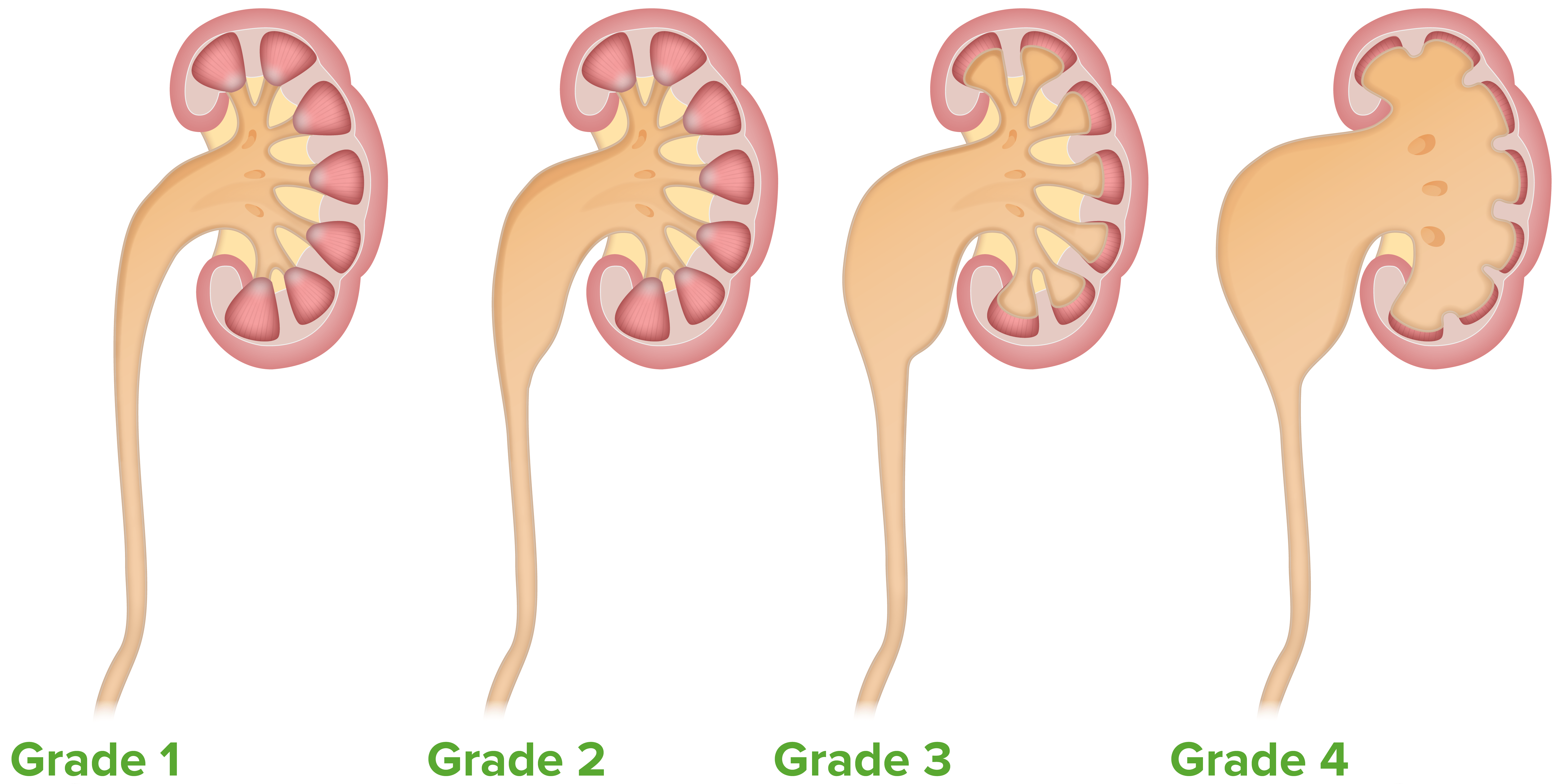
El sistema de clasificación de la hidronefrosis de Onen
Imagen por Lecturio.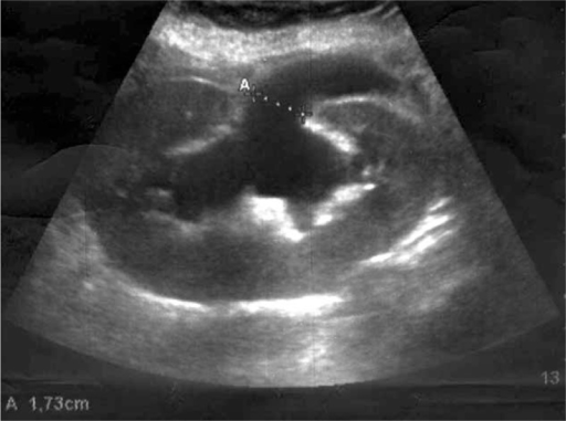
Ultrasonido renal que demuestra una hidronefrosis severa:
A: Los calipers demuestran la dilatación de la pelvis renal.
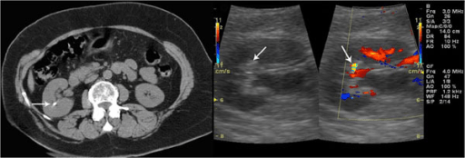
La TC sin contrasteconfirma la presencia de un cálculo renal: Izquierda: un ultrasonido en escala de grises que muestra una pequeña mancha hiperecoica sin sombra acústica posterior Derecha: una ultrasonido Doppler en color que muestra un signo de centelleo
Imagen: “F2: Unenhanced CT confirms the presence of renal stone. Grey-scale sonogram shows small hyperechoic spot without posterior acoustic shadowing. Color Doppler sonogram shows a twinkling sign.” por Gianfranco Vallone et al. Licencia: CC BY 2.0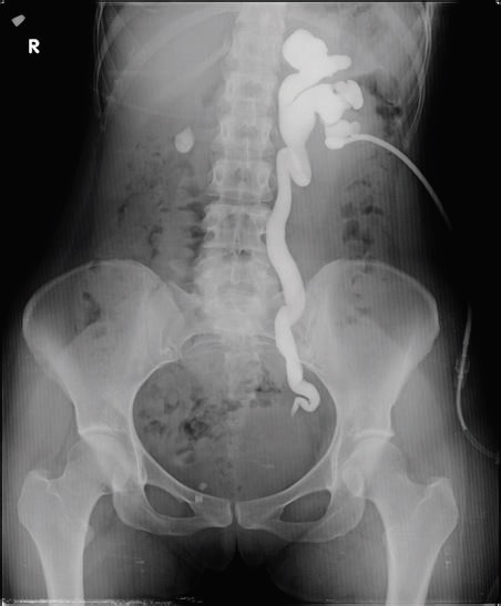
La pielografía anterógrada demuestra una hidronefrosis del lado izquierdo grado III con obstrucción en la unión ureterovesical.
Imagen: “Antegrade pyelogram of grade III hydronephrosis with obstruction at the ureterovesical junction” por Erkan Efe, Murat Bakacak, Salih Serin, Eyüp Kolus, Önder Ercan, y Sefa Resim. Licencia: CC BY 4.0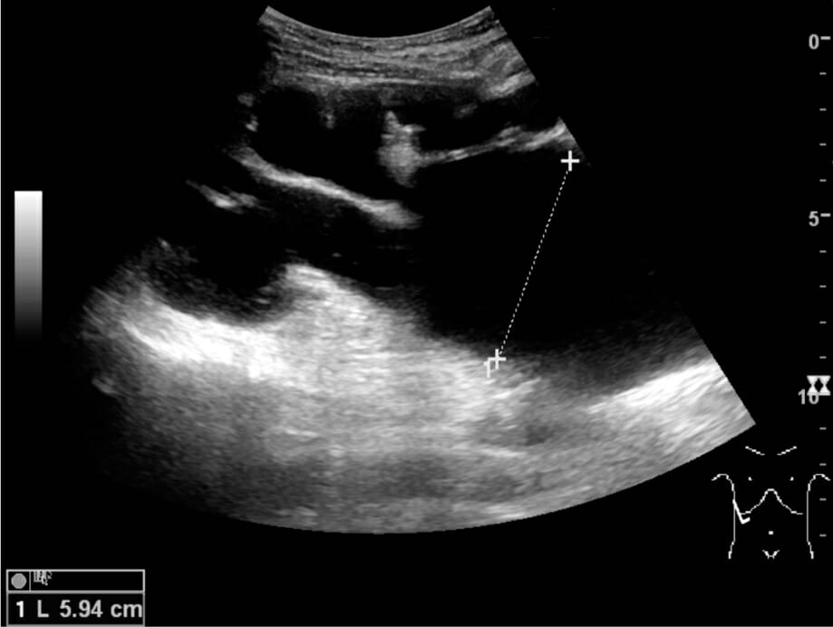
Hidronefrosis terminal con adelgazamiento cortical (grado IV): La medición de la dilatación pélvica en la imagen de US se ilustra con “+” y una línea discontinua.
Imagen: “Ultrasonography of end-stage hydronephrosis with cortical thinning” por Kristoffer Lindskov Hansen, Michael Bachmann Nielsen and Caroline Ewertsen. Licencia: CC BY 4.0El tratamiento depende de la causa de la obstrucción, el grado de anormalidades metabólicas y la presencia de infección.
El objetivo es disminuir la presión en EN Erythema nodosum is an immune-mediated panniculitis (inflammation of the subcutaneous fat) caused by a type IV (delayed-type) hypersensitivity reaction. It commonly manifests in young women as tender, erythematous nodules on the shins. Erythema Nodosum el sistema colector.