La enfermedad ósea de Paget, también conocida históricamente como osteítis deformante, es un trastorno focal del metabolismo óseo que afecta entre el 2%–9% de las personas. Las zonas más afectadas son el cráneo, la columna vertebral, la pelvis Pelvis The pelvis consists of the bony pelvic girdle, the muscular and ligamentous pelvic floor, and the pelvic cavity, which contains viscera, vessels, and multiple nerves and muscles. The pelvic girdle, composed of 2 "hip" bones and the sacrum, is a ring-like bony structure of the axial skeleton that links the vertebral column with the lower extremities. Pelvis: Anatomy y los LOS Neisseria huesos largos de las extremidades inferiores. Las dos principales manifestaciones clínicas de la enfermedad de Paget son el dolor Dolor Inflammation óseo y las consecuencias de las deformidades óseas, así como fracturas, artrosis o pinzamiento nervioso. El tratamiento incluye bifosfonatos, calcitonina y cirugía para el tratamiento de fracturas, deformidades y complicaciones. El pronóstico de la enfermedad ósea de Paget es bueno, especialmente si el tratamiento se inicia antes de que se produzcan cambios importantes en EN Erythema nodosum is an immune-mediated panniculitis (inflammation of the subcutaneous fat) caused by a type IV (delayed-type) hypersensitivity reaction. It commonly manifests in young women as tender, erythematous nodules on the shins. Erythema Nodosum los LOS Neisseria huesos.
Last updated: Dec 15, 2025
La enfermedad ósea de Paget, también conocida como osteítis deformante es un trastorno del metabolismo óseo que afecta al AL Amyloidosis envejecimiento del esqueleto.
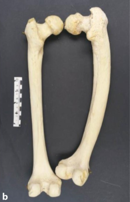
Fémur humano pagético (lado derecho), vista posterior
Fémur izquierdo sano del mismo individuo. La carga de peso sobre el hueso pagético, irregularmente engrosado pero estructuralmente débil, hace que se incline hacia delante.
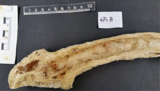
Corte longitudinal de un fémur humano pagético de una mujer de 71 años. Obsérvese la corteza irregularmente engrosada y focalmente porosa, la médula fibrótica y la forma curvada.
Imagen: “Epidemiology and pathology of Paget’s disease of bone – a review” por Wiener Medizinische Wochenschrift. Licencia: CC BY 4.0.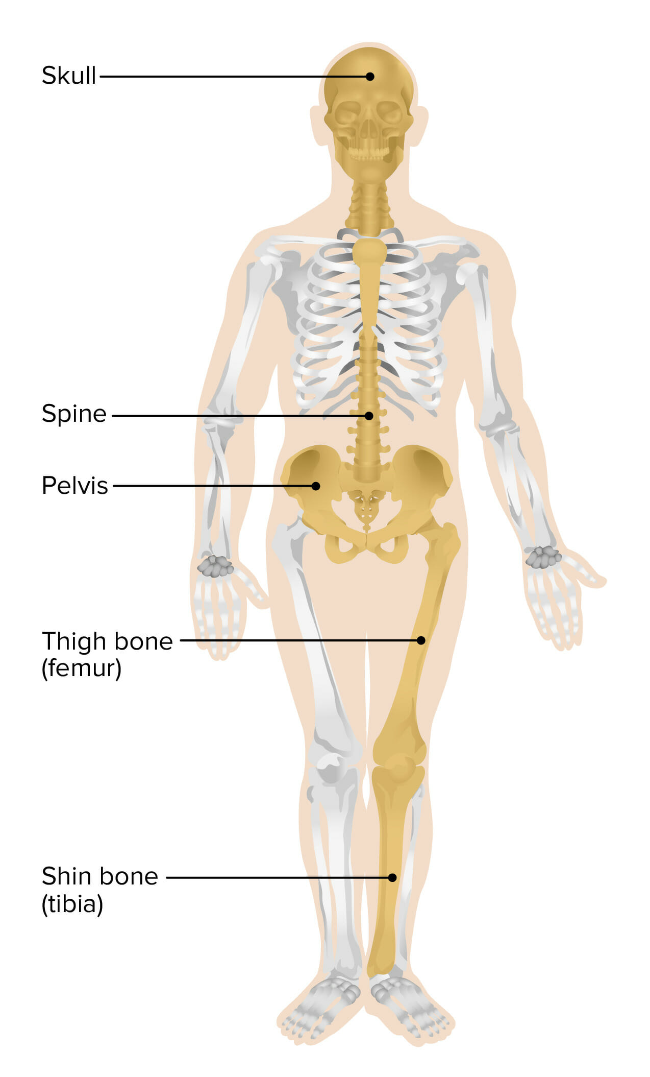
Sitios más comunmente implicados en la enfermedad ósea de Paget
Imagen por Lecturio.Más del 70% de los LOS Neisseria casos son asintomáticos.
Los LOS Neisseria lugares implicados rara vez cambian a lo largo de la vida del paciente.
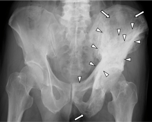
Radiografía que muestra cambios osteolíticos (flechas completas) y escleróticos (cabezas de flecha) en el acetábulo izquierdo, la mitad del ilion y el pubis
Imagen: “Five-year follow-up of Japanese patients with Paget’s disease of the bone after treatment with low-dose oral alendronate: a case series” por Iba K, Takada J, Wada T, Yamashita T. Licencia CC BY 2.0.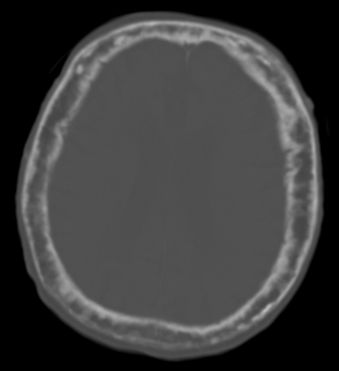
Enfermedad de Paget en el cráneo observada incidentalmente en una TC
El cráneo está notablemente engrosado con un espacio diploico ensanchado, zonas escleróticas y radiolúcidas mal definidas en toda su extensión. La corteza está engrosada y es irregular. Las zonas focalmente engrosadas corresponden con las “motas de algodón” que se observan en las placas planas de la cabeza.