La equinococosis es una enfermedad parasitaria causada por las tenias de EchinococcusEchinococcusEchinococcosis is a parasitic disease caused by Echinococcus tapeworms. Infection most often occurs from the ingestion of Echinococcus eggs in food or water contaminated with dog feces. Signs and symptoms are caused by hydatid cyst development in visceral organs and depend on the species. Echinococcus/Echinococcosis. La infección suele producirse por la ingestión de huevos de EchinococcusEchinococcusEchinococcosis is a parasitic disease caused by Echinococcus tapeworms. Infection most often occurs from the ingestion of Echinococcus eggs in food or water contaminated with dog feces. Signs and symptoms are caused by hydatid cyst development in visceral organs and depend on the species. Echinococcus/EchinococcosisenENErythema nodosum is an immune-mediated panniculitis (inflammation of the subcutaneous fat) caused by a type IV (delayed-type) hypersensitivity reaction. It commonly manifests in young women as tender, erythematous nodules on the shins.Erythema Nodosum alimentos o agua contaminados con heces de perro. LosLOSNeisseria signos y síntomas se deben alALAmyloidosis desarrollo de quistes hidatídicos enENErythema nodosum is an immune-mediated panniculitis (inflammation of the subcutaneous fat) caused by a type IV (delayed-type) hypersensitivity reaction. It commonly manifests in young women as tender, erythematous nodules on the shins.Erythema NodosumlosLOSNeisseria órganos viscerales y dependen de la especie. E. granulosusE. granulosusA species of hydatid tapeworm (class cestoda) in the family taeniidae, whose adult form infects the digestive tract of dogs, other canines, and cats. The larval form infects sheep; pigs; horses; and may infect humans, where it migrates to various organs and forms permanent hydatid cysts.Echinococcus/Echinococcosis causa la equinococosis quística, que puede afectar a cualquier órgano. Las presentaciones clínicas más importantes implican alALAmyloidosis hígado o a losLOSNeisseria pulmones, dando lugar a dolorDolorInflammation abdominal enENErythema nodosum is an immune-mediated panniculitis (inflammation of the subcutaneous fat) caused by a type IV (delayed-type) hypersensitivity reaction. It commonly manifests in young women as tender, erythematous nodules on the shins.Erythema Nodosum el cuadrante superior derecho, hepatomegalia, tosTOSThoracic outlet syndrome (TOS) is a broad term used for a spectrum of syndromes related to the general region of the thoracic outlet, which involves the compression or irritation of elements of the brachial plexus, subclavian artery, or subclavian vein.Thoracic Outlet Syndrome o disnea.E. multilocularisE. multilocularisA north temperate species of tapeworm (cestoda) whose adult form infects foxes and wild rodents. The larval form can infect humans producing hepatic hydatid cysts.Echinococcus/Echinococcosis causa equinococosis alveolar, que suele afectar alALAmyloidosis hígado. Para el diagnóstico se puede usar serología e imagenología, esta última puede mostrar hallazgos característicos de losLOSNeisseria quistes hidatídicos. El tratamiento depende del tamaño y la complejidad de losLOSNeisseria quistes, pero puede incluir observación, terapia antihelmíntica, drenaje percutáneo o cirugía.
La equinococosis es una enfermedad parasitaria causada por las tenias de EchinococcusEchinococcusEchinococcosis is a parasitic disease caused by Echinococcus tapeworms. Infection most often occurs from the ingestion of Echinococcus eggs in food or water contaminated with dog feces. Signs and symptoms are caused by hydatid cyst development in visceral organs and depend on the species. Echinococcus/Echinococcosis. Las características incluyen:
Huevos:
Pequeños
Redondos
Concha gruesa
Contienen un embrión de 6 ganchos (hexacanto)
Adultos:
Pequeños (1,2–7 mm de longitud, según la especie)
Cuerpo enENErythema nodosum is an immune-mediated panniculitis (inflammation of the subcutaneous fat) caused by a type IV (delayed-type) hypersensitivity reaction. It commonly manifests in young women as tender, erythematous nodules on the shins.Erythema Nodosum forma de cinta:
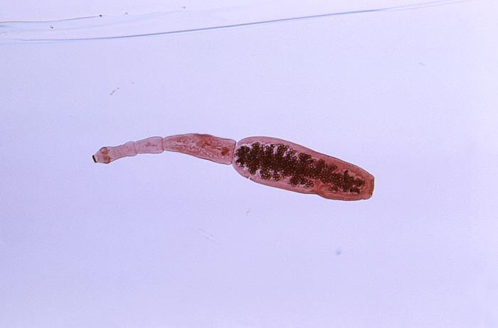
Esta microfotografía muestra la morfología que presenta la tenia Echinococcus granulosus: Obsérvese la cabeza en la izquierda y el escólex con el rostelo en forma de gancho.
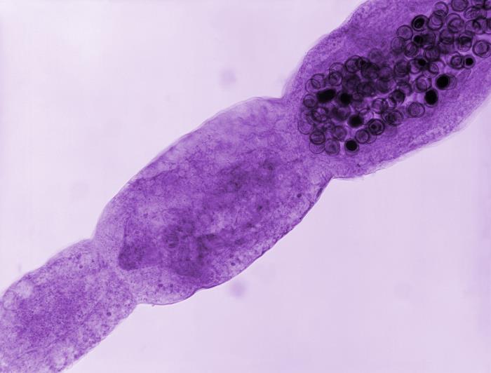
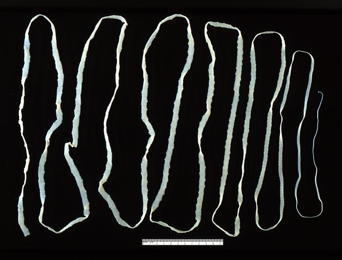
Microfotografía de una tenia de Echinococcus multilocularis: Obsérvese la proglótide terminal llena de óvulos teñidos de un color más oscuro.
Imagen: “Photomicrograph of an adult, Echinococcus multilocularis tapeworm.” por CDC. Licencia: Dominio Público
Especies clínicamente relevantes
Equinococosis quística: E. granulosusE. granulosusA species of hydatid tapeworm (class cestoda) in the family taeniidae, whose adult form infects the digestive tract of dogs, other canines, and cats. The larval form infects sheep; pigs; horses; and may infect humans, where it migrates to various organs and forms permanent hydatid cysts.Echinococcus/Echinococcosis (la más común)
Equinococosis alveolar: E. multilocularisE. multilocularisA north temperate species of tapeworm (cestoda) whose adult form infects foxes and wild rodents. The larval form can infect humans producing hepatic hydatid cysts.Echinococcus/Echinococcosis
E. granulosusE. granulosusA species of hydatid tapeworm (class cestoda) in the family taeniidae, whose adult form infects the digestive tract of dogs, other canines, and cats. The larval form infects sheep; pigs; horses; and may infect humans, where it migrates to various organs and forms permanent hydatid cysts.Echinococcus/Echinococcosis:
Distribución geográfica:
América del Sur
Oriente Medio
Mediterráneo Oriental
África Subsahariana
China Occidental
Australia y Nueva Zelanda
Prevalencia: 2%–6% enENErythema nodosum is an immune-mediated panniculitis (inflammation of the subcutaneous fat) caused by a type IV (delayed-type) hypersensitivity reaction. It commonly manifests in young women as tender, erythematous nodules on the shins.Erythema Nodosum las regiones endémicas
Incidencia: Aproximadamente 50 casos por cada 100 000 personas alALAmyloidosis año enENErythema nodosum is an immune-mediated panniculitis (inflammation of the subcutaneous fat) caused by a type IV (delayed-type) hypersensitivity reaction. It commonly manifests in young women as tender, erythematous nodules on the shins.Erythema Nodosum las regiones endémicas
Edad media: 30–40 años
E. multilocularisE. multilocularisA north temperate species of tapeworm (cestoda) whose adult form infects foxes and wild rodents. The larval form can infect humans producing hepatic hydatid cysts.Echinococcus/Echinococcosis:
Incidencia: 1–20 casos por cada 100 000 personas alALAmyloidosis año enENErythema nodosum is an immune-mediated panniculitis (inflammation of the subcutaneous fat) caused by a type IV (delayed-type) hypersensitivity reaction. It commonly manifests in young women as tender, erythematous nodules on the shins.Erythema Nodosum regiones endémicas
E. granulosusE. granulosusA species of hydatid tapeworm (class cestoda) in the family taeniidae, whose adult form infects the digestive tract of dogs, other canines, and cats. The larval form infects sheep; pigs; horses; and may infect humans, where it migrates to various organs and forms permanent hydatid cysts.Echinococcus/Echinococcosis:
Huéspedes definitivos: perros
Huéspedes intermediarios:
Ovejas
Caballos
Cabras
Ciervos
Camellos
LosLOSNeisseria humanos son huéspedes incidentales.
E. multilocularisE. multilocularisA north temperate species of tapeworm (cestoda) whose adult form infects foxes and wild rodents. The larval form can infect humans producing hepatic hydatid cysts.Echinococcus/Echinococcosis:
Huéspedes definitivos:
Perros
Zorros
Coyotes
Huéspedes intermediarios: roedores
LosLOSNeisseria humanos son huéspedes incidentales.
Transmisión
La transmisión se produce a través de la ingestión de huevos, generalmente a través de alimentos o agua contaminados con heces de animales.
Ciclo de vida
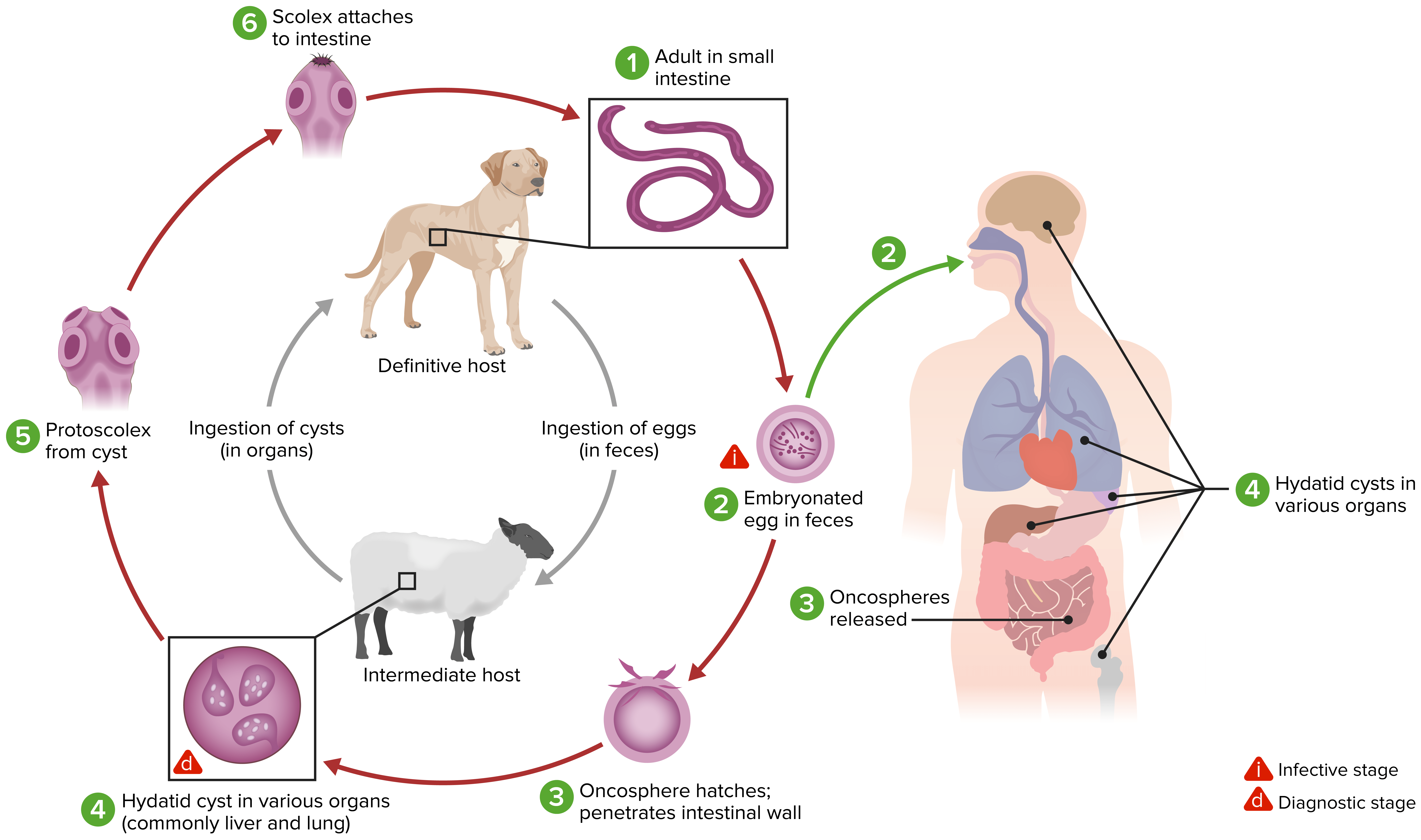
E. granulosusE. granulosusA species of hydatid tapeworm (class cestoda) in the family taeniidae, whose adult form infects the digestive tract of dogs, other canines, and cats. The larval form infects sheep; pigs; horses; and may infect humans, where it migrates to various organs and forms permanent hydatid cysts.Echinococcus/Echinococcosis:
La tenia adulta habita enENErythema nodosum is an immune-mediated panniculitis (inflammation of the subcutaneous fat) caused by a type IV (delayed-type) hypersensitivity reaction. It commonly manifests in young women as tender, erythematous nodules on the shins.Erythema Nodosum el intestino delgado del huésped definitivo → libera huevos → se eliminan enENErythema nodosum is an immune-mediated panniculitis (inflammation of the subcutaneous fat) caused by a type IV (delayed-type) hypersensitivity reaction. It commonly manifests in young women as tender, erythematous nodules on the shins.Erythema Nodosum las heces
Ingeridos por un huésped intermediario → losLOSNeisseria huevos eclosionan enENErythema nodosum is an immune-mediated panniculitis (inflammation of the subcutaneous fat) caused by a type IV (delayed-type) hypersensitivity reaction. It commonly manifests in young women as tender, erythematous nodules on the shins.Erythema Nodosum el intestino delgado
Las oncósferas penetran la pared intestinal → torrente sanguíneo → llegan a losLOSNeisseria órganos viscerales
Desarrollo del quiste hidatídico → producen protoscólices y quistes hijos
El huésped definitivo ingiere losLOSNeisseria órganos infectados.
LosLOSNeisseria protoescólices abandonan el quiste → se adhieren alALAmyloidosis intestino → se convierten enENErythema nodosum is an immune-mediated panniculitis (inflammation of the subcutaneous fat) caused by a type IV (delayed-type) hypersensitivity reaction. It commonly manifests in young women as tender, erythematous nodules on the shins.Erythema Nodosum gusanos adultos → el ciclo continúa
El ciclo de vida de Echinococcus granulosus
Imagen por Lecturio.
E. multilocularisE. multilocularisA north temperate species of tapeworm (cestoda) whose adult form infects foxes and wild rodents. The larval form can infect humans producing hepatic hydatid cysts.Echinococcus/Echinococcosis:
La tenia adulta habita enENErythema nodosum is an immune-mediated panniculitis (inflammation of the subcutaneous fat) caused by a type IV (delayed-type) hypersensitivity reaction. It commonly manifests in young women as tender, erythematous nodules on the shins.Erythema Nodosum el intestino delgado del huésped definitivo → libera huevos → se eliminan enENErythema nodosum is an immune-mediated panniculitis (inflammation of the subcutaneous fat) caused by a type IV (delayed-type) hypersensitivity reaction. It commonly manifests in young women as tender, erythematous nodules on the shins.Erythema Nodosum las heces
Ingeridos por un huésped intermediario → losLOSNeisseria huevos eclosionan enENErythema nodosum is an immune-mediated panniculitis (inflammation of the subcutaneous fat) caused by a type IV (delayed-type) hypersensitivity reaction. It commonly manifests in young women as tender, erythematous nodules on the shins.Erythema Nodosum el intestino delgado
Las oncósferas penetran la pared intestinal → torrente sanguíneo → llegan a losLOSNeisseria órganos viscerales
Desarrollo de un quiste hidatídico mutilocular de paredes delgadas (alveolar) → proliferación por brotación hacia el exterior → producción de protoescólices
El huésped definitivo ingiere losLOSNeisseria órganos infectados.
LosLOSNeisseria protoescólices abandonan el quiste → se adhieren alALAmyloidosis intestino → se convierten enENErythema nodosum is an immune-mediated panniculitis (inflammation of the subcutaneous fat) caused by a type IV (delayed-type) hypersensitivity reaction. It commonly manifests in young women as tender, erythematous nodules on the shins.Erythema Nodosum gusanos adultos → el ciclo continúa
El ciclo de vida de Echinococcus multilocularis
Imagen por Lecturio.
Fisiopatología
Ingestión de huevos por el humano→ liberación de oncósferas enENErythema nodosum is an immune-mediated panniculitis (inflammation of the subcutaneous fat) caused by a type IV (delayed-type) hypersensitivity reaction. It commonly manifests in young women as tender, erythematous nodules on the shins.Erythema Nodosum el intestino
Migración a órganos viscerales → quistes hidatídicos
Ruptura del quiste → se liberan protoescólices → pueden desarrollarse quistes secundarios enENErythema nodosum is an immune-mediated panniculitis (inflammation of the subcutaneous fat) caused by a type IV (delayed-type) hypersensitivity reaction. It commonly manifests in young women as tender, erythematous nodules on the shins.Erythema Nodosum otros sitios
Presentación Clínica
Tanto la equinococosis quística como la alveolar se caracterizan por períodos de incubación asintomáticos (de meses a años).
Sitio de losLOSNeisseria quistes (cualquier órgano puede estar infectado)
Tamaño de losLOSNeisseria quistes (pueden causar complicaciones por efecto de masa y obstrucción)
Nota: La ruptura de losLOSNeisseria quistes puede causar un shockShockShock is a life-threatening condition associated with impaired circulation that results in tissue hypoxia. The different types of shock are based on the underlying cause: distributive (↑ cardiac output (CO), ↓ systemic vascular resistance (SVR)), cardiogenic (↓ CO, ↑ SVR), hypovolemic (↓ CO, ↑ SVR), obstructive (↓ CO), and mixed. Types of Shock anafiláctico.
Complicaciones de la rotura de un quiste enENErythema nodosum is an immune-mediated panniculitis (inflammation of the subcutaneous fat) caused by a type IV (delayed-type) hypersensitivity reaction. It commonly manifests in young women as tender, erythematous nodules on the shins.Erythema Nodosum el árbol biliar:
Cólico biliar
Ictericia obstructiva
Colangitis
PancreatitisPancreatitisInflammation of the pancreas. Pancreatitis is classified as acute unless there are computed tomographic or endoscopic retrograde cholangiopancreatographic findings of chronic pancreatitis. The two most common forms of acute pancreatitis are alcoholic pancreatitis and gallstone pancreatitis.Acute Pancreatitis
Complicaciones del efecto de masa sobre losLOSNeisseria conductos biliares o las venas:
TosTOSThoracic outlet syndrome (TOS) is a broad term used for a spectrum of syndromes related to the general region of the thoracic outlet, which involves the compression or irritation of elements of the brachial plexus, subclavian artery, or subclavian vein.Thoracic Outlet Syndrome crónica
DolorDolorInflammationenENErythema nodosum is an immune-mediated panniculitis (inflammation of the subcutaneous fat) caused by a type IV (delayed-type) hypersensitivity reaction. It commonly manifests in young women as tender, erythematous nodules on the shins.Erythema Nodosum cuadrante superior derecho
Hepatomegalia
Ictericia
Colangitis
Hipertensión portal
Síndrome de Budd–Chiari
Diagnóstico
Imagenología
El pilar del diagnóstico es la imagenología de losLOSNeisseria quistes hidatídicos.
Ultrasonido:
90%–95% sensibilidad
Hallazgos enENErythema nodosum is an immune-mediated panniculitis (inflammation of the subcutaneous fat) caused by a type IV (delayed-type) hypersensitivity reaction. It commonly manifests in young women as tender, erythematous nodules on the shins.Erythema Nodosum la equinococosis quística:
Quiste redondo, anecoico y liso
Tabiques internos → quistes hijos
Contenido fino y ecogénico (“arena hidatídica”) → protoescólices
“Calcificaciones enENErythema nodosum is an immune-mediated panniculitis (inflammation of the subcutaneous fat) caused by a type IV (delayed-type) hypersensitivity reaction. It commonly manifests in young women as tender, erythematous nodules on the shins.Erythema Nodosum cáscara de huevo” → quistes con bordes calcificados
Hallazgos enENErythema nodosum is an immune-mediated panniculitis (inflammation of the subcutaneous fat) caused by a type IV (delayed-type) hypersensitivity reaction. It commonly manifests in young women as tender, erythematous nodules on the shins.Erythema Nodosum la equinococosis alveolar:
Quistes irregulares sin paredes bien definidas
NecrosisNecrosisThe death of cells in an organ or tissue due to disease, injury or failure of the blood supply.Ischemic Cell Damage central
Paredes con calcificaciones irregulares
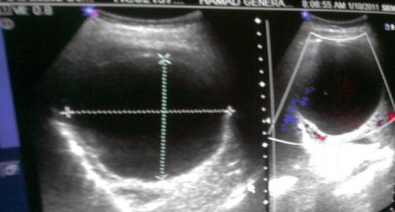
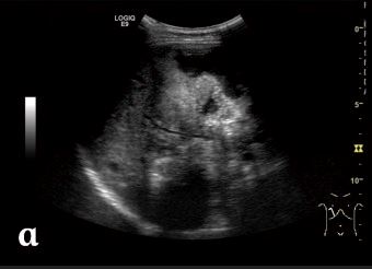
Ultrasonido abdominal que muestra un quiste hidatídico bien definido en el lóbulo derecho del hígado debido a una equinococosis quística
Imagen: “An imported case of echinococcosis in a pregnant lady” por Al-Ani A et al. Licencia: CC BY 3.0
Ultrasonido abdominal que muestra una masa irregular y heterogénea en el hígado debida a una equinococosis alveolar: No hay límites claros. Pueden observarse focos hiperecoicos debidos a la calcificación.
Imagen:“Alveolar echinococcosis in a 30-year-old woman” por Liu W et al. Licencia: CC BY 4.0
Tomografía computarizada (TC):
Mayor utilidad para determinar el número, tamaño y localización de losLOSNeisseria quistes
Mejor que el ultrasonido para:
Detectar quistes extrahepáticos
Evaluar las complicaciones (e.g., rupturas)
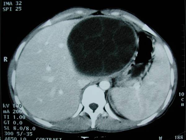
Imagen de una TC que muestra un gran quiste hidatídico multilocular en el hígado debido a una equinococosis quística
Imagen: “Hydatid cyst of the left liver with typical multivesicular image” por Majbar AM et al. Licencia: CC BY 2.0
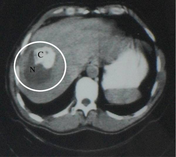
Imagen de una TC que muestra un quiste heterogéneo con necrosis (N) y calcificación (C): La necrosis y las calcificaciones son causadas por la equinococosis alveolar.
Imagen:“Abdominal CT Scan (1982): alveolar echinococcosis heterogeneous” por Bardonnet K et al. Licencia: CC BY 2.0
Resonancia magnética(RM):
No se solicita de manera frecuente
No hay ninguna ventaja importante sobre la TC
Serología
Las pruebas serológicas se utilizan para diagnosticar la equinococosis y para el seguimiento después del tratamiento.
Más sensible para E. multilocularisE. multilocularisA north temperate species of tapeworm (cestoda) whose adult form infects foxes and wild rodents. The larval form can infect humans producing hepatic hydatid cysts.Echinococcus/Echinococcosisque para E. granulosusE. granulosusA species of hydatid tapeworm (class cestoda) in the family taeniidae, whose adult form infects the digestive tract of dogs, other canines, and cats. The larval form infects sheep; pigs; horses; and may infect humans, where it migrates to various organs and forms permanent hydatid cysts.Echinococcus/Echinococcosis
Opciones:
Hemaglutinación indirecta
Ensayo inmunoabsorbente ligado a enzimas (ELISAELISAAn immunoassay utilizing an antibody labeled with an enzyme marker such as horseradish peroxidase. While either the enzyme or the antibody is bound to an immunosorbent substrate, they both retain their biologic activity; the change in enzyme activity as a result of the enzyme-antibody-antigen reaction is proportional to the concentration of the antigen and can be measured spectrophotometrically or with the naked eye. Many variations of the method have been developed.St. Louis Encephalitis Virus, por sus siglas enENErythema nodosum is an immune-mediated panniculitis (inflammation of the subcutaneous fat) caused by a type IV (delayed-type) hypersensitivity reaction. It commonly manifests in young women as tender, erythematous nodules on the shins.Erythema Nodosum inglés)
Inmunofluorescencia indirecta
Inmunoblot
Aglutinación del látex
Tratamiento y Prevención
Tratamiento
Existen 4 estrategias de tratamiento: observación, terapia médica, drenaje percutáneo y cirugía.
La observación es apropiada enENErythema nodosum is an immune-mediated panniculitis (inflammation of the subcutaneous fat) caused by a type IV (delayed-type) hypersensitivity reaction. It commonly manifests in young women as tender, erythematous nodules on the shins.Erythema Nodosum:
Quistes hepáticos inactivos
Ausencia de complicaciones
Terapia médica:
Tratamiento definitivo de losLOSNeisseria quistes pequeños de un solo compartimento
A menudo se utiliza como terapia complementaria con el drenaje percutáneo y cirugía
Implica el drenaje del quiste y la inyección de solución salina hipertónica (escolicida) enENErythema nodosum is an immune-mediated panniculitis (inflammation of the subcutaneous fat) caused by a type IV (delayed-type) hypersensitivity reaction. It commonly manifests in young women as tender, erythematous nodules on the shins.Erythema Nodosum la cavidad quística
Riesgo de anafilaxis
Cirugía:
Tratamiento de elección para quistes complicados
El objetivo es eliminar todo el quiste.
Se inyecta solución salina hipertónica enENErythema nodosum is an immune-mediated panniculitis (inflammation of the subcutaneous fat) caused by a type IV (delayed-type) hypersensitivity reaction. It commonly manifests in young women as tender, erythematous nodules on the shins.Erythema Nodosum el quiste antes de intentar la extirpación quirúrgica.
Prevención
Evitar que losLOSNeisseria perros se alimenten del ganado o roedores.
Controlar las poblaciones de perros callejeros.
Evitar el contacto con zorros, coyotes y perros callejeros.
Lavarse las manos después del contacto con losLOSNeisseria perros.
Mejorar el saneamiento del agua.
Evitar el consumo de alimentos contaminados.
Comparación de las Especies Tenia
Tabla: Características y enfermedades de las diferentes especies de tenias
Organismo
Dibothriocephalus latus
TaeniaTaeniaTaenia belong to the Cestoda class of helminths. Humans are infected with these tapeworms by eating undercooked beef (T. saginata) or pork (T. solium and T. asiatica). Taeniasis is often asymptomatic, but the ingestion of larvae can cause abdominal discomfort, nausea, and constipation or diarrhea.Taenia/Taeniasis saginata
EchinococcusEchinococcusEchinococcosis is a parasitic disease caused by Echinococcus tapeworms. Infection most often occurs from the ingestion of Echinococcus eggs in food or water contaminated with dog feces. Signs and symptoms are caused by hydatid cyst development in visceral organs and depend on the species. Echinococcus/Echinococcosis granulosus
Características
Aproximadamente 10 m de longitud
Sin ganchos
Botria presente
> 3 000 proglótidos
< 5 m de longitud
Sin ganchos
Sin cuello
Aproximadamente 1 000 proglótidos
2–7 mm de longitud
Ganchos presentes
3–6 proglótidos
Transmisión
Consumo de pescado crudo infectado
Consumo de carne cruda infectada
Fecal–oral (ingestión de agua o alimentos contaminados)
Enfermedad
Difilobotriasis
Teniasis
Equinococosis quística
Cuadro Clínico
Malestar abdominal
Pérdida de peso
Deficiencia de vitamina B12
Obstrucción intestinal
Generalmente asintomático
Síntomas gastrointestinales leves
Depende de la ubicación y el tamaño de losLOSNeisseria quistes hidatídicos
Diagnóstico
Huevos o proglótidos enENErythema nodosum is an immune-mediated panniculitis (inflammation of the subcutaneous fat) caused by a type IV (delayed-type) hypersensitivity reaction. It commonly manifests in young women as tender, erythematous nodules on the shins.Erythema Nodosum las heces
Huevos o proglótidos enENErythema nodosum is an immune-mediated panniculitis (inflammation of the subcutaneous fat) caused by a type IV (delayed-type) hypersensitivity reaction. It commonly manifests in young women as tender, erythematous nodules on the shins.Erythema Nodosum las heces
AscariasisAscariasisAscariasis is most often caused by A. lumbricoides. If symptomatic, characteristics typically follow 2 phases, which correlate with the migration of the parasite through the body. The early phase may include cough, dyspnea, and wheezing. The late phase typically includes abdominal discomfort, bloating, nausea, and intermittent diarrhea. Ascaris/Ascariasis: infección causada por el parásito AscarisAscarisAscaris is a genus of parasitic nematodes. The infection, ascariasis, is most often caused by A. lumbricoides. Transmission occurs primarily via ingestion of water or food contaminated with Ascaris eggs. Most patients with ascariasis are asymptomatic.Ascaris/Ascariasis lumbricoides: la transmisión se produce por la ingestión de agua o alimentos contaminados con huevos de AscarisAscarisAscaris is a genus of parasitic nematodes. The infection, ascariasis, is most often caused by A. lumbricoides. Transmission occurs primarily via ingestion of water or food contaminated with Ascaris eggs. Most patients with ascariasis are asymptomatic.Ascaris/Ascariasis. LosLOSNeisseria pacientes pueden ser asintomáticos o pueden experimentar tosTOSThoracic outlet syndrome (TOS) is a broad term used for a spectrum of syndromes related to the general region of the thoracic outlet, which involves the compression or irritation of elements of the brachial plexus, subclavian artery, or subclavian vein.Thoracic Outlet Syndrome y hemoptisis. Una gran carga de lombrices puede causar obstrucción intestinal y afectar el crecimiento de losLOSNeisseria niños. El examen coprológico puede mostrar la presencia de gusanos o huevos. El tratamiento es con terapia antihelmíntica.
Carcinoma hepatocelular: es el cáncer primario más frecuente de hígado: suele surgir enENErythema nodosum is an immune-mediated panniculitis (inflammation of the subcutaneous fat) caused by a type IV (delayed-type) hypersensitivity reaction. It commonly manifests in young women as tender, erythematous nodules on the shins.Erythema Nodosum un hígado crónicamente enfermo o cirrótico: losLOSNeisseria síntomas constitucionales son raros y el dolorDolorInflammationenENErythema nodosum is an immune-mediated panniculitis (inflammation of the subcutaneous fat) caused by a type IV (delayed-type) hypersensitivity reaction. It commonly manifests in young women as tender, erythematous nodules on the shins.Erythema Nodosum el cuadrante superior derecho no suele aparecer. La imagenología mostrará una masa bien definida con realce durante la fase arterial y desvanecimiento durante la fase venosa. El pilar del tratamiento es la resección hepática.
Absceso hepático piogénico: infección polimicrobiana que surge de la diseminación contigua o hematógena: losLOSNeisseria pacientes pueden presentar una tríada de fiebre, malestar general y dolorDolorInflammationenENErythema nodosum is an immune-mediated panniculitis (inflammation of the subcutaneous fat) caused by a type IV (delayed-type) hypersensitivity reaction. It commonly manifests in young women as tender, erythematous nodules on the shins.Erythema Nodosum el cuadrante superior derecho. La imagenología revelará lesiones solitarias o múltiples enENErythema nodosum is an immune-mediated panniculitis (inflammation of the subcutaneous fat) caused by a type IV (delayed-type) hypersensitivity reaction. It commonly manifests in young women as tender, erythematous nodules on the shins.Erythema Nodosum el ultrasonido o la TC. Estas lesiones suelen estar bien definidas y con realce enENErythema nodosum is an immune-mediated panniculitis (inflammation of the subcutaneous fat) caused by a type IV (delayed-type) hypersensitivity reaction. It commonly manifests in young women as tender, erythematous nodules on the shins.Erythema NodosumlosLOSNeisseria bordes enENErythema nodosum is an immune-mediated panniculitis (inflammation of the subcutaneous fat) caused by a type IV (delayed-type) hypersensitivity reaction. It commonly manifests in young women as tender, erythematous nodules on the shins.Erythema Nodosum imagenología con contraste. El diagnóstico requiere aspiración con cultivo y tinción de Gram. El método principal de tratamiento es una combinación de drenaje y terapia antibiótica intravenosa.
Cirrosis: fase tardía de necrosisNecrosisThe death of cells in an organ or tissue due to disease, injury or failure of the blood supply.Ischemic Cell Damage y cicatrización hepática. La etiología varía desde la infecciosa (virusVirusViruses are infectious, obligate intracellular parasites composed of a nucleic acid core surrounded by a protein capsid. Viruses can be either naked (non-enveloped) or enveloped. The classification of viruses is complex and based on many factors, including type and structure of the nucleoid and capsid, the presence of an envelope, the replication cycle, and the host range. Virology de la hepatitis) hasta la inducida por toxinas (alcohol). LosLOSNeisseria síntomas de la cirrosis suelen ser inespecíficos (e.g., fatiga, anorexiaAnorexiaThe lack or loss of appetite accompanied by an aversion to food and the inability to eat. It is the defining characteristic of the disorder anorexia nervosa.Anorexia Nervosa, pérdida de peso). La descompensación se produce enENErythema nodosum is an immune-mediated panniculitis (inflammation of the subcutaneous fat) caused by a type IV (delayed-type) hypersensitivity reaction. It commonly manifests in young women as tender, erythematous nodules on the shins.Erythema Nodosum una fase tardía de la enfermedad, con manifestaciones como ictericia, ascitis, hipertensión portal e insuficiencia hepática. A diferencia de la equinococosis, el ultrasonido mostrará nodularidades del hígado. Para el diagnóstico se suele requerir una biopsia de hígado. El tratamiento es principalmente de soporte, siendo el trasplante de hígado el único tratamiento curativo.
Cáncer de pulmón: transformación maligna del tejido pulmonar: losLOSNeisseria síntomas incluyen tosTOSThoracic outlet syndrome (TOS) is a broad term used for a spectrum of syndromes related to the general region of the thoracic outlet, which involves the compression or irritation of elements of the brachial plexus, subclavian artery, or subclavian vein.Thoracic Outlet Syndrome, disnea, pérdida de peso y molestias enENErythema nodosum is an immune-mediated panniculitis (inflammation of the subcutaneous fat) caused by a type IV (delayed-type) hypersensitivity reaction. It commonly manifests in young women as tender, erythematous nodules on the shins.Erythema Nodosum el pecho. La diseminación regional y metastásica provocan síntomas y complicaciones adicionales según la localización y losLOSNeisseria órganos afectados. El diagnóstico definitivo y la estadificación se realizan mediante biopsia, identificación de mutaciones genéticas con pruebas de biomarcadores e imagenología. El tratamiento se basa enENErythema nodosum is an immune-mediated panniculitis (inflammation of the subcutaneous fat) caused by a type IV (delayed-type) hypersensitivity reaction. It commonly manifests in young women as tender, erythematous nodules on the shins.Erythema Nodosum el estadio del cáncer y el perfil molecular asociado.
TuberculosisTuberculosisTuberculosis (TB) is an infectious disease caused by Mycobacterium tuberculosis complex bacteria. The bacteria usually attack the lungs but can also damage other parts of the body. Approximately 30% of people around the world are infected with this pathogen, with the majority harboring a latent infection. Tuberculosis spreads through the air when a person with active pulmonary infection coughs or sneezes. Tuberculosis: enfermedad causada por Mycobacterium tuberculosisMycobacterium tuberculosisTuberculosis (TB) is an infectious disease caused by Mycobacterium tuberculosis complex bacteria. The bacteria usually attack the lungs but can also damage other parts of the body. Approximately 30% of people around the world are infected with this pathogen, with the majority harboring a latent infection. Tuberculosis spreads through the air when a person with active pulmonary infection coughs or sneezes.Tuberculosis: losLOSNeisseria síntomas incluyen fiebre, tosTOSThoracic outlet syndrome (TOS) is a broad term used for a spectrum of syndromes related to the general region of the thoracic outlet, which involves the compression or irritation of elements of the brachial plexus, subclavian artery, or subclavian vein.Thoracic Outlet Syndrome productiva, sudores nocturnos y pérdida de peso. Las lesiones pulmonares cavitatorias, que podrían parecerse a un quiste hidatídico, pueden verse enENErythema nodosum is an immune-mediated panniculitis (inflammation of the subcutaneous fat) caused by a type IV (delayed-type) hypersensitivity reaction. It commonly manifests in young women as tender, erythematous nodules on the shins.Erythema Nodosum la imagenología. El diagnóstico se realiza mediante la identificación de bacilos ácido-resistentes enENErythema nodosum is an immune-mediated panniculitis (inflammation of the subcutaneous fat) caused by a type IV (delayed-type) hypersensitivity reaction. It commonly manifests in young women as tender, erythematous nodules on the shins.Erythema Nodosum el cultivo de esputo. Se requieren múltiples medicamentos antimicrobianos para su tratamiento, incluyendo isoniazida, rifampicina, pirazinamida y etambutol.
Wang, N., Zhong, X., Song, X., et al. (2017). Molecular and biochemical characterization of calmodulin from Echinococcus granulosus. Parasit Vectors 10(1):597. https://www.ncbi.nlm.nih.gov/pmc/articles/PMC5716380/
Siracusano, A., Delunardo, F., Teggi, A., Ortona, E. (2011). Host-parasite relationship in cystic echinococcosis: an evolving story. Clin Dev Immunol 2012:639362. http://www.ncbi.nlm.nih.gov/pmc/articles/PMC3206507/
Wang, K., Zhang, X., Jin, Z., Ma, H., Teng, Z., Wang, L. (2013). Modeling and analysis of the transmission of Echinococcosis with application to Xinjiang Uygur Autonomous Region of China. J Theor Biol 333:78–90. http://reference.medscape.com/medline/abstract/23669505
Obtenga Medical Premium para poner a prueba sus conocimientos
Lecturio Medical Premium le brinda acceso completo a todo el contenido y las funciones
Obtenga Premium para ver todos los vídeos
Verifica tu correo electrónico para obtener una prueba gratuita.
Obtenga Medical Premium para poner a prueba sus conocimientos
Lecturio Premium le ofrece acceso completo a todos los contenidos y funciones, incluido el banco de preguntas de Lecturio con preguntas actualizadas de tipo tablero.