Domina Conceptos Médicos

Estudia para la escuela de medicina y tus examenes con Lecturio.

Cáncer de Testículo

El cáncer de testículo es la neoplasia sólida más frecuente que afecta a los LOS Neisseria hombres de 15–40 años de edad. La mayoría de los LOS Neisseria cánceres testiculares son tumores de células germinales, y pueden clasificarse como seminomas y tumores no seminomatosos. La presentación más común del cáncer testicular es una masa testicular indolora. El diagnóstico se realiza a través de la exploración física, el ultrasonido testicular y los LOS Neisseria marcadores tumorales séricos. Los LOS Neisseria estudios de imagen adicionales ayudan a la estadificación y a la evaluación de enfermedad metastásica. El tratamiento consiste en EN Erythema nodosum is an immune-mediated panniculitis (inflammation of the subcutaneous fat) caused by a type IV (delayed-type) hypersensitivity reaction. It commonly manifests in young women as tender, erythematous nodules on the shins. Erythema Nodosum una orquiectomía inguinal quirúrgica, y la terapia adyuvante posterior se basa en EN Erythema nodosum is an immune-mediated panniculitis (inflammation of the subcutaneous fat) caused by a type IV (delayed-type) hypersensitivity reaction. It commonly manifests in young women as tender, erythematous nodules on the shins. Erythema Nodosum la patología y el estadio de la enfermedad. Las estrategias posteriores incluyen vigilancia de la enfermedad, radioterapia y quimioterapia. El pronóstico de la enfermedad es excelente, ya que el cáncer de testículo es una de las neoplasias sólidas más curables.

Last updated: Sep 10, 2026

Editorial responsibility: Stanley Oiseth, Lindsay Jones, Evelin Maza

Descripción General

Epidemiología

  • Las neoplasias testiculares son los LOS Neisseria tumores más frecuentes en EN Erythema nodosum is an immune-mediated panniculitis (inflammation of the subcutaneous fat) caused by a type IV (delayed-type) hypersensitivity reaction. It commonly manifests in young women as tender, erythematous nodules on the shins. Erythema Nodosum los LOS Neisseria hombres de 15–40 años de edad:
    • Los LOS Neisseria tumores de células germinales constituyen el 90%–95% de todos los LOS Neisseria cánceres testiculares.
    • Se presenta más comúnmente como un seminoma Seminoma A radiosensitive, malignant neoplasm of the testis, thought to be derived from primordial germ cells of the sexually undifferentiated embryonic gonad. There are three variants: classical (typical), the most common type; anaplastic; and spermatocytic. The classical seminoma is composed of fairly well differentiated sheets or cords of uniform polygonal or round cells (seminoma cells), each cell having abundant clear cytoplasm, distinct cell membranes, a centrally placed round nucleus, and one or more nucleoli. In the female, a grossly and histologically identical neoplasm, known as dysgerminoma, occurs. Testicular Cancer localizado en EN Erythema nodosum is an immune-mediated panniculitis (inflammation of the subcutaneous fat) caused by a type IV (delayed-type) hypersensitivity reaction. It commonly manifests in young women as tender, erythematous nodules on the shins. Erythema Nodosum alrededor del 50% de los LOS Neisseria casos
    • Alrededor del 25%–30% de los LOS Neisseria pacientes presenta enfermedad metastásica al AL Amyloidosis momento del diagnóstico.
  • Incidencia anual en EN Erythema nodosum is an immune-mediated panniculitis (inflammation of the subcutaneous fat) caused by a type IV (delayed-type) hypersensitivity reaction. It commonly manifests in young women as tender, erythematous nodules on the shins. Erythema Nodosum Estados Unidos: 6,4 de cada 100 000 hombres:
    • La incidencia es mayor entre los LOS Neisseria caucásicos no hispanos.
    • Menor incidencia entre los LOS Neisseria afroamericanos

Factores de riesgo

  • Antecedentes de un tumor Tumor Inflammation de células germinales o de una neoplasia de células germinales in situ
  • Antecedentes de criptorquidia (el factor de riesgo conocido más fuerte (aumento del riesgo de 4 a 8 veces)
  • Antecedentes familiares de cáncer testicular
  • VIH
  • Síndrome de Klinefelter, síndrome de Down
  • Síndrome de Peutz-Jeghers
  • Complejo de Carney
  • Infertilidad
  • Disgenesia testicular
  • Hipospadias
  • Disgenesia gonadal/trastornos del desarrollo sexual
  • Consumo de cannabis

Clasificación

  • Tumor Tumor Inflammation de células germinales(90%–95%):
    • Seminoma Seminoma A radiosensitive, malignant neoplasm of the testis, thought to be derived from primordial germ cells of the sexually undifferentiated embryonic gonad. There are three variants: classical (typical), the most common type; anaplastic; and spermatocytic. The classical seminoma is composed of fairly well differentiated sheets or cords of uniform polygonal or round cells (seminoma cells), each cell having abundant clear cytoplasm, distinct cell membranes, a centrally placed round nucleus, and one or more nucleoli. In the female, a grossly and histologically identical neoplasm, known as dysgerminoma, occurs. Testicular Cancer
    • Tumor Tumor Inflammation de células germinales no seminomatosos:
      • Carcinoma embrionario
      • Tumor Tumor Inflammation del saco vitelino
      • Coriocarcinoma
      • Teratoma Teratoma A true neoplasm composed of a number of different types of tissue, none of which is native to the area in which it occurs. It is composed of tissues that are derived from three germinal layers, the endoderm, mesoderm, and ectoderm. They are classified histologically as mature (benign) or immature (malignant). Imaging of the Mediastinum
      • Tumor Tumor Inflammation mixto de células germinales
  • Tumores no germinales (5%):
    • Tumores de células de Leydig
    • Tumores de células de Sertoli
    • Linfoma

Fisiopatología

Los LOS Neisseria mecanismos no se conocen del todo, pero parece que hay diferentes factores que intervienen en EN Erythema nodosum is an immune-mediated panniculitis (inflammation of the subcutaneous fat) caused by a type IV (delayed-type) hypersensitivity reaction. It commonly manifests in young women as tender, erythematous nodules on the shins. Erythema Nodosum el desarrollo del cáncer testicular.

Síndrome de disgenesia testicular

  • Los LOS Neisseria factores ambientales (i.e., exposiciones intrauterinas) y las mutaciones genéticas afectan a las células germinales durante el desarrollo temprano.
  • Las alteraciones funcionales dan lugar a diferentes fenotipos, que van desde la criptorquidia, el hipospadias y otras malformaciones genitales hasta los LOS Neisseria tumores de células germinales/cáncer de testículo.

Factores genéticos

  • Riesgo 4× mayor en EN Erythema nodosum is an immune-mediated panniculitis (inflammation of the subcutaneous fat) caused by a type IV (delayed-type) hypersensitivity reaction. It commonly manifests in young women as tender, erythematous nodules on the shins. Erythema Nodosum los LOS Neisseria hijos de los LOS Neisseria afectados
  • Riesgo 8–10× mayor en EN Erythema nodosum is an immune-mediated panniculitis (inflammation of the subcutaneous fat) caused by a type IV (delayed-type) hypersensitivity reaction. It commonly manifests in young women as tender, erythematous nodules on the shins. Erythema Nodosum los LOS Neisseria hermanos de los LOS Neisseria individuos afectados
  • El locus Locus Specific regions that are mapped within a genome. Genetic loci are usually identified with a shorthand notation that indicates the chromosome number and the position of a specific band along the P or Q arm of the chromosome where they are found. For example the locus 6p21 is found within band 21 of the P-arm of chromosome 6. Many well known genetic loci are also known by common names that are associated with a genetic function or hereditary disease. Basic Terms of Genetics KITLG (ligando de KIT) en EN Erythema nodosum is an immune-mediated panniculitis (inflammation of the subcutaneous fat) caused by a type IV (delayed-type) hypersensitivity reaction. It commonly manifests in young women as tender, erythematous nodules on the shins. Erythema Nodosum el cromosoma 12 tiene una fuerte asociación con el riesgo de tumor Tumor Inflammation de células germinales en EN Erythema nodosum is an immune-mediated panniculitis (inflammation of the subcutaneous fat) caused by a type IV (delayed-type) hypersensitivity reaction. It commonly manifests in young women as tender, erythematous nodules on the shins. Erythema Nodosum los LOS Neisseria testículos.
  • Alteración genética característica: ganancia del cromosoma 12p, a menudo en EN Erythema nodosum is an immune-mediated panniculitis (inflammation of the subcutaneous fat) caused by a type IV (delayed-type) hypersensitivity reaction. It commonly manifests in young women as tender, erythematous nodules on the shins. Erythema Nodosum forma de isocromosoma i(12p).
  • Patogénesis:
    • El desarrollo anormal de las células germinales progresa desde una lesión precursora, la neoplasia de células germinales in situ (GCNIS), hasta tumores de células germinales invasivos.
    • La GCNIS representa células germinales detenidas que no lograron diferenciarse normalmente durante el desarrollo fetal.
    • La GCNIS es la lesión precursora de prácticamente todos los LOS Neisseria tumores de células germinales del adulto.

Presentación Clínica

Hallazgos clásicos

  • Genitales:
    • La masa testicular indolora es la presentación más común.
    • Algunos hombres pueden presentar aumento de volumen, dolor Dolor Inflammation o molestias testiculares.
    • Pesadez o dolor Dolor Inflammation sordo
    • Eritema, dolor Dolor Inflammation y aumento de volumen escrotal (se parecen a los LOS Neisseria síntomas de la epididimitis aguda)
  • La enfermedad metastásica puede asociarse a dolor Dolor Inflammation abdominal, dolor Dolor Inflammation de espalda y dificultad para respirar.
  • Puede presentarse ginecomastia, especialmente cuando la β-hCG está elevada.
  • Seminomas: comúnmente hacen metástasis a través del sistema linfático → se presentan con signos de enfermedad diseminada
  • En EN Erythema nodosum is an immune-mediated panniculitis (inflammation of the subcutaneous fat) caused by a type IV (delayed-type) hypersensitivity reaction. It commonly manifests in young women as tender, erythematous nodules on the shins. Erythema Nodosum la exploración: masa firme, intratesticular y que no transiluminable

Subtipos histológicos

Tablea: Tumores de células germinales
Tipo Subtipo Características
Seminoma Seminoma A radiosensitive, malignant neoplasm of the testis, thought to be derived from primordial germ cells of the sexually undifferentiated embryonic gonad. There are three variants: classical (typical), the most common type; anaplastic; and spermatocytic. The classical seminoma is composed of fairly well differentiated sheets or cords of uniform polygonal or round cells (seminoma cells), each cell having abundant clear cytoplasm, distinct cell membranes, a centrally placed round nucleus, and one or more nucleoli. In the female, a grossly and histologically identical neoplasm, known as dysgerminoma, occurs. Testicular Cancer
  • Corresponde a ~mitad de los LOS Neisseria tumores de células germinales
  • Edad promedio de presentación: 40 años
  • Causa un agrandamiento testicular homogéneo
  • Células grandes en EN Erythema nodosum is an immune-mediated panniculitis (inflammation of the subcutaneous fat) caused by a type IV (delayed-type) hypersensitivity reaction. It commonly manifests in young women as tender, erythematous nodules on the shins. Erythema Nodosum lóbulos con aspecto de huevo frito
  • Marcadores séricos: ↑ β-hCG
  • ↑ Fosfatasa alcalina placentaria
  • Pronóstico:
    • Excelente
    • Metástasis linfática tardía y radiosensible
Tumor Tumor Inflammation no seminomatoso Tumor Tumor Inflammation del saco vitelino o del seno endodérmico testicular
  • Común en EN Erythema nodosum is an immune-mediated panniculitis (inflammation of the subcutaneous fat) caused by a type IV (delayed-type) hypersensitivity reaction. It commonly manifests in young women as tender, erythematous nodules on the shins. Erythema Nodosum niños < 3 años
  • Morfología: presencia de cuerpos de Schiller-Duval.
  • Marcadores séricos: ↑ AFP AFP The first alpha-globulins to appear in mammalian sera during fetal development and the dominant serum proteins in early embryonic life. Hepatocellular Carcinoma (HCC) and Liver Metastases (bastante característico)
  • Agresivo
Coriocarcinoma
  • Propagación hematógena temprana; puede incluir el cerebro
  • Alteración de las células trofoblásticas
  • Marcadores séricos: ↑ β-hCG, sin producción de AFP AFP The first alpha-globulins to appear in mammalian sera during fetal development and the dominant serum proteins in early embryonic life. Hepatocellular Carcinoma (HCC) and Liver Metastases
  • Muy agresivo; mal pronóstico
  • Puede causar ginecomastia o síntomas de hipertiroidismo
Carcinoma embrionario
  • Edad promedio de presentación: 30 años
  • Grupos y láminas de células con atipia
  • Localmente agresivo
  • Marcadores séricos: puede presentar ↑ AFP AFP The first alpha-globulins to appear in mammalian sera during fetal development and the dominant serum proteins in early embryonic life. Hepatocellular Carcinoma (HCC) and Liver Metastases y/o ↑ β-hCG
Teratoma Teratoma A true neoplasm composed of a number of different types of tissue, none of which is native to the area in which it occurs. It is composed of tissues that are derived from three germinal layers, the endoderm, mesoderm, and ectoderm. They are classified histologically as mature (benign) or immature (malignant). Imaging of the Mediastinum
  • Contiene tejidos maduros o inmaduros de múltiples capas germinales
  • Los LOS Neisseria que presentan transformación maligna son resistentes a la quimioterapia
  • Benigno en EN Erythema nodosum is an immune-mediated panniculitis (inflammation of the subcutaneous fat) caused by a type IV (delayed-type) hypersensitivity reaction. It commonly manifests in young women as tender, erythematous nodules on the shins. Erythema Nodosum niños
Tumor Tumor Inflammation mixto de células germinales
  • Compuesto por > 1 tipo
  • Aproximadamente el 30% de los LOS Neisseria TCG
AFP: alfa-fetoproteina
Tabla: Tumores no germinales
Tipo Características
Tumor Tumor Inflammation de células de Leydig
  • Color marrón dorado en EN Erythema nodosum is an immune-mediated panniculitis (inflammation of the subcutaneous fat) caused by a type IV (delayed-type) hypersensitivity reaction. It commonly manifests in young women as tender, erythematous nodules on the shins. Erythema Nodosum su apariencia macroscópica
  • Contiene cristales de Reinke
  • Produce andrógenos o estrógenos → ginecomastia en EN Erythema nodosum is an immune-mediated panniculitis (inflammation of the subcutaneous fat) caused by a type IV (delayed-type) hypersensitivity reaction. It commonly manifests in young women as tender, erythematous nodules on the shins. Erythema Nodosum hombres y pubertad precoz en EN Erythema nodosum is an immune-mediated panniculitis (inflammation of the subcutaneous fat) caused by a type IV (delayed-type) hypersensitivity reaction. It commonly manifests in young women as tender, erythematous nodules on the shins. Erythema Nodosum niños
  • En EN Erythema nodosum is an immune-mediated panniculitis (inflammation of the subcutaneous fat) caused by a type IV (delayed-type) hypersensitivity reaction. It commonly manifests in young women as tender, erythematous nodules on the shins. Erythema Nodosum algunos, puede producir corticosteroides
Tumor Tumor Inflammation de células de Sertoli
  • Masa testicular (hormonalmente silenciosa)
  • Disposición de las células: forman cordones y túbulos
Linfoma testicular
  • Cáncer testicular más común en EN Erythema nodosum is an immune-mediated panniculitis (inflammation of the subcutaneous fat) caused by a type IV (delayed-type) hypersensitivity reaction. It commonly manifests in young women as tender, erythematous nodules on the shins. Erythema Nodosum hombres mayores (> 60 años)
  • No es un cáncer primario
  • Surge de un linfoma metastásico a los LOS Neisseria testículos
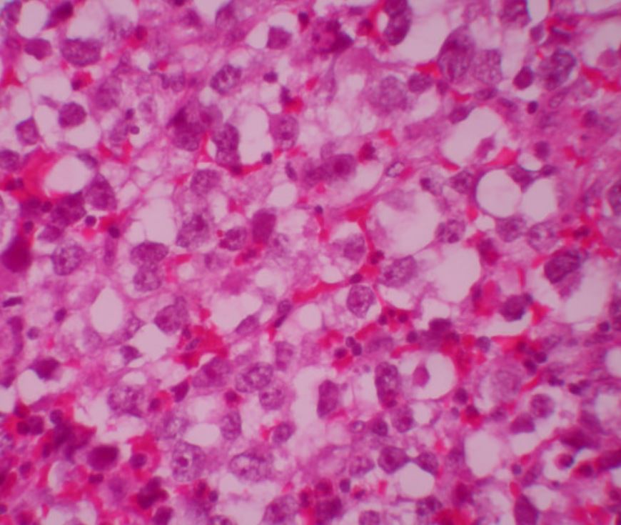
Biopsia de masa retroperitoneal compatible con un seminoma clásico

Tinción H&E de una muestra de biopsia de una masa retroperitoneal consistente con un seminoma clásico

Imagen: “Testicular seminoma presenting with duodenal perforation: a case report” por Miocinovic R, Abaza R. Licencia: CC BY 2.0, recortado por Lecturio.

Diagnóstico

Examen físico

Examen genitourinario específico:

  • Examen testicular con palpación en EN Erythema nodosum is an immune-mediated panniculitis (inflammation of the subcutaneous fat) caused by a type IV (delayed-type) hypersensitivity reaction. It commonly manifests in young women as tender, erythematous nodules on the shins. Erythema Nodosum busca de masas:
    • Palpar suavemente el testículo, el epidídimo y el escroto.
    • Evaluar la presencia de masas, lesiones y nódulos.
  • Examen abdominal general para palpar en EN Erythema nodosum is an immune-mediated panniculitis (inflammation of the subcutaneous fat) caused by a type IV (delayed-type) hypersensitivity reaction. It commonly manifests in young women as tender, erythematous nodules on the shins. Erythema Nodosum busca de masas
  • Examen de tórax para evaluar ginecomastia
  • Examen inguinal para palpar ganglios linfáticos inguinales agrandados
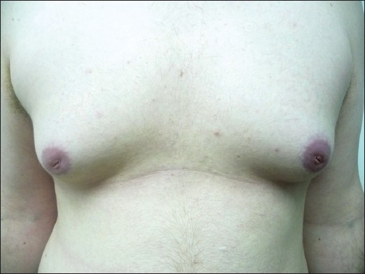
Ginecomastia bilateral

Ginecomastia bilateral en un paciente

Imagen: “Finasteride induced Gynecomastia: Case report and Review of the Literature” por Ramot Y, Czarnowicki T, Zlotogorski A. Licencia: CC BY 2.0

Pruebas de laboratorio

Obtención de marcadores tumorales:

  • Alfa-fetoproteína ( AFP AFP The first alpha-globulins to appear in mammalian sera during fetal development and the dominant serum proteins in early embryonic life. Hepatocellular Carcinoma (HCC) and Liver Metastases):
    • Vida media: 5–7 días
    • Elevado en EN Erythema nodosum is an immune-mediated panniculitis (inflammation of the subcutaneous fat) caused by a type IV (delayed-type) hypersensitivity reaction. It commonly manifests in young women as tender, erythematous nodules on the shins. Erythema Nodosum aproximadamente 50%–80% de los LOS Neisseria tumores de células germinales no seminomatosos
    • La AFP AFP The first alpha-globulins to appear in mammalian sera during fetal development and the dominant serum proteins in early embryonic life. Hepatocellular Carcinoma (HCC) and Liver Metastases no debe estar elevada en EN Erythema nodosum is an immune-mediated panniculitis (inflammation of the subcutaneous fat) caused by a type IV (delayed-type) hypersensitivity reaction. It commonly manifests in young women as tender, erythematous nodules on the shins. Erythema Nodosum el seminoma Seminoma A radiosensitive, malignant neoplasm of the testis, thought to be derived from primordial germ cells of the sexually undifferentiated embryonic gonad. There are three variants: classical (typical), the most common type; anaplastic; and spermatocytic. The classical seminoma is composed of fairly well differentiated sheets or cords of uniform polygonal or round cells (seminoma cells), each cell having abundant clear cytoplasm, distinct cell membranes, a centrally placed round nucleus, and one or more nucleoli. In the female, a grossly and histologically identical neoplasm, known as dysgerminoma, occurs. Testicular Cancer puro; una AFP AFP The first alpha-globulins to appear in mammalian sera during fetal development and the dominant serum proteins in early embryonic life. Hepatocellular Carcinoma (HCC) and Liver Metastases elevada argumenta en EN Erythema nodosum is an immune-mediated panniculitis (inflammation of the subcutaneous fat) caused by a type IV (delayed-type) hypersensitivity reaction. It commonly manifests in young women as tender, erythematous nodules on the shins. Erythema Nodosum contra del diagnóstico de seminoma Seminoma A radiosensitive, malignant neoplasm of the testis, thought to be derived from primordial germ cells of the sexually undifferentiated embryonic gonad. There are three variants: classical (typical), the most common type; anaplastic; and spermatocytic. The classical seminoma is composed of fairly well differentiated sheets or cords of uniform polygonal or round cells (seminoma cells), each cell having abundant clear cytoplasm, distinct cell membranes, a centrally placed round nucleus, and one or more nucleoli. In the female, a grossly and histologically identical neoplasm, known as dysgerminoma, occurs. Testicular Cancer puro
    • La AFP AFP The first alpha-globulins to appear in mammalian sera during fetal development and the dominant serum proteins in early embryonic life. Hepatocellular Carcinoma (HCC) and Liver Metastases también puede estar elevada en EN Erythema nodosum is an immune-mediated panniculitis (inflammation of the subcutaneous fat) caused by a type IV (delayed-type) hypersensitivity reaction. It commonly manifests in young women as tender, erythematous nodules on the shins. Erythema Nodosum las enfermedades hepáticas y en EN Erythema nodosum is an immune-mediated panniculitis (inflammation of the subcutaneous fat) caused by a type IV (delayed-type) hypersensitivity reaction. It commonly manifests in young women as tender, erythematous nodules on the shins. Erythema Nodosum los LOS Neisseria cánceres abdominales, como el carcinoma hepatocelular.
    • Los LOS Neisseria lactantes tendrán una elevación temporal de la AFP AFP The first alpha-globulins to appear in mammalian sera during fetal development and the dominant serum proteins in early embryonic life. Hepatocellular Carcinoma (HCC) and Liver Metastases que se normalizará.
  • β-hCG:
    • Vida media: 24–36 horas
    • Elevado en EN Erythema nodosum is an immune-mediated panniculitis (inflammation of the subcutaneous fat) caused by a type IV (delayed-type) hypersensitivity reaction. It commonly manifests in young women as tender, erythematous nodules on the shins. Erythema Nodosum el 20%–60% de los LOS Neisseria tumores de células germinales no seminomatosos y en EN Erythema nodosum is an immune-mediated panniculitis (inflammation of the subcutaneous fat) caused by a type IV (delayed-type) hypersensitivity reaction. It commonly manifests in young women as tender, erythematous nodules on the shins. Erythema Nodosum el 15% de los LOS Neisseria seminomas
    • Pueden verse falsas elevaciones con el consumo de marihuana
    • El hipogonadismo puede causar niveles bajos de β-hCG.
  • LDH LDH Osteosarcoma:
    • Vida media: 24 horas
    • La isoenzima más comúnmente elevada en EN Erythema nodosum is an immune-mediated panniculitis (inflammation of the subcutaneous fat) caused by a type IV (delayed-type) hypersensitivity reaction. It commonly manifests in young women as tender, erythematous nodules on the shins. Erythema Nodosum los LOS Neisseria tumores de células germinales
    • Se utiliza como indicador de la carga de enfermedad o tumor Tumor Inflammation voluminoso

Imagenología

  • Ultrasonido:
    • Seminomas: lesiones testiculares sólidas hipoecoicas (sin componente quístico)
    • Tumores no seminomatosos: lesiones con calcificaciones y áreas quísticas
  • Radiografía de tórax para descartar metástasis pulmonar
  • TC abdominal y pélvico: útil para la estadificación de la enfermedad
  • Otros estudios de imagen (e.g., TC cerebral): evaluar metástasis

Estadificación

El cáncer de testículo se clasifica según el Sistema de Estadificación TNM del American Joint Committee on Cancer y la Union for International Cancer Control.

  • Componentes de la estadificación:
    • Patología del tumor Tumor Inflammation primario
    • Estudios de imagen para estadificación
    • Marcadores tumorales en EN Erythema nodosum is an immune-mediated panniculitis (inflammation of the subcutaneous fat) caused by a type IV (delayed-type) hypersensitivity reaction. It commonly manifests in young women as tender, erythematous nodules on the shins. Erythema Nodosum suero tras la orquiectomía
  • Tumor Tumor Inflammation (T):
    • pTx: no se puede evaluar
    • pT0: sin evidencia de tumor Tumor Inflammation primario
    • pTis: neoplasia intratubular de células germinales
    • pT1: tumor Tumor Inflammation limitado a los LOS Neisseria testículos y al AL Amyloidosis epidídimo sin invasión linfovascular; puede invadir la túnica albugínea pero no la túnica vaginal
    • pT2: tumor Tumor Inflammation limitado a los LOS Neisseria testículos y al AL Amyloidosis epidídimo con invasión linfovascular o tumor Tumor Inflammation que afecta a la túnica vaginal
    • pT3: invasión tumoral del cordón espermático con o sin invasión linfovascular
    • pT4: invasión tumoral del escroto con o sin invasión linfovascular
  • Nódulo (N):
    • Nx: no se puede evaluar
    • N0: sin metástasis en EN Erythema nodosum is an immune-mediated panniculitis (inflammation of the subcutaneous fat) caused by a type IV (delayed-type) hypersensitivity reaction. It commonly manifests in young women as tender, erythematous nodules on the shins. Erythema Nodosum los LOS Neisseria ganglios linfáticos regionales
    • N1–3: metástasis en EN Erythema nodosum is an immune-mediated panniculitis (inflammation of the subcutaneous fat) caused by a type IV (delayed-type) hypersensitivity reaction. It commonly manifests in young women as tender, erythematous nodules on the shins. Erythema Nodosum los LOS Neisseria ganglios linfáticos (el número y el tamaño de los LOS Neisseria ganglios linfáticos varían)
  • Metástasis (M):
    • Mx: no se puede evaluar
    • M0: sin metástasis a distancia
    • M1: metástasis
  • Marcadores tumorales en EN Erythema nodosum is an immune-mediated panniculitis (inflammation of the subcutaneous fat) caused by a type IV (delayed-type) hypersensitivity reaction. It commonly manifests in young women as tender, erythematous nodules on the shins. Erythema Nodosum suero (S):
Tabla: Estadificación del cáncer de testículo
Estadio Descripción
I Limitado a los LOS Neisseria testículos, sin ganglios linfáticos, sin metástasis
II Testículo + ganglios linfáticos, sin metástasis:
  • IIA: 1–5 ganglios linfáticos, todas las masas ganglionares < 2 cm en EN Erythema nodosum is an immune-mediated panniculitis (inflammation of the subcutaneous fat) caused by a type IV (delayed-type) hypersensitivity reaction. It commonly manifests in young women as tender, erythematous nodules on the shins. Erythema Nodosum tamaño
  • IIB ganglios linfáticos de 2–5 cm o > 5 ganglios linfáticos (todos ≤ 5 cm)
  • IIIC: ganglios linfáticos > 5 cm
III Metástasis a distancia, marcadores tumorales significativamente elevados
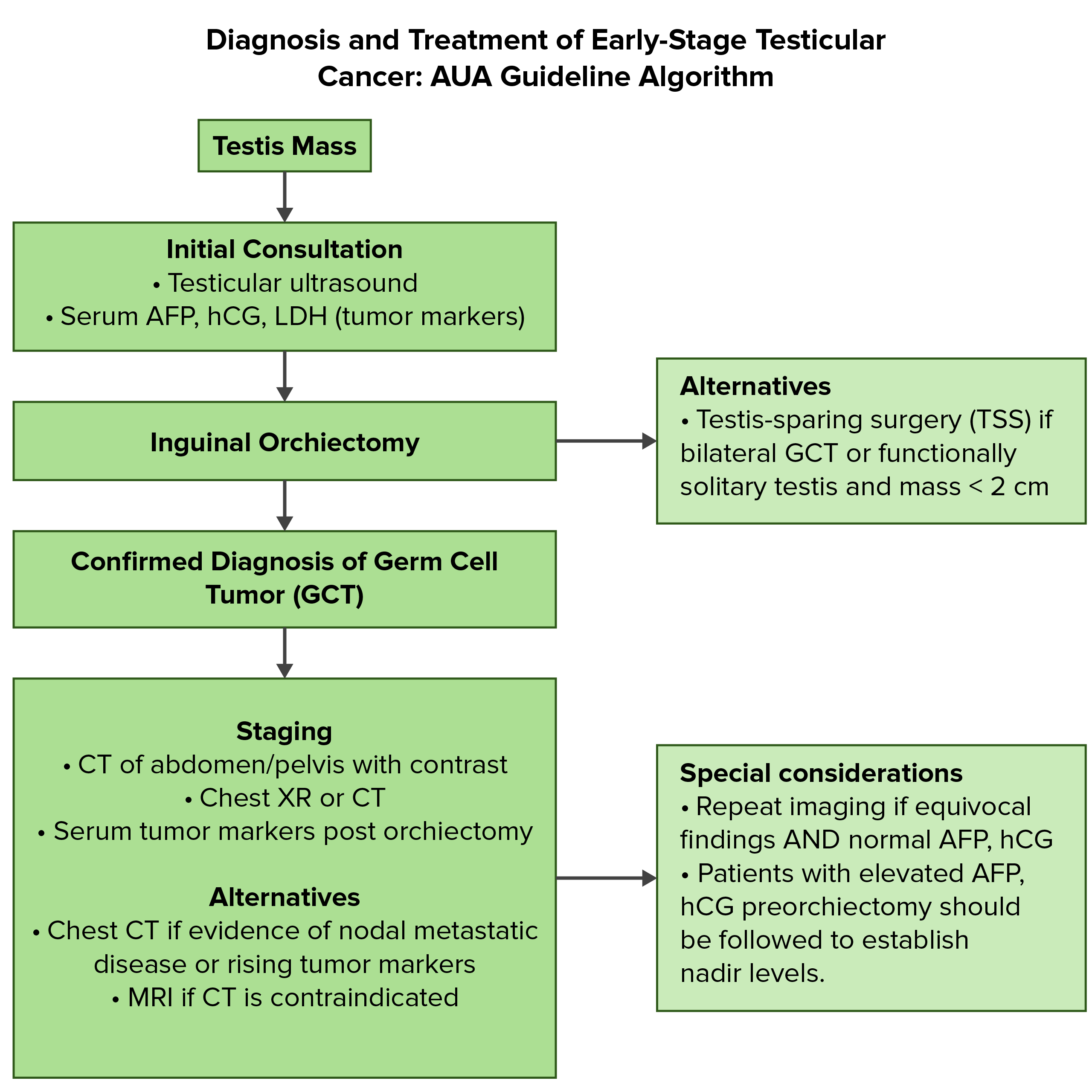
Algoritmo de la guía de la AUA

Resumen del diagnóstico y tratamiento inicial del cáncer de testículo:
AFP: alfa-fetoproteína
AUA: American Urological Association

Imagen por Lecturio. Licencia: CC BY-NC-SA 4.0

Tratamiento

Tratamiento de los LOS Neisseria seminomas

La preservación de la fertilidad del paciente debe discutirse ampliamente con los LOS Neisseria hombres en EN Erythema nodosum is an immune-mediated panniculitis (inflammation of the subcutaneous fat) caused by a type IV (delayed-type) hypersensitivity reaction. It commonly manifests in young women as tender, erythematous nodules on the shins. Erythema Nodosum edad reproductiva antes de cualquier tratamiento. El tratamiento primario es la orquiectomía inguinal radical, seguida de terapia, según el estadio:

  • Seminomas en EN Erythema nodosum is an immune-mediated panniculitis (inflammation of the subcutaneous fat) caused by a type IV (delayed-type) hypersensitivity reaction. It commonly manifests in young women as tender, erythematous nodules on the shins. Erythema Nodosum estadio I (alta tasa de curación): el tratamiento primario es la orquiectomía inguinal, seguida de:
    • Vigilancia postorquiectomía con exámenes físicos seriados, marcadores tumorales en EN Erythema nodosum is an immune-mediated panniculitis (inflammation of the subcutaneous fat) caused by a type IV (delayed-type) hypersensitivity reaction. It commonly manifests in young women as tender, erythematous nodules on the shins. Erythema Nodosum suero, imagenología abdominopélvica según se indique
    • Radioterapia: para los LOS Neisseria pacientes que no pueden adherirse a las medidas de vigilancia; se necesita una dosis terapéutica baja, ya que el seminoma Seminoma A radiosensitive, malignant neoplasm of the testis, thought to be derived from primordial germ cells of the sexually undifferentiated embryonic gonad. There are three variants: classical (typical), the most common type; anaplastic; and spermatocytic. The classical seminoma is composed of fairly well differentiated sheets or cords of uniform polygonal or round cells (seminoma cells), each cell having abundant clear cytoplasm, distinct cell membranes, a centrally placed round nucleus, and one or more nucleoli. In the female, a grossly and histologically identical neoplasm, known as dysgerminoma, occurs. Testicular Cancer es muy radiosensible.
    • Carboplatino en EN Erythema nodosum is an immune-mediated panniculitis (inflammation of the subcutaneous fat) caused by a type IV (delayed-type) hypersensitivity reaction. It commonly manifests in young women as tender, erythematous nodules on the shins. Erythema Nodosum monoterapia: también es una opción para quienes rechazan la vigilancia
  • Seminomas en EN Erythema nodosum is an immune-mediated panniculitis (inflammation of the subcutaneous fat) caused by a type IV (delayed-type) hypersensitivity reaction. It commonly manifests in young women as tender, erythematous nodules on the shins. Erythema Nodosum estadio II:
    • IIA:
      • Radioterapia
      • Quimioterapia para recaídas
    • IIB o IIC:
      • Quimioterapia con bleomicina, etopósido y cisplatino (BEP), o
      • Etopósido y cisplatino solos ( EP EP Ectopic pregnancy refers to the implantation of a fertilized egg (embryo) outside the uterine cavity. The main cause is disruption of the normal anatomy of the fallopian tube. Ectopic Pregnancy)
      • Bleomicina: asociada a toxicidad pulmonar relacionada con la dosis
      • La quimioterapia también se asocia con trombocitopenia a corto plazo y complicaciones gastrointestinales
  • Estadio III (seminomas con metástasis pulmonares o no pulmonares): quimioterapia con BEP o EP EP Ectopic pregnancy refers to the implantation of a fertilized egg (embryo) outside the uterine cavity. The main cause is disruption of the normal anatomy of the fallopian tube. Ectopic Pregnancy

Tratamiento de los LOS Neisseria tumores no seminomatosos

La preservación de la fertilidad del paciente debe discutirse ampliamente con los LOS Neisseria hombres en EN Erythema nodosum is an immune-mediated panniculitis (inflammation of the subcutaneous fat) caused by a type IV (delayed-type) hypersensitivity reaction. It commonly manifests in young women as tender, erythematous nodules on the shins. Erythema Nodosum edad reproductiva antes de cualquier tratamiento. El tratamiento primario es la orquiectomía inguinal, seguida de terapia, según el estadio:

  • Estadio I de los LOS Neisseria tumores no seminomatosos:
    • Vigilancia postorquiectomía con exámenes físicos seriados, marcadores tumorales en EN Erythema nodosum is an immune-mediated panniculitis (inflammation of the subcutaneous fat) caused by a type IV (delayed-type) hypersensitivity reaction. It commonly manifests in young women as tender, erythematous nodules on the shins. Erythema Nodosum suero, imagenología abdominopélvica según se indique
    • Disección de ganglios linfáticos retroperitoneales: para hombres con características patológicas de alto riesgo y alto riesgo de recurrencia de la enfermedad
    • Quimioterapia adyuvante: BEP
  • Estadio IIA y IIB de los LOS Neisseria tumores no seminomatosos:
    • Quimioterapia con BEP o EP EP Ectopic pregnancy refers to the implantation of a fertilized egg (embryo) outside the uterine cavity. The main cause is disruption of the normal anatomy of the fallopian tube. Ectopic Pregnancy o
    • Disección primaria de ganglios linfáticos retroperitoneales
  • Estadio IIC y III de los LOS Neisseria tumores no seminomatosos: regímenes estándar para tumores avanzados (incluye BEP o EP EP Ectopic pregnancy refers to the implantation of a fertilized egg (embryo) outside the uterine cavity. The main cause is disruption of the normal anatomy of the fallopian tube. Ectopic Pregnancy)

Pronóstico

  • Tumores de células germinales testiculares: tasas de supervivencia a los LOS Neisseria 5 años de aproximadamente el 95%.
  • Alrededor del 1%–2% presentan metástasis cerebrales.
  • La recaída después de 2 años es rara, aunque puede ocurrir recaída tardía, principalmente en EN Erythema nodosum is an immune-mediated panniculitis (inflammation of the subcutaneous fat) caused by a type IV (delayed-type) hypersensitivity reaction. It commonly manifests in young women as tender, erythematous nodules on the shins. Erythema Nodosum los LOS Neisseria no seminomas.
  • El hipogonadismo es una complicación potencial (puede asociarse a disfunción sexual e infertilidad).
  • Existe el riesgo de una 2da neoplasia en EN Erythema nodosum is an immune-mediated panniculitis (inflammation of the subcutaneous fat) caused by a type IV (delayed-type) hypersensitivity reaction. It commonly manifests in young women as tender, erythematous nodules on the shins. Erythema Nodosum aquellos tratados con radioterapia y quimioterapia.
  • El monitoreo a largo plazo también incluye disfunción sexual, infertilidad, enfermedad cardiovascular y desafíos psicosociales.

Diagnóstico Diferencial

  • Varicocele: dilatación del plexo venoso pampiniforme. El varicocele se presenta sin dolor, como el cáncer de testículo. Los varicoceles tienen el aspecto característico de “bolsa de gusanos” en EN Erythema nodosum is an immune-mediated panniculitis (inflammation of the subcutaneous fat) caused by a type IV (delayed-type) hypersensitivity reaction. It commonly manifests in young women as tender, erythematous nodules on the shins. Erythema Nodosum el examen físico. El ultrasonido muestra una transiluminación negativa y un complejo venoso dilatado con flujo sanguíneo retrógrado.
  • Espermatocele: se presenta como un quiste epididimario, que suele surgir de la cabeza del epidídimo. Al igual que el cáncer de testículo, el espermatocele también es una masa indolora que suele detectarse de forma incidental. Además, en la imagenología se observan lesiones quísticas; en el Doppler color se observa un aspecto/signo característico de “tormenta de nieve” (movimiento de los LOS Neisseria ecos internos lejos del transductor, lo que indica partículas sólidas). El espermatocele requiere un tratamiento conservador.
  • Hidrocele: representa un exceso de líquido en EN Erythema nodosum is an immune-mediated panniculitis (inflammation of the subcutaneous fat) caused by a type IV (delayed-type) hypersensitivity reaction. It commonly manifests in young women as tender, erythematous nodules on the shins. Erythema Nodosum la túnica vaginal que da lugar a un escroto hinchado. La afección se debe a diferentes factores anatómicos y relacionados con los LOS Neisseria fluidos. Los LOS Neisseria hallazgos incluyen una masa escrotal indolora que puede aumentar de tamaño, o pesadez o dolor Dolor Inflammation progresivo en EN Erythema nodosum is an immune-mediated panniculitis (inflammation of the subcutaneous fat) caused by a type IV (delayed-type) hypersensitivity reaction. It commonly manifests in young women as tender, erythematous nodules on the shins. Erythema Nodosum el escroto. En EN Erythema nodosum is an immune-mediated panniculitis (inflammation of the subcutaneous fat) caused by a type IV (delayed-type) hypersensitivity reaction. It commonly manifests in young women as tender, erythematous nodules on the shins. Erythema Nodosum comparación con el cáncer de testículo, un hidrocele tendrá una prueba de transiluminación positiva en EN Erythema nodosum is an immune-mediated panniculitis (inflammation of the subcutaneous fat) caused by a type IV (delayed-type) hypersensitivity reaction. It commonly manifests in young women as tender, erythematous nodules on the shins. Erythema Nodosum el ultrasonido.
  • Hernia Hernia Protrusion of tissue, structure, or part of an organ through the bone, muscular tissue, or the membrane by which it is normally contained. Hernia may involve tissues such as the abdominal wall or the respiratory diaphragm. Hernias may be internal, external, congenital, or acquired. Abdominal Hernias inguinal indirecta: la hernia Hernia Protrusion of tissue, structure, or part of an organ through the bone, muscular tissue, or the membrane by which it is normally contained. Hernia may involve tissues such as the abdominal wall or the respiratory diaphragm. Hernias may be internal, external, congenital, or acquired. Abdominal Hernias inguinal congénita se produce cuando el proceso vaginal no se cierra tras el descenso de los LOS Neisseria testículos. La hernia Hernia Protrusion of tissue, structure, or part of an organ through the bone, muscular tissue, or the membrane by which it is normally contained. Hernia may involve tissues such as the abdominal wall or the respiratory diaphragm. Hernias may be internal, external, congenital, or acquired. Abdominal Hernias inguinal indirecta permite el paso del contenido abdominal. A diferencia del cáncer testicular, la hernia Hernia Protrusion of tissue, structure, or part of an organ through the bone, muscular tissue, or the membrane by which it is normally contained. Hernia may involve tissues such as the abdominal wall or the respiratory diaphragm. Hernias may be internal, external, congenital, or acquired. Abdominal Hernias se presenta como un abultamiento en EN Erythema nodosum is an immune-mediated panniculitis (inflammation of the subcutaneous fat) caused by a type IV (delayed-type) hypersensitivity reaction. It commonly manifests in young women as tender, erythematous nodules on the shins. Erythema Nodosum la ingle que puede llegar al AL Amyloidosis escroto, que se hace HACE Altitude Sickness más prominente con el aumento de la presión intraabdominal (esfuerzos, tos TOS Thoracic outlet syndrome (TOS) is a broad term used for a spectrum of syndromes related to the general region of the thoracic outlet, which involves the compression or irritation of elements of the brachial plexus, subclavian artery, or subclavian vein. Thoracic Outlet Syndrome). El tratamiento es la reparación quirúrgica.

Referencias

  1. Gaddam, S., Chesnut, G. (2021). Testicle cancer. StatPearls. Retrieved May 21, 2021, from https://www.ncbi.nlm.nih.gov/books/NBK563159/
  2. Oh, W. (2021). Overview of the treatment of testicular germ cell tumors. UpToDate. Retrieved May 21, 2021, from https://www.uptodate.com/contents/overview-of-the-treatment-of-testicular-germ-cell-tumors
  3. Rose, T. (2021). Testis neoplasms. AUA Core Curriculum. Retrieved May 22, 2021, from https://auau.auanet.org/core
  4. Steele, G. (2021). Clinical manifestations, diagnosis, and staging of testicular germ cell tumors. UpToDate. Retrieved May 21, 2021, from https://www.uptodate.com/contents/clinical-manifestations-diagnosis-and-staging-of-testicular-germ-cell-tumors
  5. Rajpert-De Meyts, E., Skakkebaek, N.E., Toppari, J. (2018). Testicular Cancer Pathogenesis, Diagnosis and Endocrine Aspects. Feingold, K.R., et al. (Ed.). Endotext. South Dartmouth (MA): MDText.com, Inc. https://www.ncbi.nlm.nih.gov/books/NBK278992/

¡Crea tu cuenta gratis o inicia una sesión para seguir leyendo!

Regístrate ahora y obtén acceso gratuito a Lecturio con páginas de concepto, videos médicos y cuestionarios para tu educación médica.

User Reviews

Details