


Uma vez que as células humanas dependem principalmente do metabolismo aeróbico, é de vital importância obter eficientemente oxigénio do ambiente e trazê-lo para os tecidos enquanto se excreta o subproduto da respiração celular (CO2). A respiração envolve os sistemas respiratório e circulatório. Existem 4 processos que fornecem O2 ao corpo e eliminam o CO2. O sistema respiratório está envolvido na ventilação pulmonar e na respiração externa, enquanto o sistema circulatório é responsável pelo transporte e respiração interna. A ventilação pulmonar (respiração) representa o movimento do ar AR Aortic regurgitation (AR) is a cardiac condition characterized by the backflow of blood from the aorta to the left ventricle during diastole. Aortic regurgitation is associated with an abnormal aortic valve and/or aortic root stemming from multiple causes, commonly rheumatic heart disease as well as congenital and degenerative valvular disorders. Aortic Regurgitation para dentro e para fora dos pulmões. A respiração externa, ou trocas gasosas, é representada pela troca de O2 e CO2 entre os pulmões e o sangue.
Last updated: Dec 15, 2025
As trocas gasosas ocorrem ao nível dos alvéolos nos pulmões e nos capilares da circulação pulmonar.
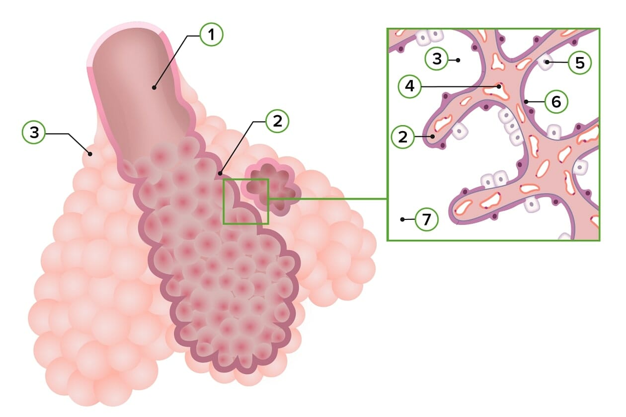
Diagrama esquemático da estrutura da zona respiratória do trato respiratório inferior:
Observar a composição da parede alveolar:
1) Bronquíolo respiratório
2) Septo interalveolar primário
3) Saco alveolar
4) Capilares
5) Pneumócito tipo II
6) Pneumócito tipo I
7) Ducto alveolar
Durante as trocas gasosas, O₂ e CO₂ devem atravessar a membrana pulmonar. Este processo é impulsionado por múltiplas forças complexas determinadas pelas propriedades físicas desses gases.
A taxa de troca gasosa é determinada pela eficiência da troca através da membrana pulmonar e pela velocidade com que ela pode ser trazida do ar AR Aortic regurgitation (AR) is a cardiac condition characterized by the backflow of blood from the aorta to the left ventricle during diastole. Aortic regurgitation is associated with an abnormal aortic valve and/or aortic root stemming from multiple causes, commonly rheumatic heart disease as well as congenital and degenerative valvular disorders. Aortic Regurgitation (para O₂) ou do corpo (para CO₂).
O₂ e CO₂ devem ser transportados pela corrente sanguínea para alcançar os locais de troca gasosa.
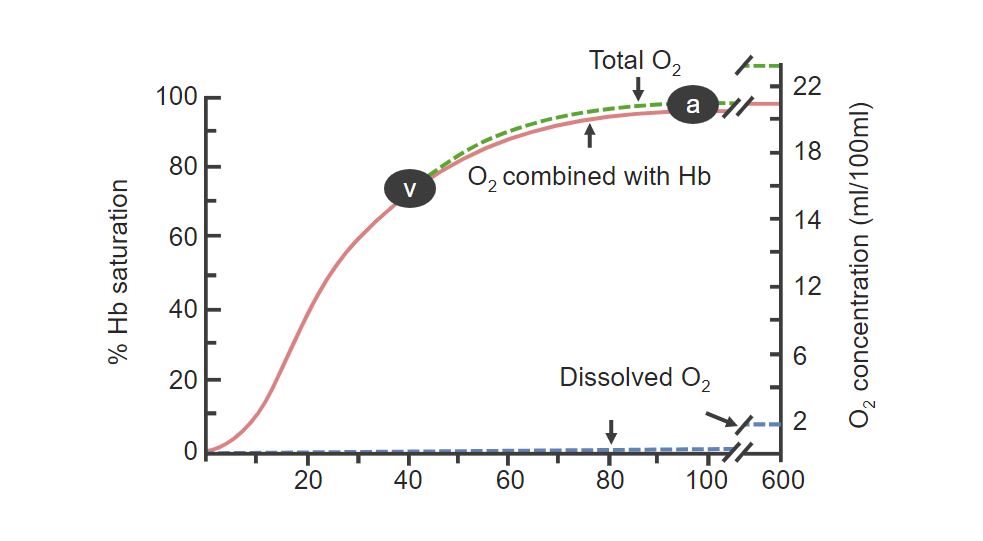
Saturação da hemoglobina:
A percentagem de hemoglobina ligada aumenta com a concentração de O2 . Observe a letra A, que significa a saturação da hemoglobina no sangue arterial (próximo de 99%). A letra V significa a saturação de hemoglobina do sangue venoso.
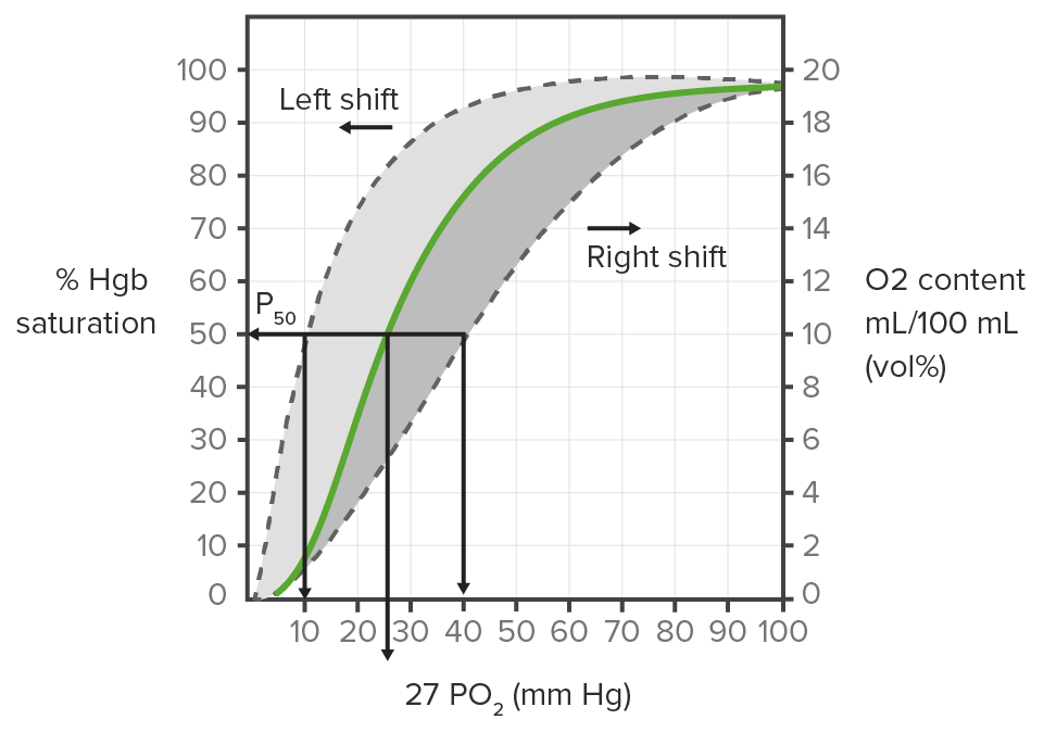
Curva de dissociação da hemoglobina:
Mostra os deslocamentos para a direita e para a esquerda que podem ocorrer quando o fornecimento de oxigénio aos tecidos aumenta ou diminui, respetivamente.
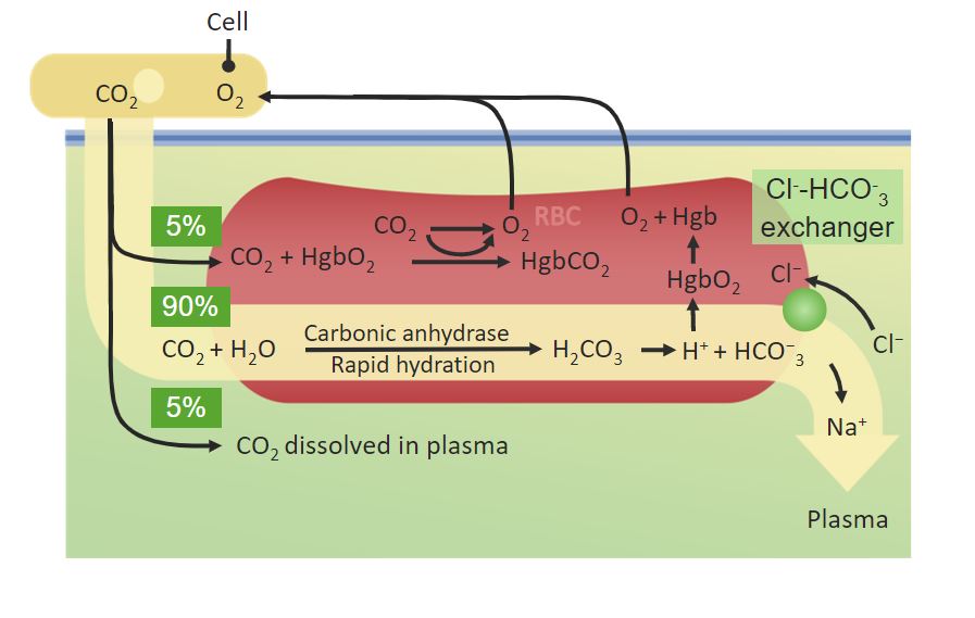
Percentagens de CO2 transportado no plasma:
Observe como 90% é convertido pela anidrase carbónica em HCO 3 – dentro dos glóbulos vermelhos.
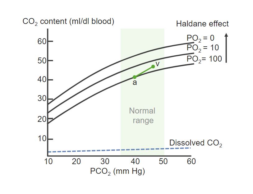
Curva de dissociação de Hb e CO2 :
Observe como a PCO 2 aumenta do lado arterial (a) para o lado venoso (v). A curva de dissociação é elevada com a diminuição da PO2 , referindo-se ao facto de que uma baixa concentração de O2 permite que mais CO 2 se ligue à Hb.
Ventilação e perfusão são os mecanismos que transportam O₂ e CO₂ entre a membrana pulmonar e os tecidos do corpo.
Perfusão é o fluxo de sangue para a vasculatura pulmonar.
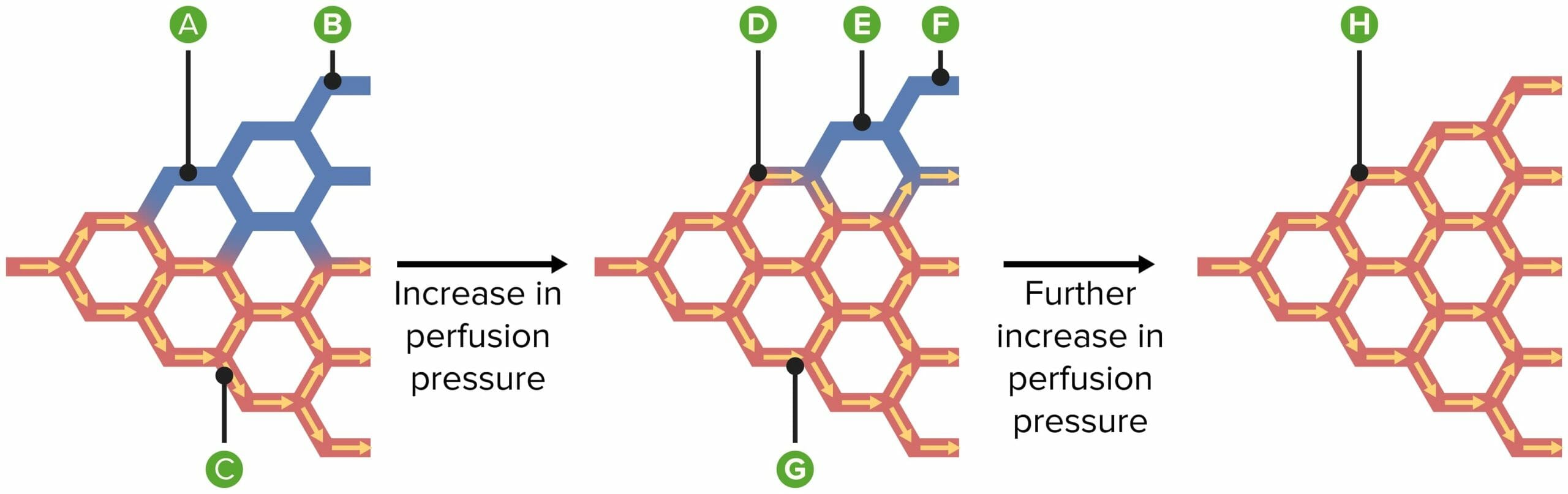
O diagrama explica os mecanismos de recrutamento e distensão dos vasos sanguíneos, quando a pressão arterial pulmonar média é aumentada:
A) Alguns vasos estão abertos, mas não conduzem sangue.
B) Alguns vasos estão colapsados.
C) Outros vasos estão abertos e conduzem sangue.
D) Os vasos anteriormente abertos, não condutores, agora conduzem o sangue.
E) No 1º fase de recrutamento, vasos previamente colapsados tornam-se patentes, mas não conduzem sangue.
F) Mais tarde, durante o recrutamento, os vasos previamente colapsados agora conduzem sangue.
G) Enquanto isso, a distensão alarga vasos, condutores de sangue, previamente abertos.
H) Agora, todos os vasos dilatam-se, levando a uma redução da resistência.
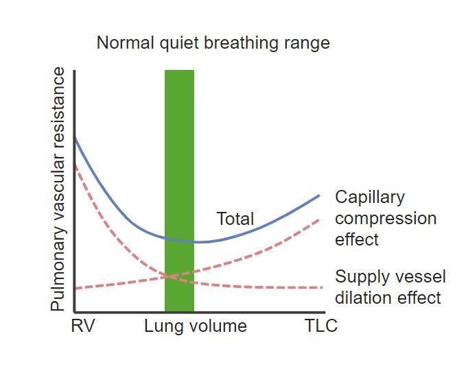
Gráfico que mostra a relação entre o volume pulmonar (eixo dos x) e a resistência vascular pulmonar (eixo dos y):
Num volume pulmonar baixo (VR: volume residual) e volume pulmonar muito alto (Capacidade Pulmonar Total: CPT) a resistência vascular aumenta.
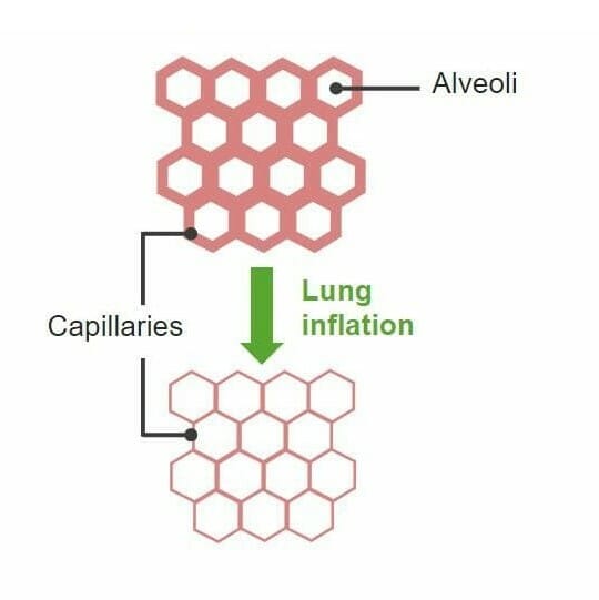
Diagrama eque explica os mecanismos de recrutamento e distensão dos vasos sanguíneos quando a pressão arterial pulmonar média é aumentada
Imagem por Lecturio. Licença: CC BY-NC-SA 4.0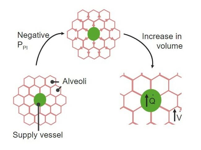
Efeito de dilatação do vaso:
A pressão pleural negativa exercida sobre os alvéolos distende as paredes dos vasos sanguíneos e aumenta o seu diâmetro.
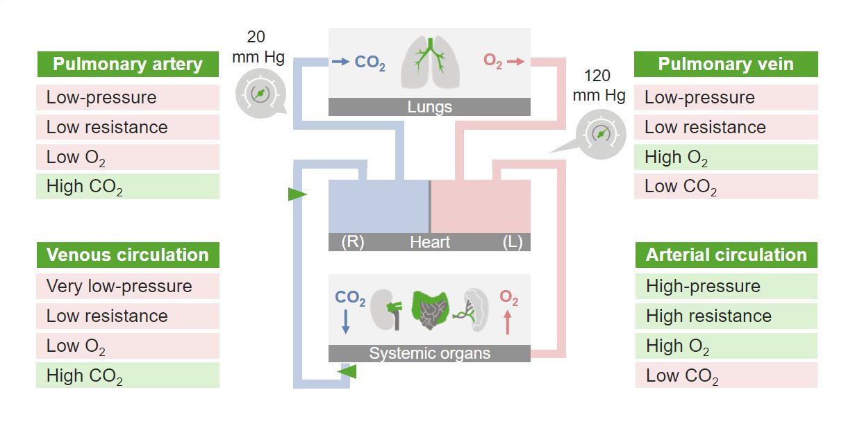
Visão geral do fluxo sanguíneo pulmonar
Imagem por Lecturio. Licença: CC BY-NC-SA 4.0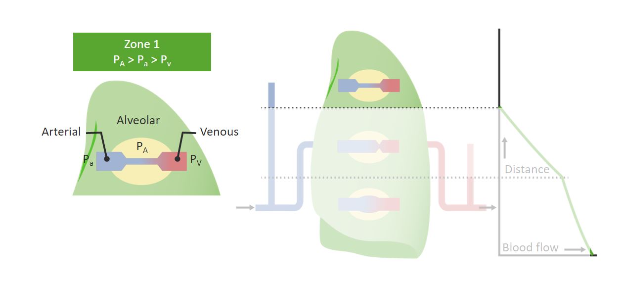
Zona 1 (Apex) do pulmão e o efeito da gravidade sobre ele:
A pressão arterial (Pa) é menor que a pressão alveolar (PA), tornando o fluxo de sangue difícil, se não impossível. A relação entre as pressões pode ser vista na caixa verde.
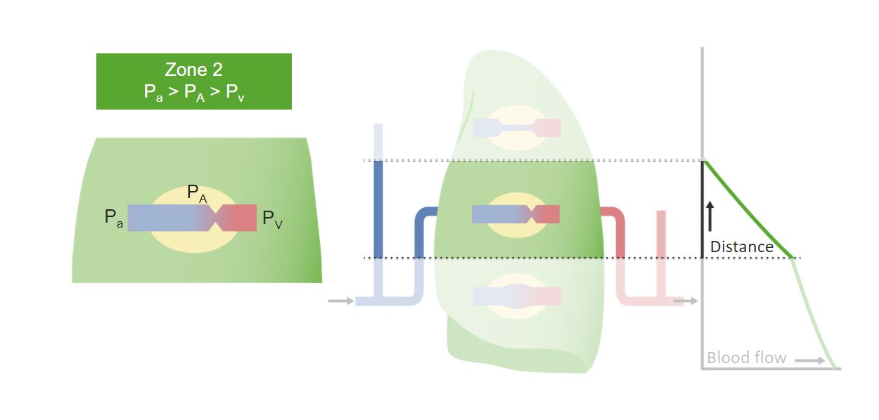
Zona 2 do pulmão e o efeito da gravidade sobre ele:
Há mais fluxo sanguíneo permitido pelo aumento da pressão arterial. A relação entre as pressões pode ser vista na caixa verde.
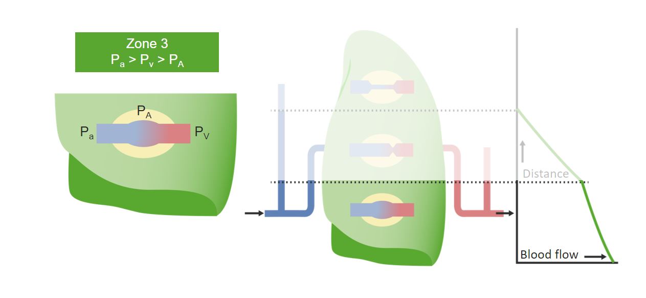
Zona 3 (Base) do pulmão e o efeito da gravidade sobre ele:
É a zona com maior fluxo sanguíneo no pulmão. A força da gravidade torna mais sangue disponível e a pressão arterial maior do que a pressão alveolar. A relação entre as pressões pode ser vista na caixa verde.
Existem 2 diferenças importantes de PO₂ (gradientes):

Diferença arteriovenosa (av) na PO 2 entre sangue venoso e arterial:
Pressões de O2 e CO2 nos alvéolos e na circulação sistémica antes e após as trocas gasosas.
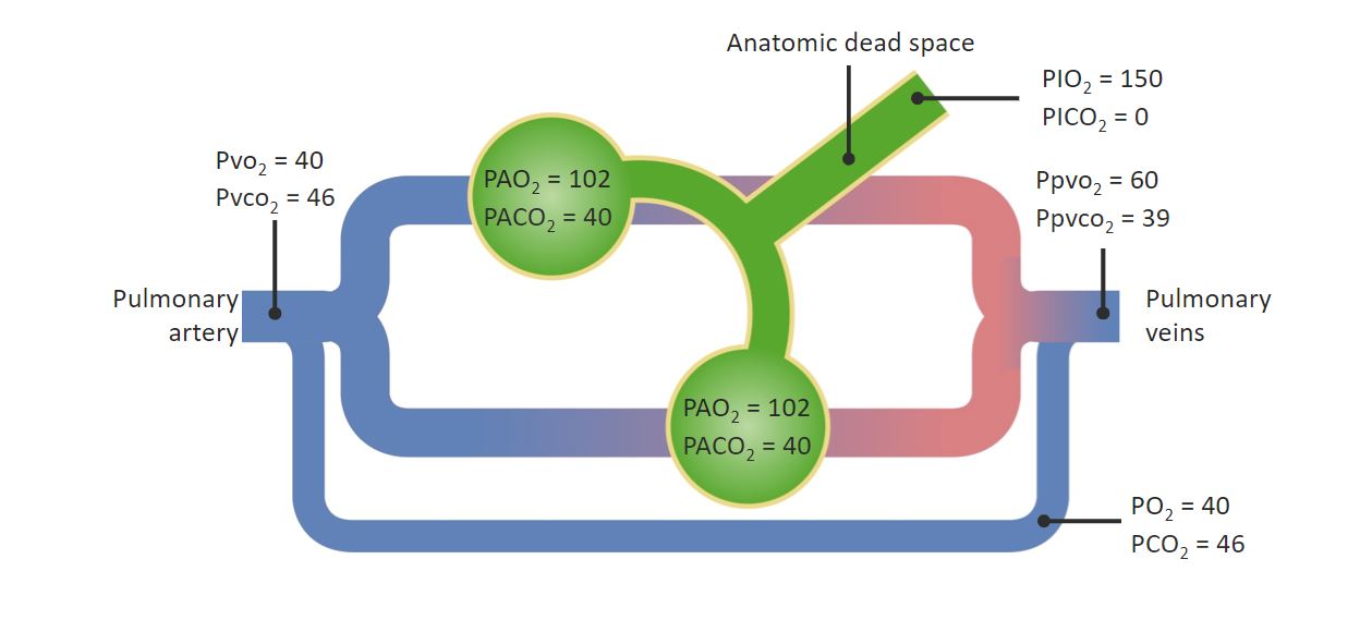
Diagrama de uma derivação DIREITA PARA ESQUERDA:
Veja a comunicação que permite ao sangue não fazer as trocas gasosas e diminuir a pressão arterial de O2.
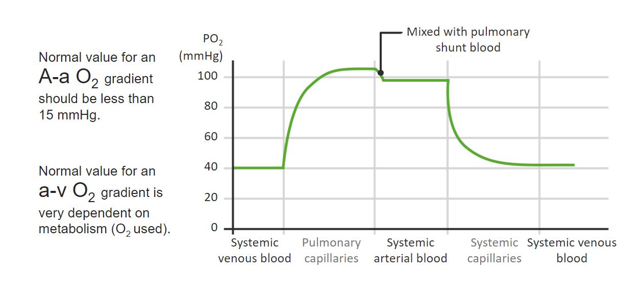
Gradientes de PO 2 nas circulações pulmonar e sistémica:
Veja o gradiente de Aa no ponto de comunicação entre os capilares pulmonares e a circulação arterial sistémica. O gradiente av seria a diferença entre a PO2 da circulação arterial sistémica antes dos capilares e a PO2 da circulação venosa sistémica após os capilares.
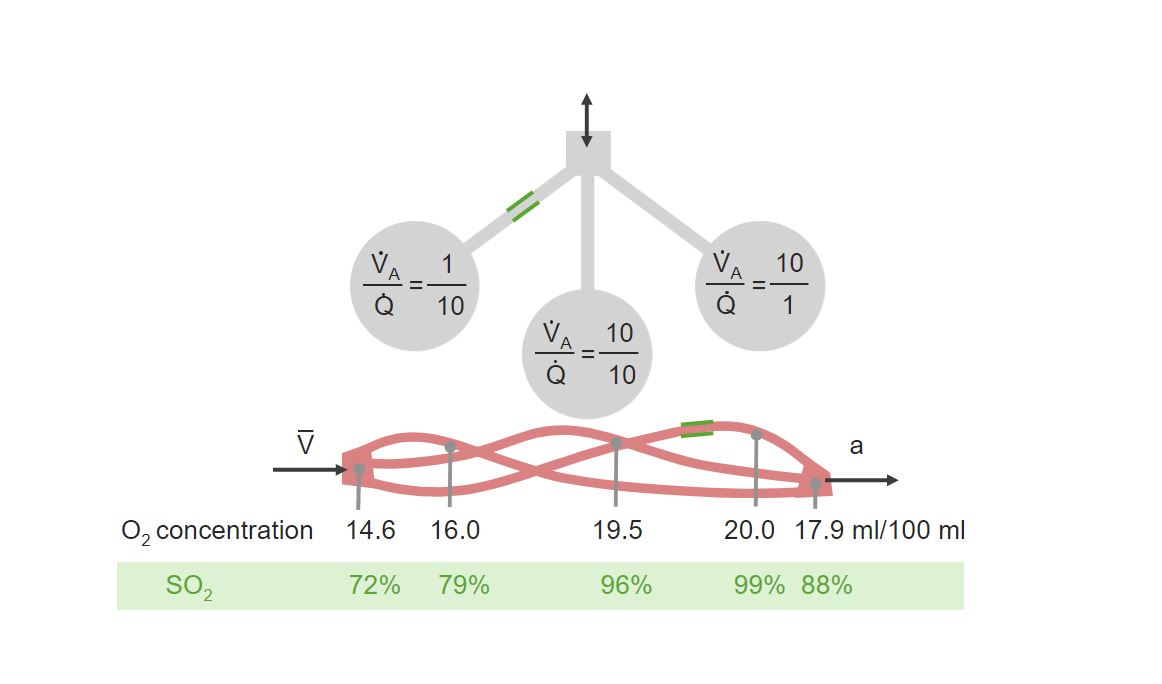
Exemplos esquemáticos de incompatibilidade de ventilação (V) para perfusão (Q):
À direita, pode ser visto um exemplo de baixa ventilação para alta perfusão. À esquerda, pode ser visto um exemplo de alta ventilação para baixa perfusão. No centro encontra-se uma situação normal de ventilação a perfusão com uma relação de 1 para 1.
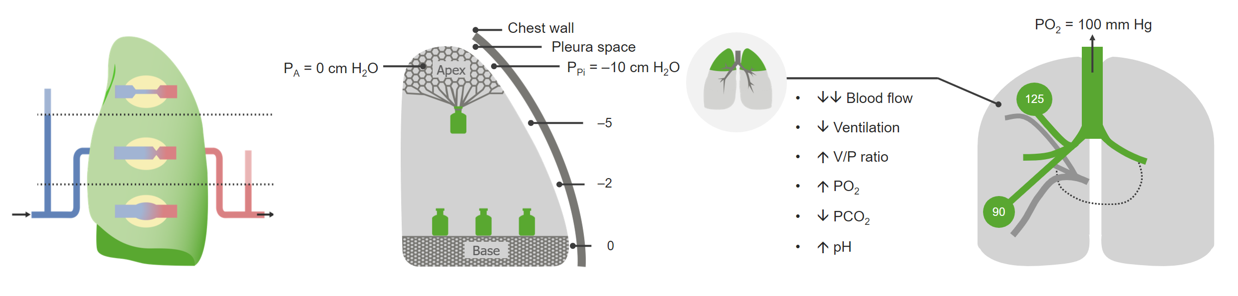
Relação ventilação/perfusão aplicada ao ápice do pulmão ereto de um indivíduo saudável:
Perfusão reduzida do ápice (direita), o efeito inflador da gravidade devido ao peso do pulmão (centro) e os parâmetros resultantes durante a inspiração (direita). Observar o aumento da relação V/Q (V/P) devido ao aumento da ventilação e à redução da perfusão.
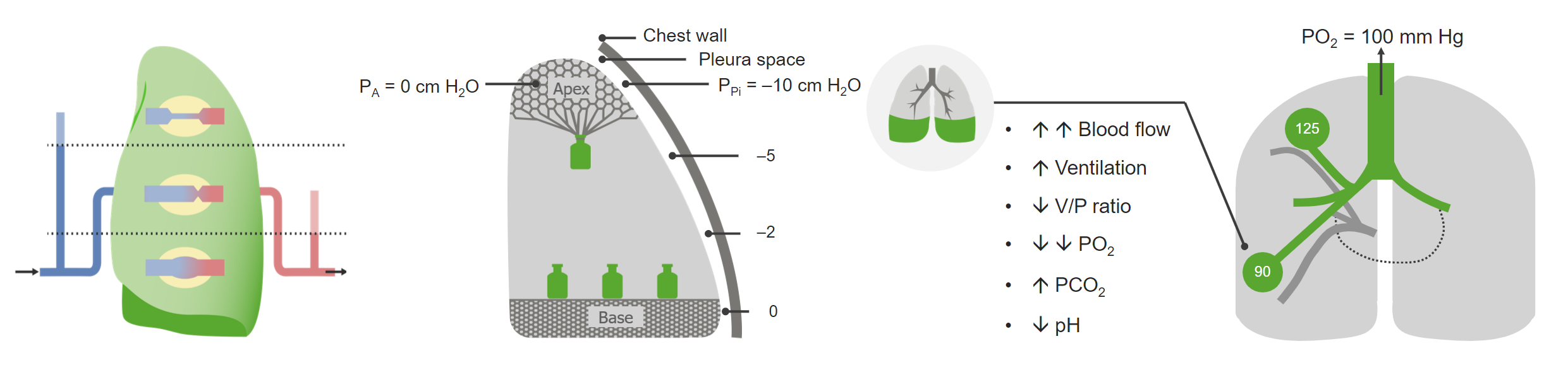
Relação ventilação/perfusão aplicada à base do pulmão ereto de um indivíduo saudável:
Veja o aumento da perfusão da base (direita), o efeito compressivo da gravidade devido ao peso do pulmão (centro) e os parâmetros resultantes durante a inspiração (direita). A relação V/Q (V/P) está diminuída devido ao aumento da perfusão.