


A uveíte é uma inflamação da úvea, a camada média pigmentada do olho, que consiste na íris, corpo ciliar e coróide. Esta patologia é caracterizada com base no local afetado; a uveíte anterior é a mais MAIS Androgen Insensitivity Syndrome comum. A uveíte pode ser causada por uma infeção ou doença sistémica, no entanto, em alguns casos, a causa é idiopática. Os pacientes apresentam visão turva, hiperemia ocular e dor (frequentemente na uveíte anterior) ou diminuição da visão e moscas volantes (na uveíte intermédia e posterior). O diagnóstico é realizado através de fundoscopia e exame com lâmpada de fenda. O tratamento para a uveíte anterior é realizado com corticóides tópicos, enquanto a uveíte em locais mais MAIS Androgen Insensitivity Syndrome profundos necessita de injeção. A uveíte por infeções ou doenças sistémicas requer tratamento dirigido à causa.
Last updated: Dec 15, 2025
A uveíte é uma inflamação da úvea, a camada média do olho, constituída pela íris, corpo ciliar e coróide.

Esta imagem representa a anatomia do olho. Na catarata, existe turvação do cristalino, que opacifica a luz quando esta se projeta na retina; assim, quando os níveis de luz se encontram reduzidos, essencialmente à noite, existe diminuição da visão.
Imagem por Lecturio.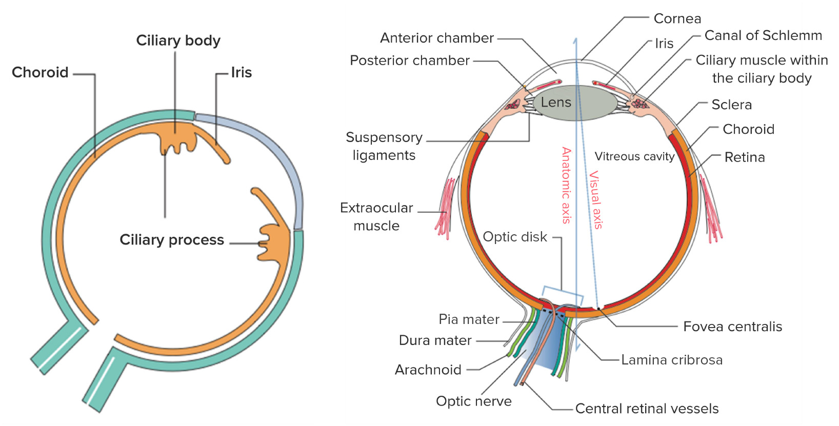
Esquerda: os componentes da úvea, a camada média vascular do olho
Direita: estrutura geral do olho
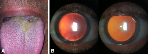
Uveíte anterior bilateral não granulomatosa associada à sífilis. (A): Lesão compatível com cancro sifilítico na base da língua; (B): Exame com lâmpada de fenda: pequenos precipitados queráticos (direita) e depósitos de fibrina a cobrir o endotélio corneano (esquerda) na retroiluminação
Imagem: “initial presentation of syphilis” por Hospital de Olhos Santa Luzia, Estrada do Encanamento 909, CEP 52070-000, Recife-Pernambuco, Brazil. Licença: CC BY 2.0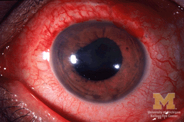
Imagem representativa de uveíte anterior, com hiperemia conjuntival e pupila irregular
Imagem: “Anterior-uveitis” por Jonathan Trobe, M.D. Licença: CC BY 3.0O diagnóstico é realizado através de fundoscopia e exame com lâmpada de fenda. A depressão escleral é uma manobra adicional utilizada para visualizar estruturas posteriores ao cristalino.
Achados
Exames laboratoriais e exames de imagem adicionais estão indicados quando existem sinais e sintomas extraoculares que podem ser atribuídos a uma doença específica (e.g., sarcoidose, sífilis).
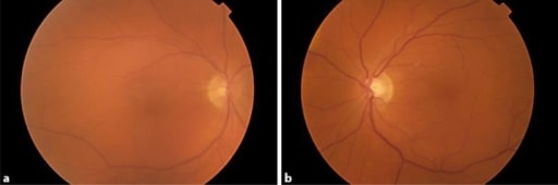
Imagens do fundo ocular com vitrite em ambos os olhos. O paciente desenvolveu uveíte bilateral após tratamento com dabrafenibe e trametinibe.
Imagem: “Uveitis as a Result of MAP Kinase Pathway Inhibition” por Royal Surrey County Hospital NHS Foundation Trust, Guildford, Surrey, London, UK. Licença: CC BY 3.0
Grávida de 27 anos com panuveíte por doença de Behçet, com rubor ciliar e hipópion (camadas de leucócitos na parte inferior da câmara anterior)
Imagem: “Behçet’s Uveitis” por Department of Ophthalmology, Istanbul University, Istanbul Faculty of Medicine, Istanbul, Turkey. Licença: CC BY 2.0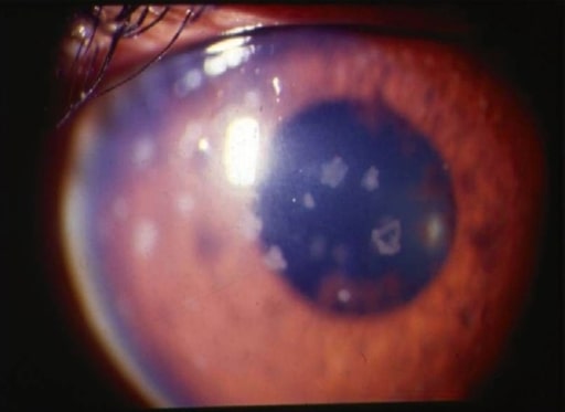
Uveíte anterior: imagem com lâmpada de fenda a demonstrar precipitados queráticos grandes e antigos
Imagem: “anterior uveitis” por L. V. Prasad Eye Institute, Kallam Anji Reddy Campus, Hyderabad, India. Licença: CC BY 2.0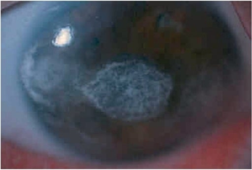
A iluminação difusa na lâmpada de fenda mostra queratopatia em banda grave no olho esquerdo em rapaz com uveíte anterior (associada a artrite idiopática juvenil).
Imagem: “keratopathy” por Department of Ophthalmology, Faculty of Medicine, Istanbul University, Istanbul, Turkey. Licença: CC BY 2.5