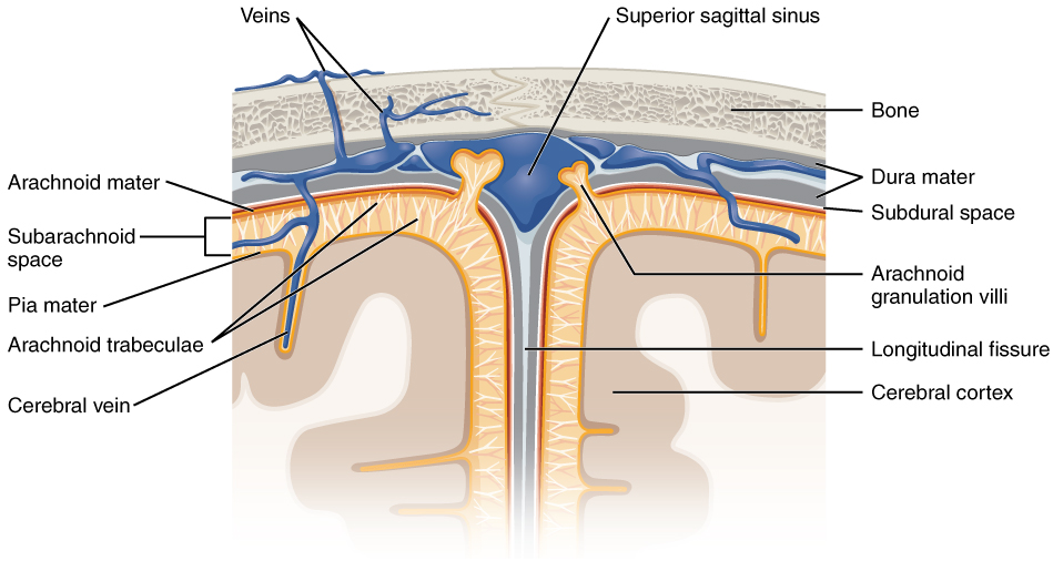
El cerebro y la médula espinal están envueltos por 3 capas superpuestas de tejido conectivo llamadas meninges Meninges The brain and the spinal cord are enveloped by 3 overlapping layers of connective tissue called the meninges. The layers are, from the most external layer to the most internal layer, the dura mater, arachnoid mater, and pia mater. Between these layers are 3 potential spaces called the epidural, subdural, and subarachnoid spaces. Meninges: Anatomy. Las capas son, desde la más externa a la más interna; la duramadre, la aracnoides y la piamadre. Entre estas capas hay 3 espacios potenciales llamados; espacio epidural, subdural y subaracnoideo. Las meninges Meninges The brain and the spinal cord are enveloped by 3 overlapping layers of connective tissue called the meninges. The layers are, from the most external layer to the most internal layer, the dura mater, arachnoid mater, and pia mater. Between these layers are 3 potential spaces called the epidural, subdural, and subarachnoid spaces. Meninges: Anatomy tienen la función de proteger el contenido del cerebro y la médula espinal. La infección del SNC se presenta con una inflamación de las meninges Meninges The brain and the spinal cord are enveloped by 3 overlapping layers of connective tissue called the meninges. The layers are, from the most external layer to the most internal layer, the dura mater, arachnoid mater, and pia mater. Between these layers are 3 potential spaces called the epidural, subdural, and subarachnoid spaces. Meninges: Anatomy, y la etiología se puede investigar examinando el LCR, que está contenido en EN Erythema nodosum is an immune-mediated panniculitis (inflammation of the subcutaneous fat) caused by a type IV (delayed-type) hypersensitivity reaction. It commonly manifests in young women as tender, erythematous nodules on the shins. Erythema Nodosum el espacio subaracnoideo.
Last updated: Dec 15, 2025
Las meninges Meninges The brain and the spinal cord are enveloped by 3 overlapping layers of connective tissue called the meninges. The layers are, from the most external layer to the most internal layer, the dura mater, arachnoid mater, and pia mater. Between these layers are 3 potential spaces called the epidural, subdural, and subarachnoid spaces. Meninges: Anatomy son capas de tejido conectivo que protegen el cerebro y la médula espinal.
Las meninges Meninges The brain and the spinal cord are enveloped by 3 overlapping layers of connective tissue called the meninges. The layers are, from the most external layer to the most internal layer, the dura mater, arachnoid mater, and pia mater. Between these layers are 3 potential spaces called the epidural, subdural, and subarachnoid spaces. Meninges: Anatomy están formadas por 3 capas de tejido conectivo, con espacios potenciales entre ellas:
| Capa | Origen | Características |
|---|---|---|
| Espacio epidural | NA |
|
| Duramadre | Mesodermo |
|
| Espacio subdural | NA |
|
| Aracnoides ( leptomeninges Leptomeninges Meninges: Anatomy) | Cresta neural |
|
| Espacio subaracnoideo | Plexo coroideo |
|
| Piamadre ( leptomeninges Leptomeninges Meninges: Anatomy) | Cresta neural |
|
Capas de las meninges y sus relaciones bajo el cráneo
Imagen: “Diagram of section of top of brain showing the meninges and subarachnoid space” por OpenStax. Licencia: CC BY 4.0La duramadre es la capa más gruesa de las meninges Meninges The brain and the spinal cord are enveloped by 3 overlapping layers of connective tissue called the meninges. The layers are, from the most external layer to the most internal layer, the dura mater, arachnoid mater, and pia mater. Between these layers are 3 potential spaces called the epidural, subdural, and subarachnoid spaces. Meninges: Anatomy y provee estructura al AL Amyloidosis cerebro.
Los LOS Neisseria pliegues durales y los LOS Neisseria senos durales proceden de la duramadre.
| Estructura | Características |
|---|---|
| Hoz del cerebro | Separación de los LOS Neisseria hemisferios cerebrales derecho e izquierdo |
| Hoz del cerebelo | Separa los LOS Neisseria hemisferios cerebelosos derecho e izquierdo |
| Tienda del cerebelo | Tienda, o techo, sobre el cerebelo |
| Diafragma estriado | El techo de la glándula hipófisis |
| Senos durales | Las dos capas de la duramadre se extienden juntas por la mayor parte del cráneo. Cuando se separan, el hueco entre ellos se llama seno venoso dural. Estos senos drenan la sangre y el líquido cefalorraquídeo del cerebro y desembocan en EN Erythema nodosum is an immune-mediated panniculitis (inflammation of the subcutaneous fat) caused by a type IV (delayed-type) hypersensitivity reaction. It commonly manifests in young women as tender, erythematous nodules on the shins. Erythema Nodosum la vena yugular interna. |
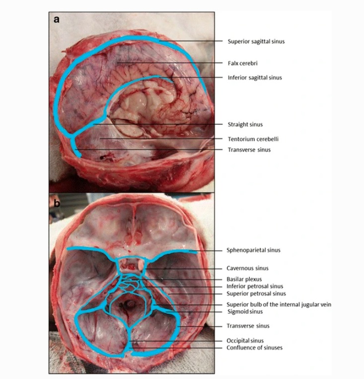
Senos venosos durales
Imagen: “Dural venous sinuses” por Jmarchn. Licencia: CC BY-SA 3.0, editado por Emma C. Cheshire et al. (2017).Trastornos neoplásicos:
Trastornos infecciosos:
Trastornos traumáticos: