La cavidad abdominal tiene una anatomía compleja e intrincada. El médico debe saber en EN Erythema nodosum is an immune-mediated panniculitis (inflammation of the subcutaneous fat) caused by a type IV (delayed-type) hypersensitivity reaction. It commonly manifests in young women as tender, erythematous nodules on the shins. Erythema Nodosum qué zona del abdomen se encuentra cada una de las estructuras principales para comprender la presentación clínica de las patologías abdominales y/o en EN Erythema nodosum is an immune-mediated panniculitis (inflammation of the subcutaneous fat) caused by a type IV (delayed-type) hypersensitivity reaction. It commonly manifests in young women as tender, erythematous nodules on the shins. Erythema Nodosum situaciones de traumatismo para estimar qué órganos probablemente fueron lesionados. El cirujano general, especialmente en EN Erythema nodosum is an immune-mediated panniculitis (inflammation of the subcutaneous fat) caused by a type IV (delayed-type) hypersensitivity reaction. It commonly manifests in young women as tender, erythematous nodules on the shins. Erythema Nodosum situaciones de emergencia, utiliza este conocimiento para ejecutar el enfoque quirúrgico más ventajoso para una situación particular.
Last updated: Dec 15, 2025
Superior:
Inferior:
Lateral:
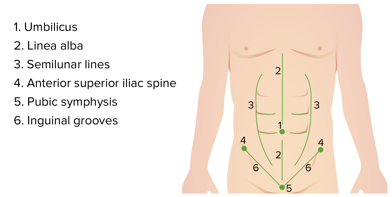
Puntos de referencia anatómicos superficiales del abdomen
Imagen por Lecturio. Licencia: CC BY-NC-SA 4.0Lo siguiente divide el abdomen en EN Erythema nodosum is an immune-mediated panniculitis (inflammation of the subcutaneous fat) caused by a type IV (delayed-type) hypersensitivity reaction. It commonly manifests in young women as tender, erythematous nodules on the shins. Erythema Nodosum sus 9 regiones:
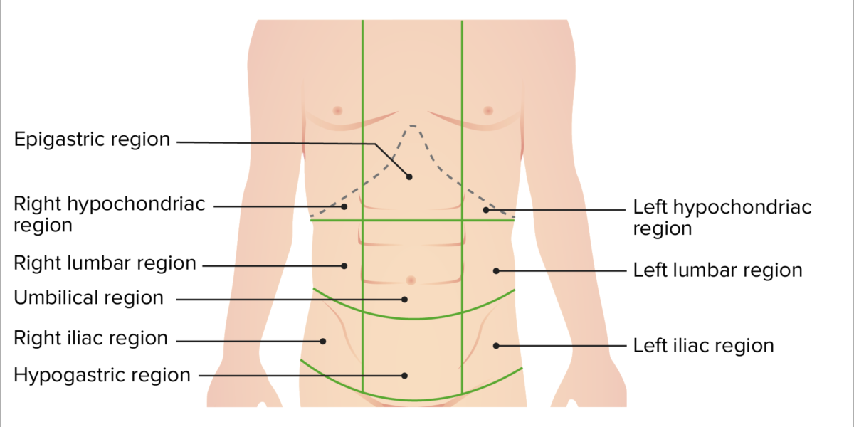
Regiones del abdomen
Imagen por Lecturio. Licencia: CC BY-NC-SA 4.0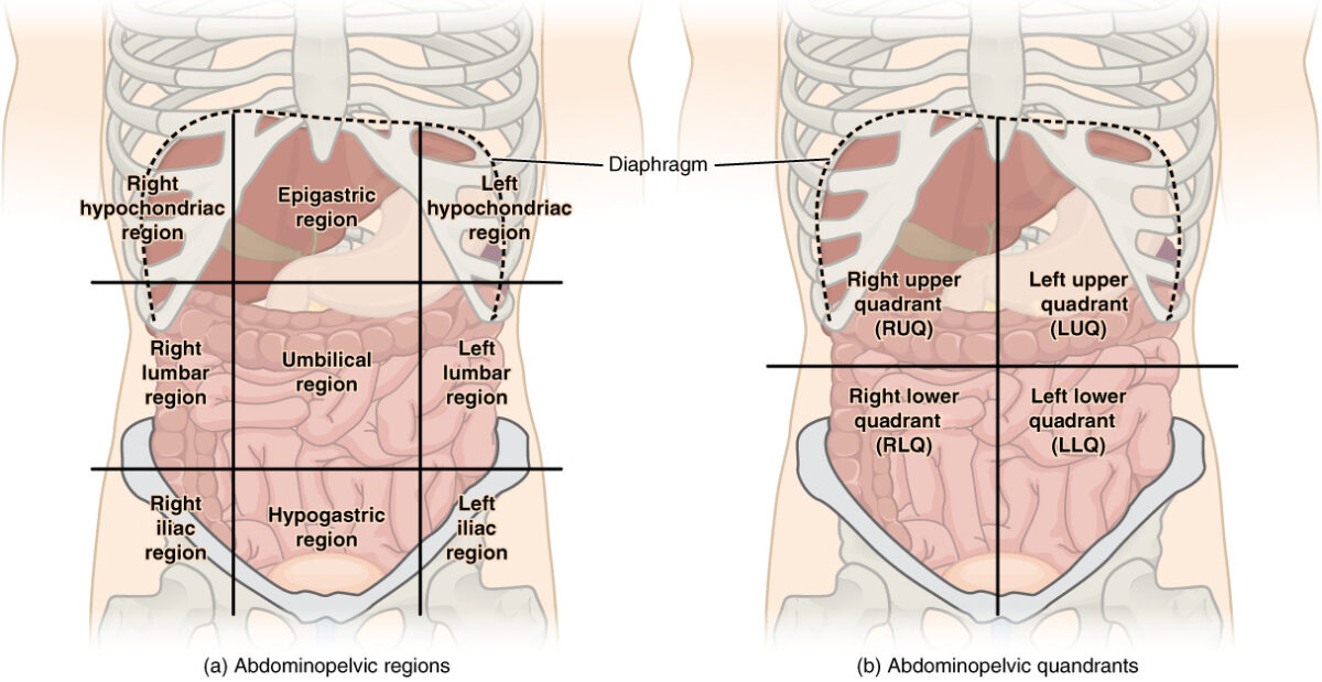
Cuadrantes abdominales:
Hay (a) 9 regiones abdominales y (b) 4 cuadrantes abdominales en la cavidad peritoneal.
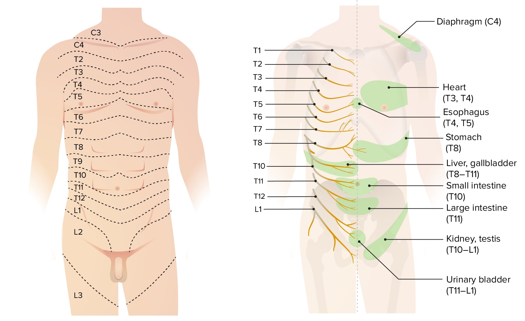
Dermatomas del tórax, abdomen y pelvis
Imagen por Lecturio.
Capas de la pared abdominal
Imagen: “Gray399” por Henry Gray. Licencia: Dominio Público, editado por Lecturio.Herniorrafia ventral: reparación quirúrgica de las hernias de la pared abdominal.
Las estructuras quirúrgicas relevantes del hipocondrio derecho incluyen el hígado y el árbol biliar.
Localización:

Localización del hígado en el hipocondrio derecho y epigastrio
Imagen por Lecturio. Licencia: CC BY-NC-SA 4.0Límites:
Superficies:
Vista anterior de la superficie diafragmática del hígado, con los ligamentos falciformes, triangulares, redondos y coronarios:
Obsérvese que el ligamento redondo se extiende desde el borde libre del ligamento falciforme.
Imagen por Lecturio. Licencia: CC BY-NC-SA 4.0
Vista inferior de la superficie visceral del hígado:
Obsérvese la estructura irregular que resulta de las impresiones de los órganos vecinos. La impresión cólica es causada por la flexura hepática del colon. La porción descendente del duodeno forma la impresión duodenal.
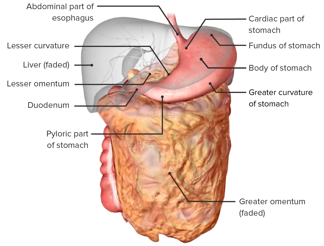
Vista anterior del hígado:
En esta imagen, el hígado se ha levantado para mostrar el epiplón menor, que consta de los ligamentos hepatogástrico y hepatoduodenal. Esta doble capa de peritoneo conecta el hígado con la curvatura menor del estómago y el duodeno.
Vesícula biliar:
Vías biliares:
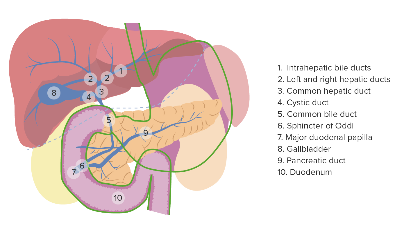
Anatomía de la vesícula biliar y las vías biliares
Imagen por Lecturio. Licencia: CC BY-NC-SA 4.0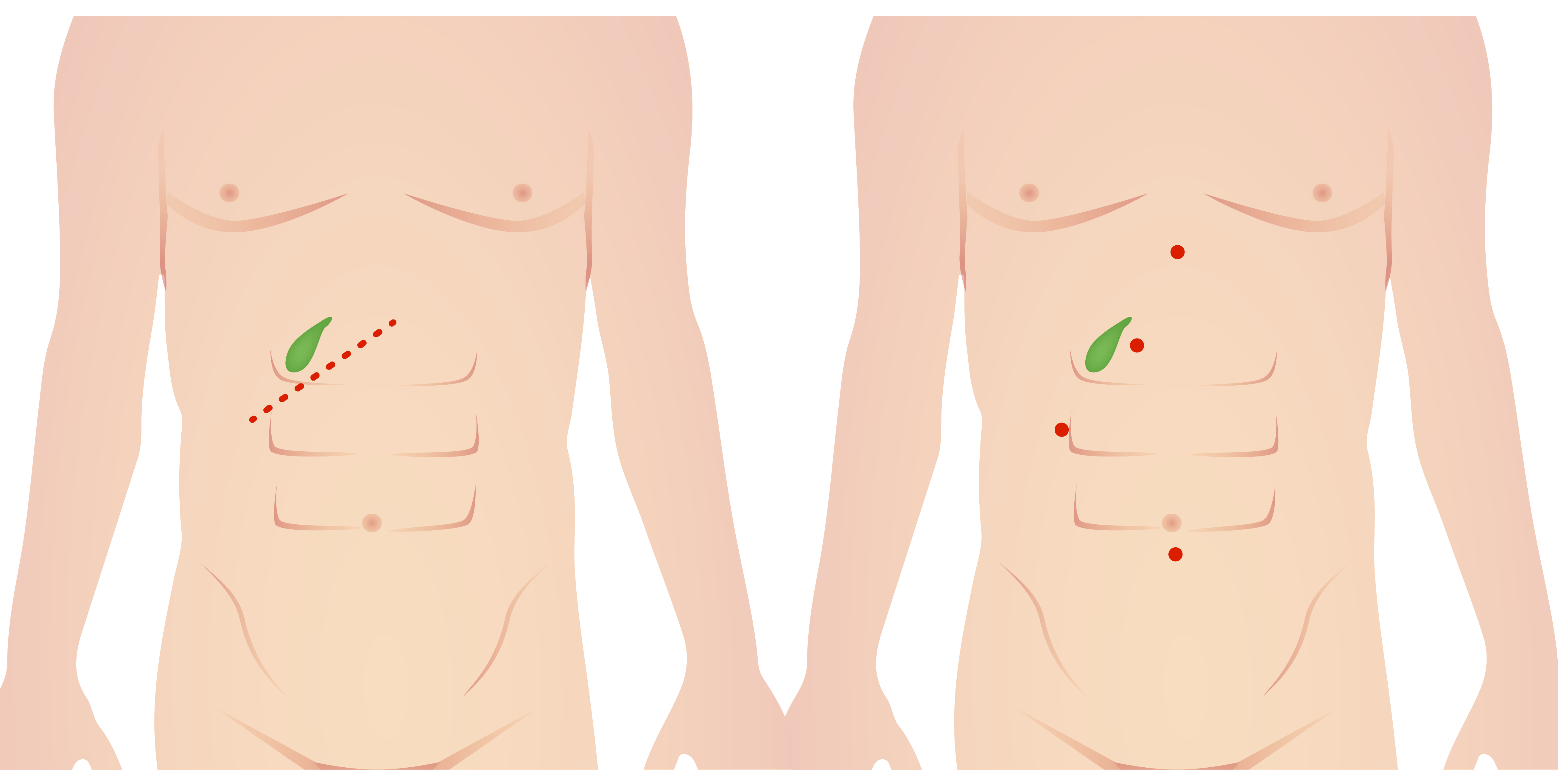
Incisiones para colecistectomía abierta (izquierda) y laparoscópica (derecha)
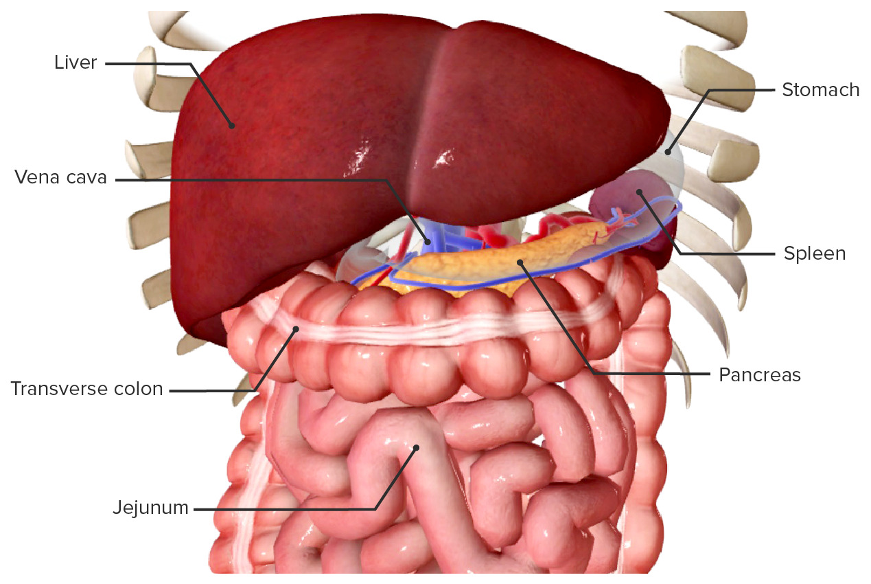
Imagen por Lecturio. Licencia: CC BY-NC-SA 4.0El epigastrio contiene el colon Colon The large intestines constitute the last portion of the digestive system. The large intestine consists of the cecum, appendix, colon (with ascending, transverse, descending, and sigmoid segments), rectum, and anal canal. The primary function of the colon is to remove water and compact the stool prior to expulsion from the body via the rectum and anal canal. Colon, Cecum, and Appendix: Anatomy transverso, el duodeno y el páncreas.
Colon in situ, cubierto por el omento mayor
Imagem por BioDigital, editada por Lecturio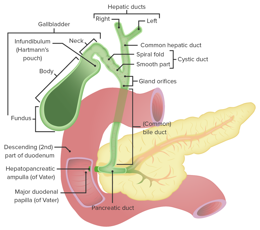
El duodeno y su relación con el páncreas y las vías biliares
Imagen por Lecturio. Licencia: CC BY-NC-SA 4.0Segmentos:
Conductos:
Diferentes partes del páncreas y sus relaciones
Imagen: “The pancreas” por OpenStax College. Licencia: CC BY 3.0Las estructuras más importantes del hipocondrio izquierdo son el bazo y el estómago.
Localización:
Relaciones:
Ligamentos:
Irrigación sanguínea: arteria y vena esplénica
Bazo in situ, vista anterior (el estómago está difuminado):
Obsérvense las relaciones espaciales con los órganos abdominales vecinos.
Bazo in situ, vista posterior:
Obsérvense las relaciones espaciales con los órganos abdominales vecinos.
Segmentos:
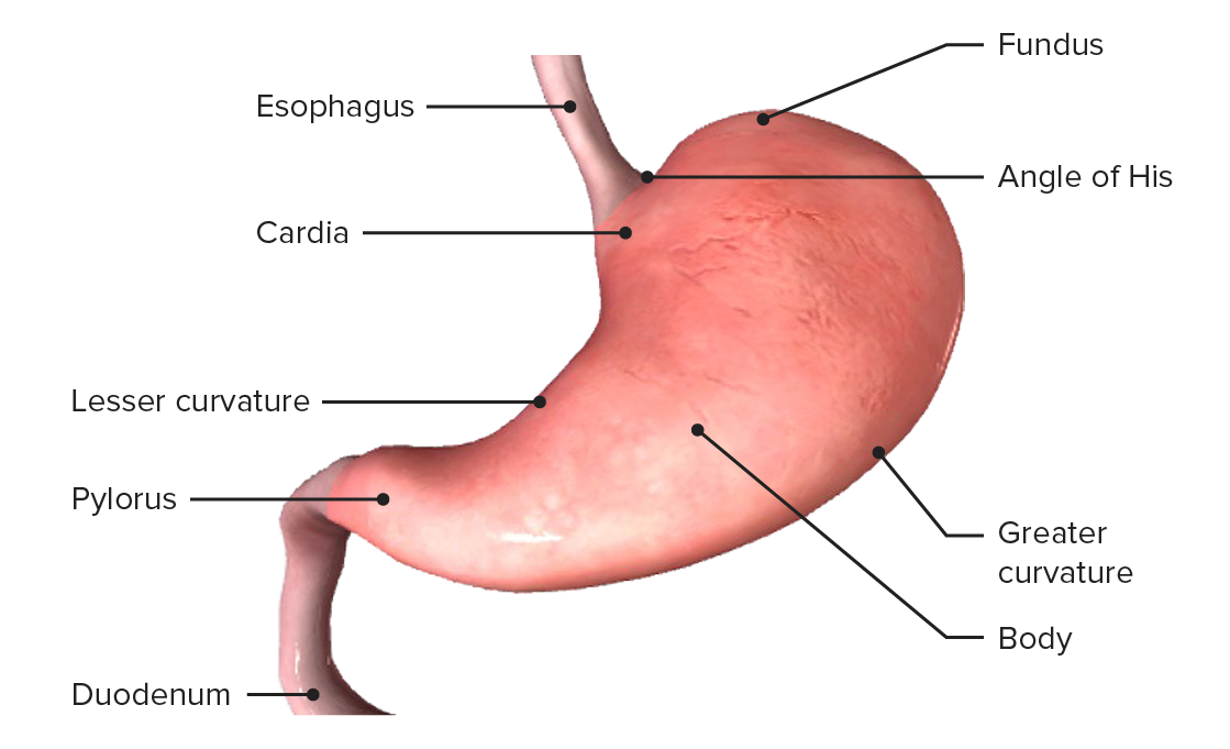
Anatomía del estómago
Imagen por BioDigital, editada por Lecturio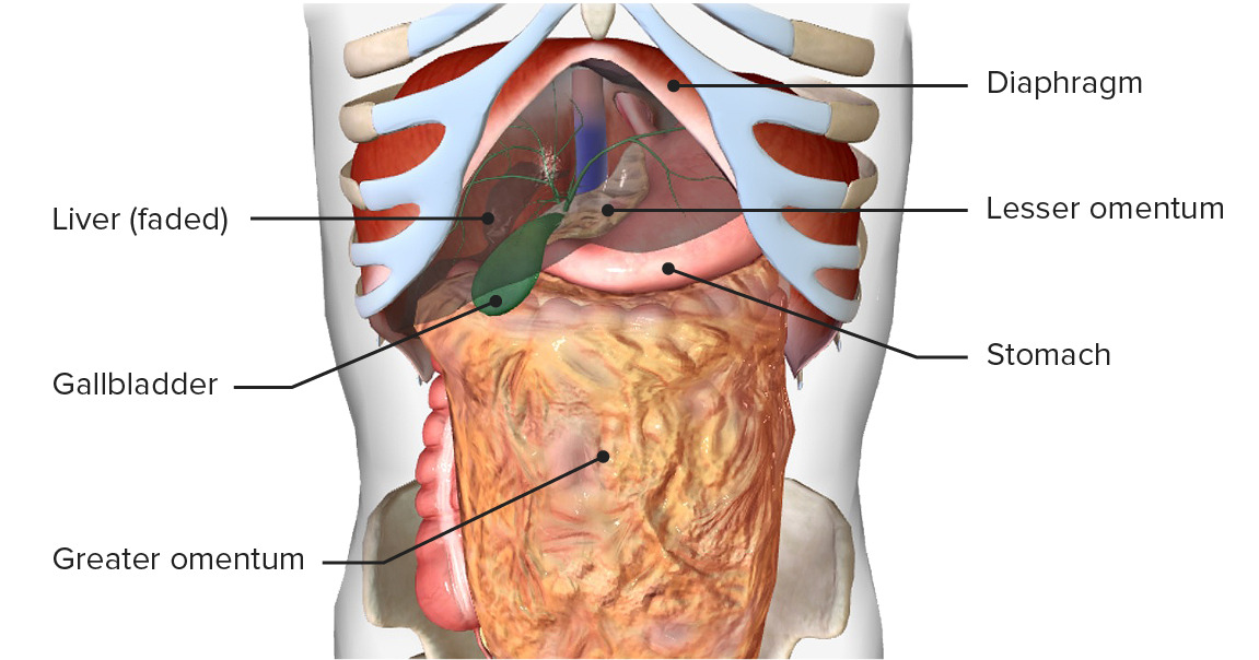
Estómago in situ
Image by BioDigital, edited by LecturioIrrigación arterial:
Drenaje venoso:
Inervación:

Irrigación e inervación del estómago
Imagen por BioDigital, editada por Lecturio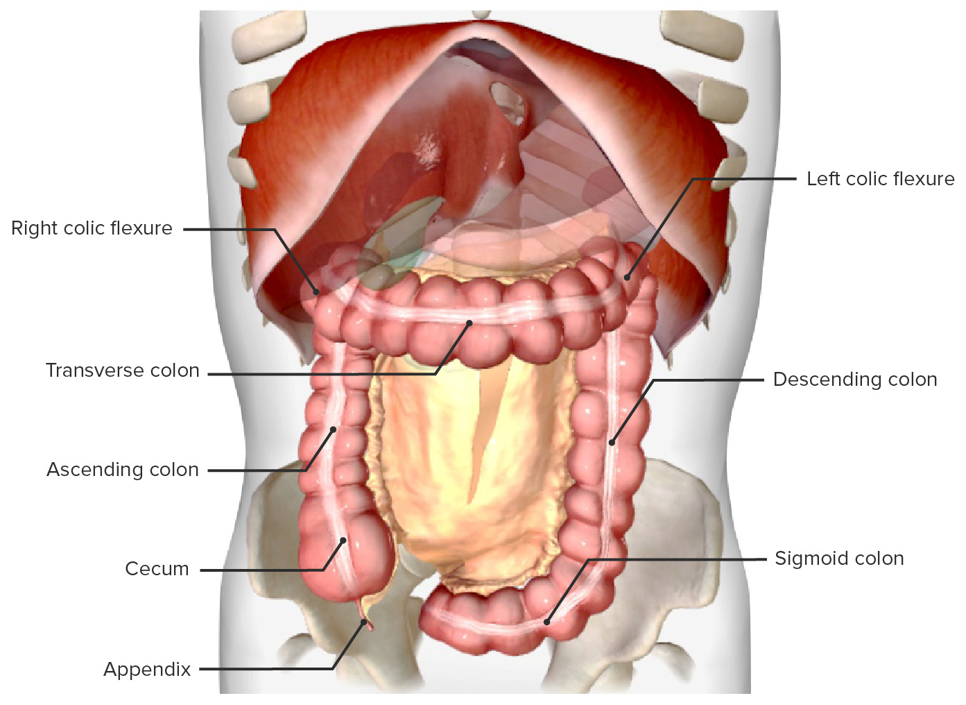
Colon in situ, vista anterior, con el epiplón mayor y el intestino delgado retirados
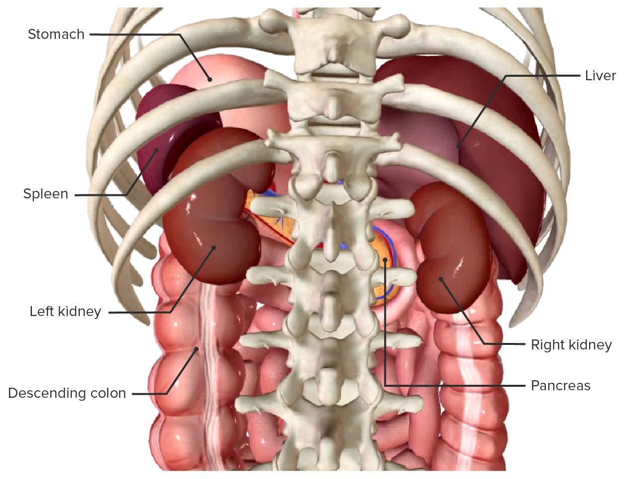
Imagen por BioDigital, editada por LecturioLocalización:
Cápsula renal:
Relaciones:
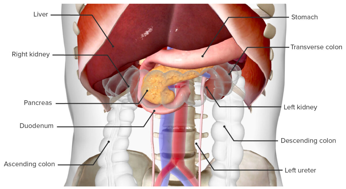
Vista anterior de los riñones y órganos vecinos
Imagen por BioDigital, editada por Lecturio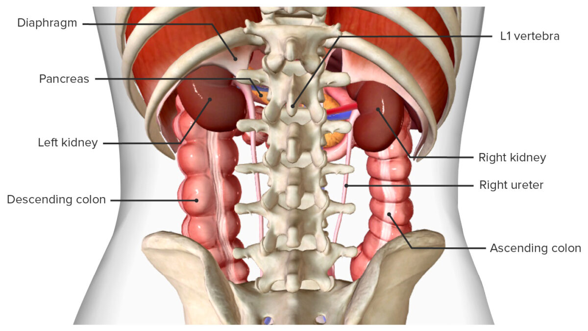
Vista posterior de los riñones y órganos vecinos
Imagen por BioDigital, editada por LecturioCaracterísticas externas:
Irrigación sanguínea: arterias y venas renales
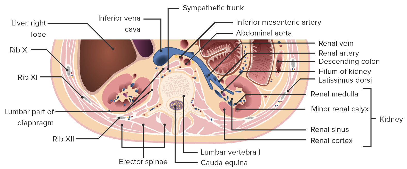
Corte transversal del abdomen enfocado en los riñones:
Obsérvese cómo el riñón está incrustado en el tejido adiposo dentro de las correderas (grasa paranéfrica y perinéfrica), que es continuo con la grasa de los senos renales.
Irrigación sanguínea
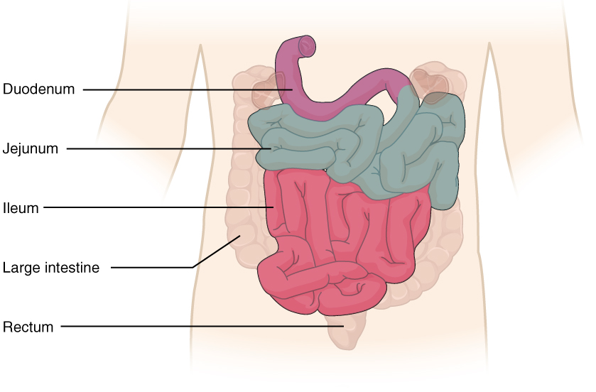
El intestino delgado y sus partes
Imagen: “2417 Small IntestineN” por OpenStax College. Licencia: CC BY 3.0Ramas vasculares:
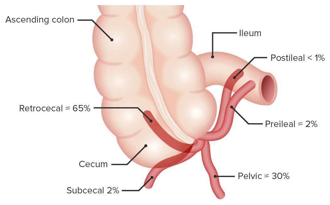
Variantes de localización del apéndice vermiforme
Imagen por Lecturio. Licencia: CC BY-NC-SA 4.0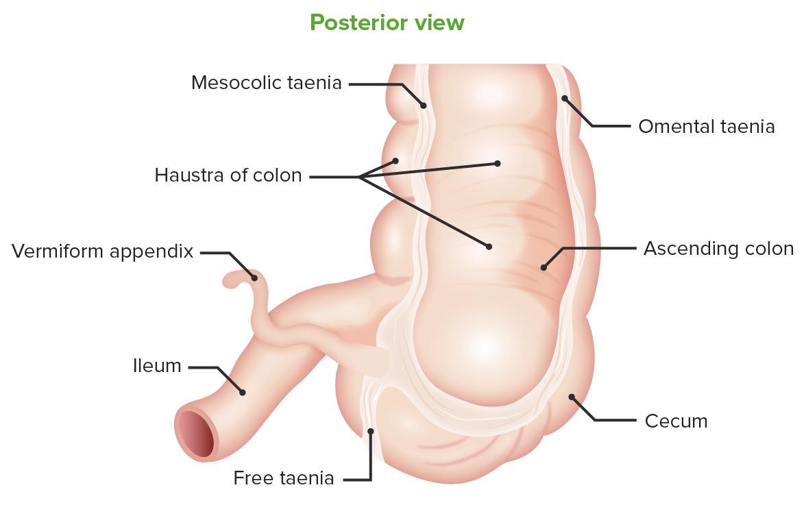
Vista posterior del ciego:
Ubicación del apéndice vermiforme en la confluencia de la tenias
Punto de McBurney:
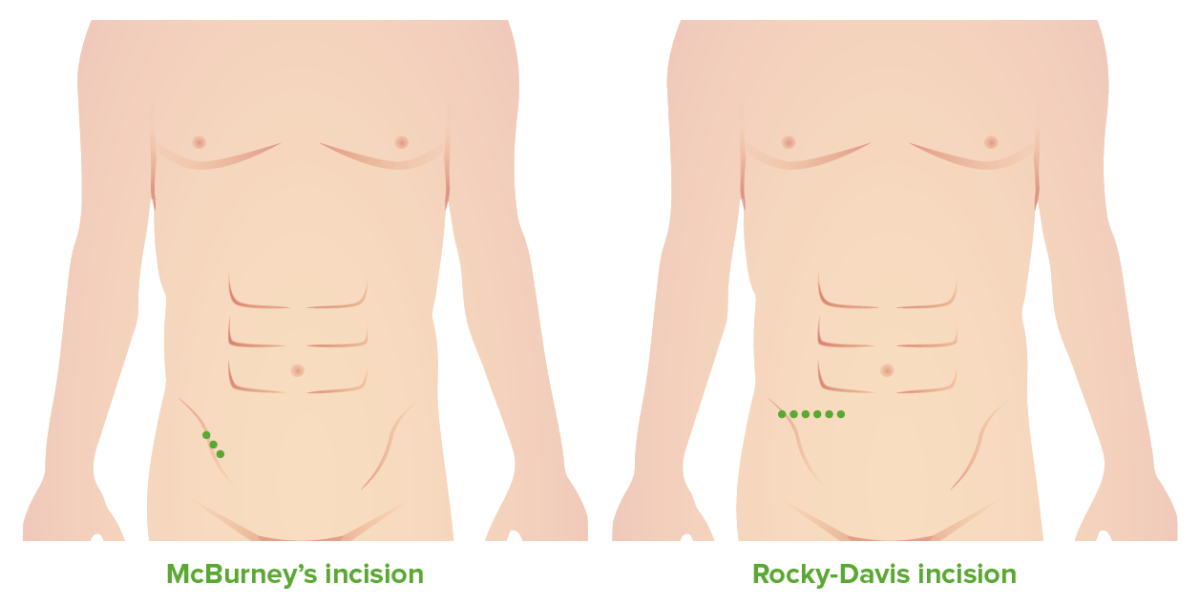
Incisiones de McBurney y Rocky-Davis
Imagen por Lecturio. Licencia: CC BY-NC-SA 4.0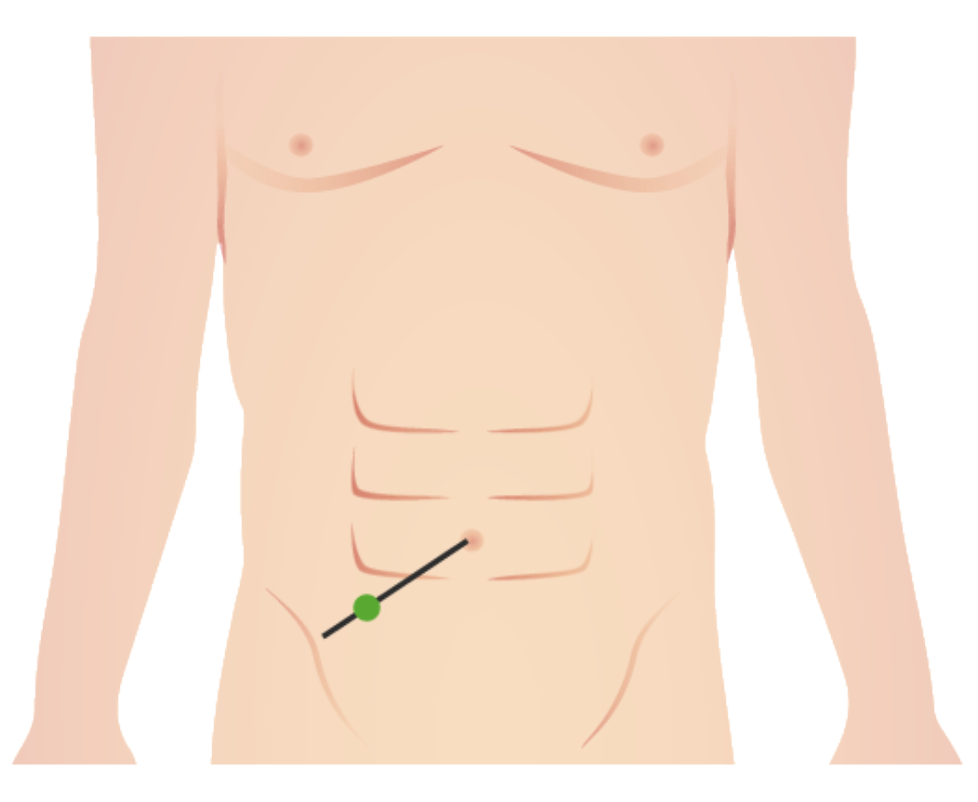
Ubicación del punto de McBurney
Imagen por Lecturio. Licencia: CC BY-NC-SA 4.0Límites:
Curso:
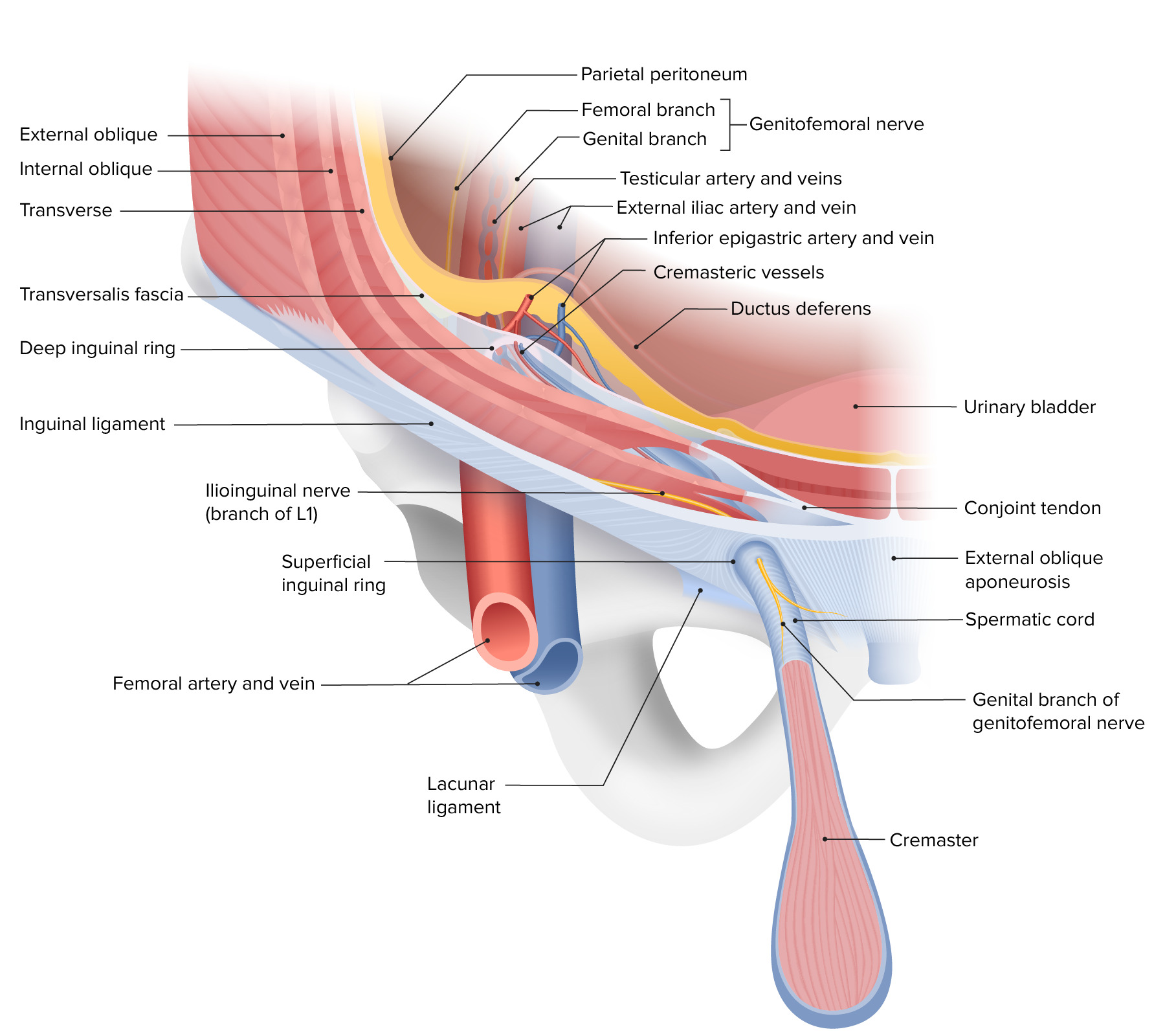
Las capas de la pared abdominal anterior, representando el trayecto del canal inguinal y la composición de los anillos inguinales profundos y superficiales
Imagen por Lecturio. Licencia: CC BY-NC-SA 4.0Contenido:
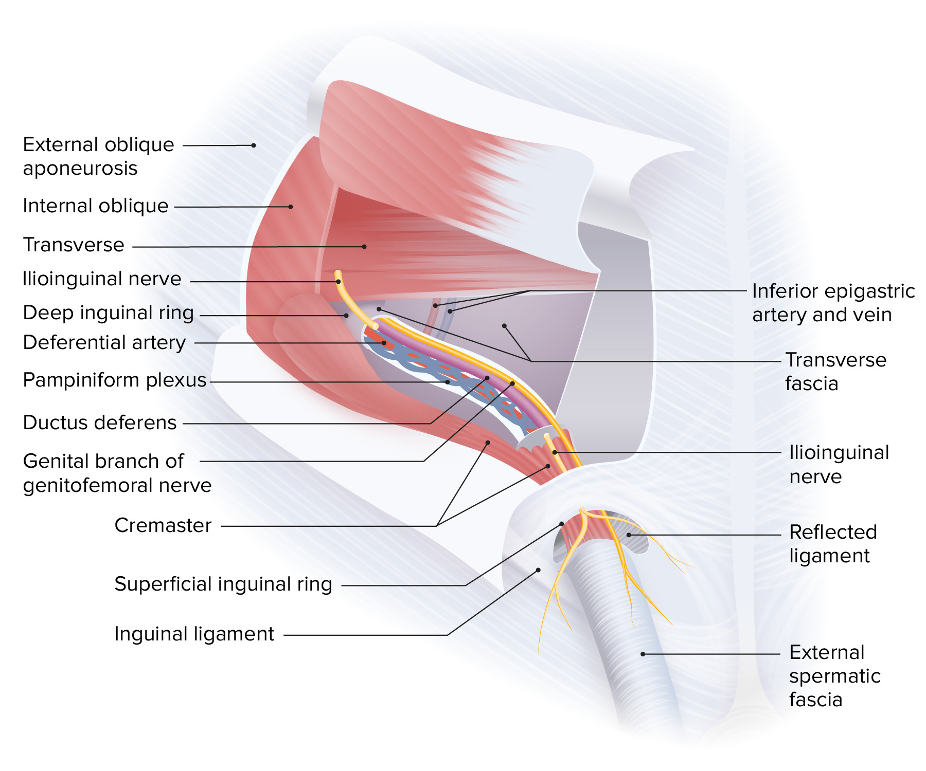
Límites y contenido del canal inguinal masculino
Obsérvese que el nervio ilioinguinal recorre el canal inguinal externamente al cordón espermático.
Segmentos:
Ligamento ancho (ligamentum latum):
Irrigación y drenaje:
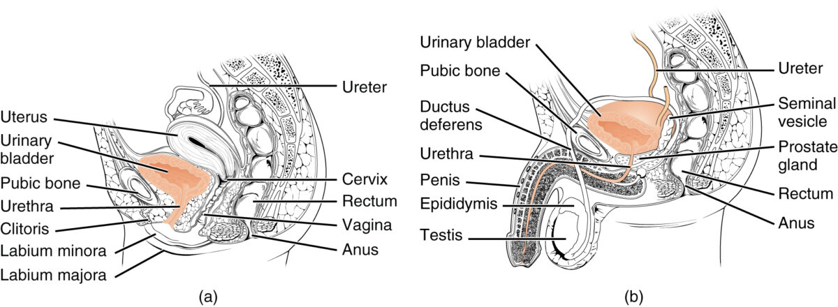
Diagrama de un corte sagital de
la pelvis en una mujer (a) y en un hombre (b):En la pelvis femenina, obsérvese la proximidad de la vejiga a la pared vaginal anterior. En la pelvis masculina, hay que destacar la proximidad de la vejiga al recto.
Imagen: “The urethra transports urine from the bladder to the outside of the body” por OpenStax College. Licencia: CC BY 4.0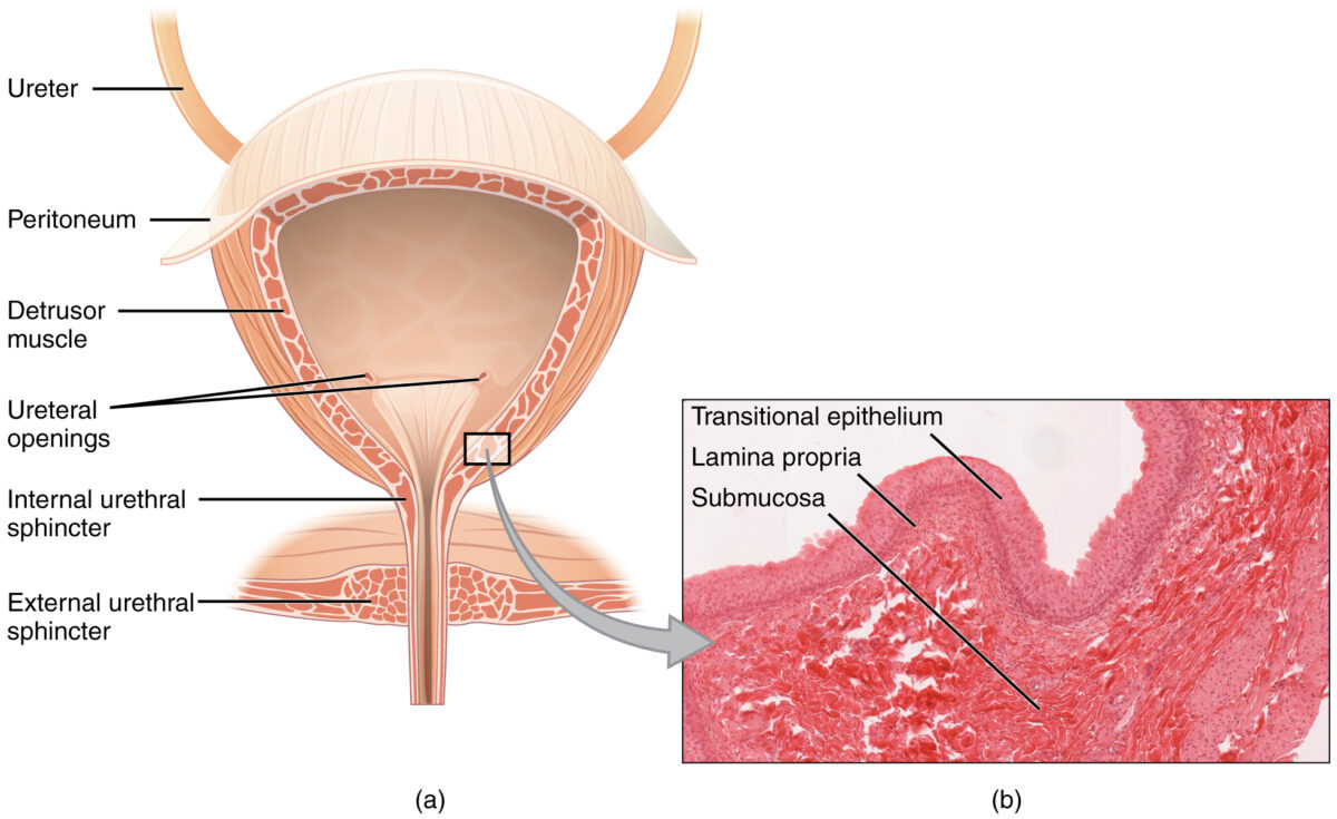
Corte coronal de la vejiga:
Este corte histológico ampliado (b) ilustra el epitelio de transición, la lámina propia y la submucosa.
Localización:
Lóbulos:
Estructuras relacionadas:
Estructuras glandulares accesorias:
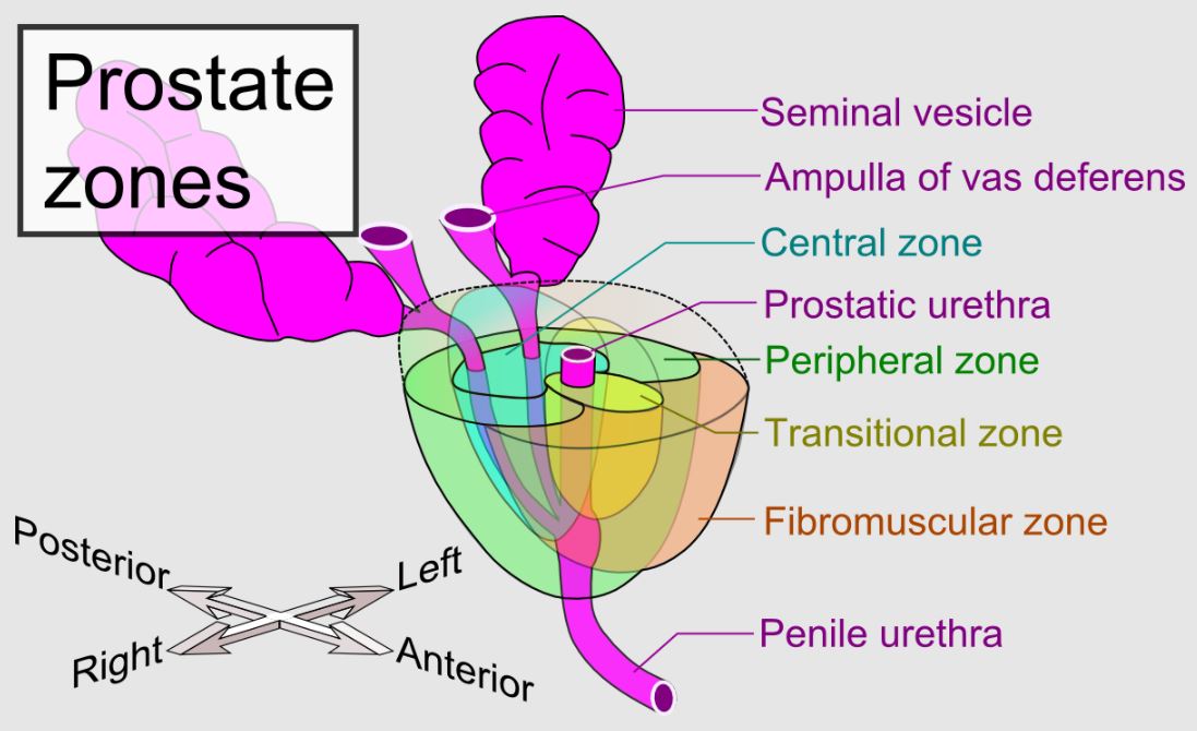
Glándula prostática y sus principales zonas:
Zonas periféricas, transicionales y centrales en relación con otras estructuras del sistema genitourinario masculino
Obsérvese cómo la próstata está posicionada alrededor de la uretra prostática.
Localización:
Estructura:
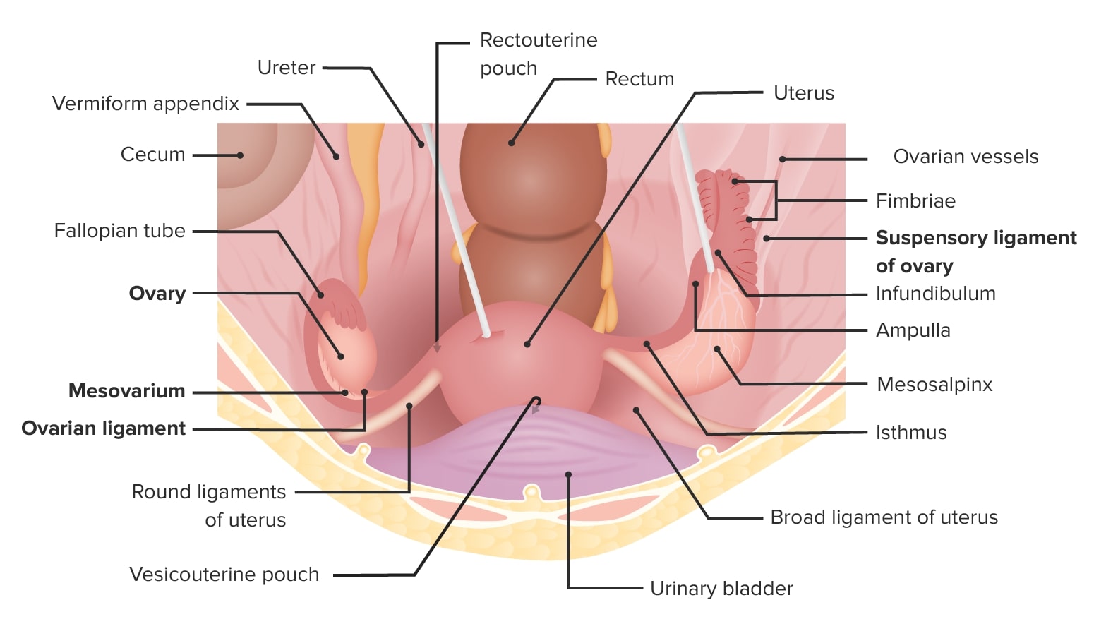
Vista superior de la pelvis femenina que muestra el útero in situ, sus ligamentos de soporte y su relación con los ovarios y los órganos vecinos.
Imagen por Lecturio. Licencia: CC BY-NC-SA 4.0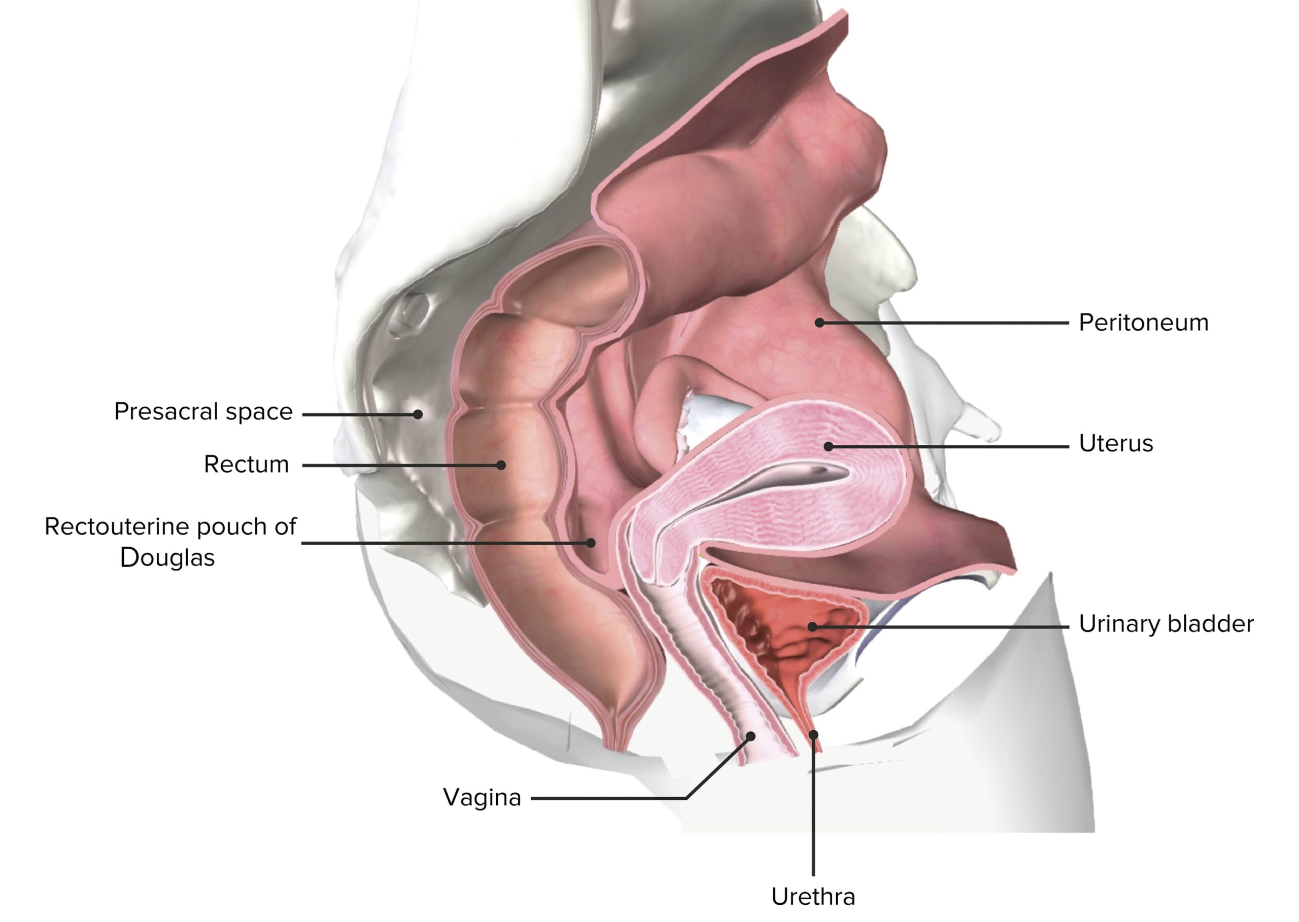
Corte de la pelvis femenina que muestra el útero in situ
Imagen de BioDigital, editada por LecturioEstructuras de soporte:
Irrigación sanguínea:
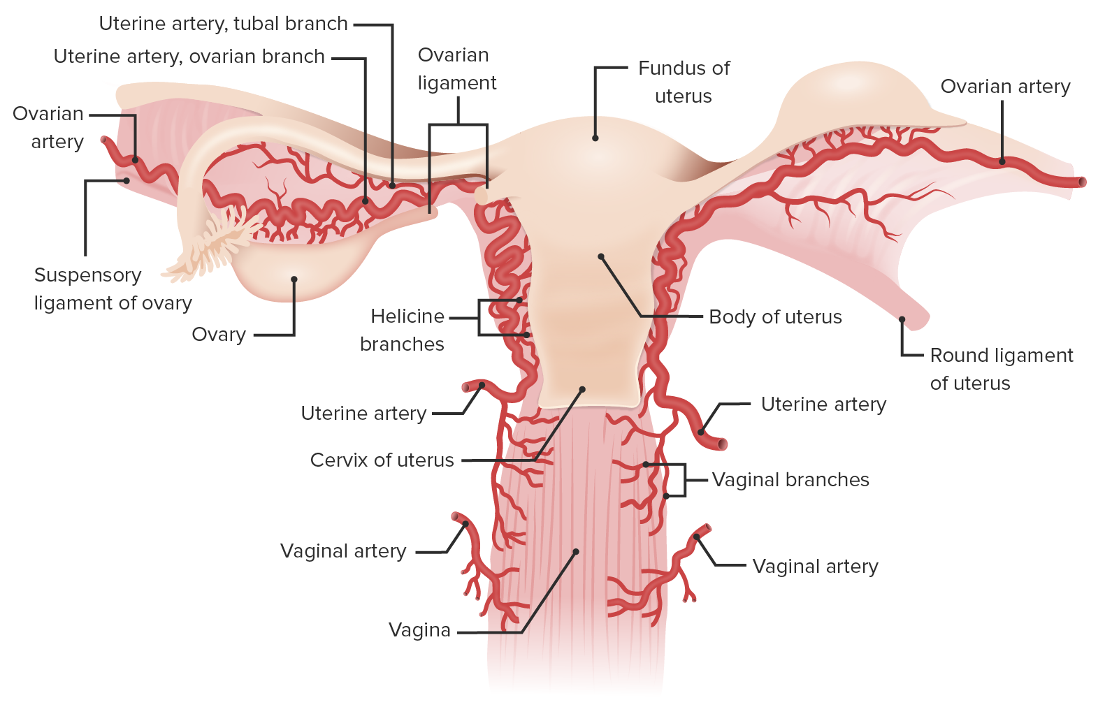
Irrigación sanguínea del útero y los ovarios:
Obsérvese la arteria ovárica que se desplaza a lo largo del ligamento suspensorio del ovario, irrigando tanto los ovarios como el tercio lateral de la trompa uterina. La arteria ovárica continúa en el mesosalpinx para anastomosarse con ramas de la arteria uterina.