


Uma oclusão vascular da retina Retina The ten-layered nervous tissue membrane of the eye. It is continuous with the optic nerve and receives images of external objects and transmits visual impulses to the brain. Its outer surface is in contact with the choroid and the inner surface with the vitreous body. The outermost layer is pigmented, whereas the inner nine layers are transparent. Eye: Anatomy é um bloqueio numa artéria ou numa veia principal da retina Retina The ten-layered nervous tissue membrane of the eye. It is continuous with the optic nerve and receives images of external objects and transmits visual impulses to the brain. Its outer surface is in contact with the choroid and the inner surface with the vitreous body. The outermost layer is pigmented, whereas the inner nine layers are transparent. Eye: Anatomy. A oclusão pode ser classificada segundo a sua localização, como oclusão da artéria central da retina Retina The ten-layered nervous tissue membrane of the eye. It is continuous with the optic nerve and receives images of external objects and transmits visual impulses to the brain. Its outer surface is in contact with the choroid and the inner surface with the vitreous body. The outermost layer is pigmented, whereas the inner nine layers are transparent. Eye: Anatomy (OACR), oclusão do ramo arterial da retina Retina The ten-layered nervous tissue membrane of the eye. It is continuous with the optic nerve and receives images of external objects and transmits visual impulses to the brain. Its outer surface is in contact with the choroid and the inner surface with the vitreous body. The outermost layer is pigmented, whereas the inner nine layers are transparent. Eye: Anatomy, oclusão da veia central da retina Retina The ten-layered nervous tissue membrane of the eye. It is continuous with the optic nerve and receives images of external objects and transmits visual impulses to the brain. Its outer surface is in contact with the choroid and the inner surface with the vitreous body. The outermost layer is pigmented, whereas the inner nine layers are transparent. Eye: Anatomy (OVCR) ou oclusão do ramo venoso da retina Retina The ten-layered nervous tissue membrane of the eye. It is continuous with the optic nerve and receives images of external objects and transmits visual impulses to the brain. Its outer surface is in contact with the choroid and the inner surface with the vitreous body. The outermost layer is pigmented, whereas the inner nine layers are transparent. Eye: Anatomy. Normalmente, a oclusão de um vaso da retina Retina The ten-layered nervous tissue membrane of the eye. It is continuous with the optic nerve and receives images of external objects and transmits visual impulses to the brain. Its outer surface is in contact with the choroid and the inner surface with the vitreous body. The outermost layer is pigmented, whereas the inner nine layers are transparent. Eye: Anatomy é resultado de um evento tromboembólico. Os fatores de risco incluem hipertensão, diabetes Diabetes Diabetes mellitus (DM) is a metabolic disease characterized by hyperglycemia and dysfunction of the regulation of glucose metabolism by insulin. Type 1 DM is diagnosed mostly in children and young adults as the result of autoimmune destruction of β cells in the pancreas and the resulting lack of insulin. Type 2 DM has a significant association with obesity and is characterized by insulin resistance. Diabetes Mellitus mellitus e doença valvular cardíaca. A oclusão de um vaso central da retina Retina The ten-layered nervous tissue membrane of the eye. It is continuous with the optic nerve and receives images of external objects and transmits visual impulses to the brain. Its outer surface is in contact with the choroid and the inner surface with the vitreous body. The outermost layer is pigmented, whereas the inner nine layers are transparent. Eye: Anatomy é caracterizada por perda de visão súbita, unilateral e indolor e/ou perda de visão transitória (amaurose fugaz). As opções de tratamento são limitadas e são geralmente pouco eficazes. Quando a região da mácula está envolvida, o prognóstico é especialmente reservado, por estar associado à perda permanente da visão.
Last updated: Dec 15, 2025
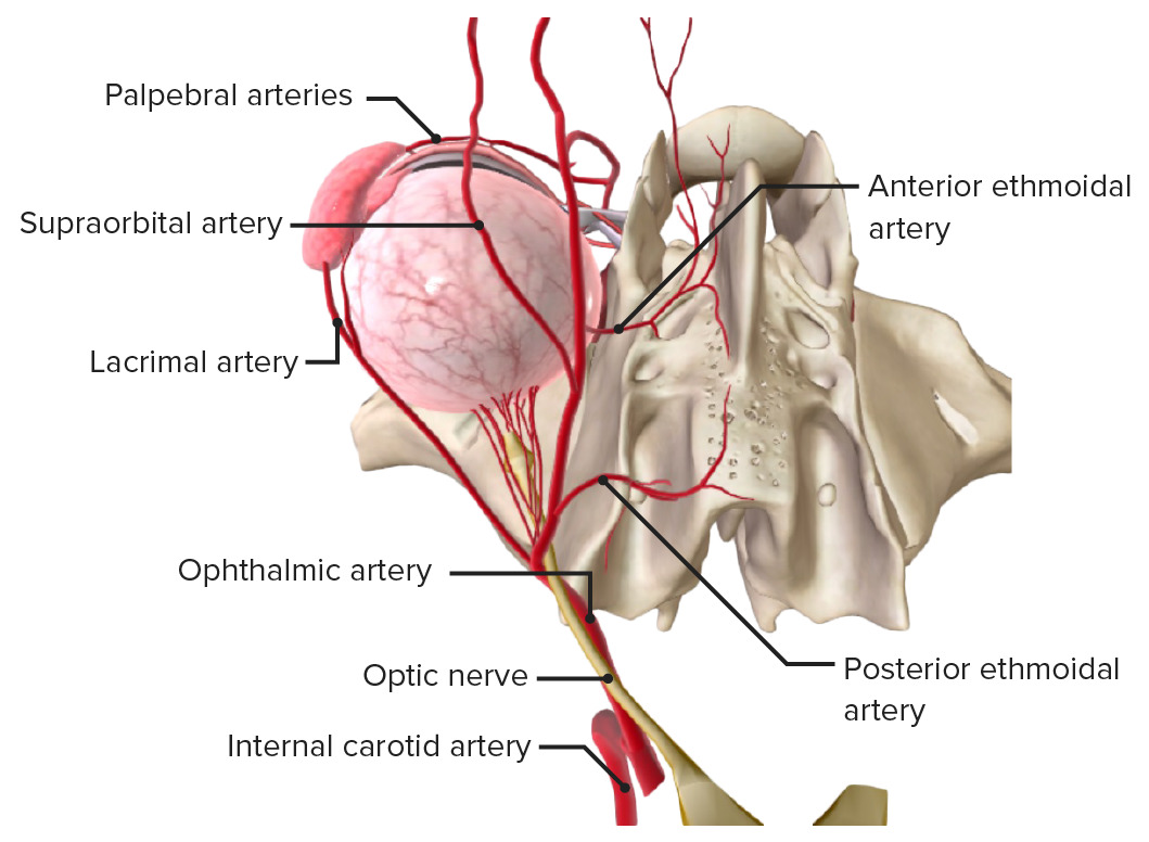
A artéria carótida com a artéria oftálmica e os seus ramos. Observe a artéria central da retina e as artérias coroideias posteriores, que entram no olho em volta do nervo ótico.
Imagem por BioDigital, editado por Lecturio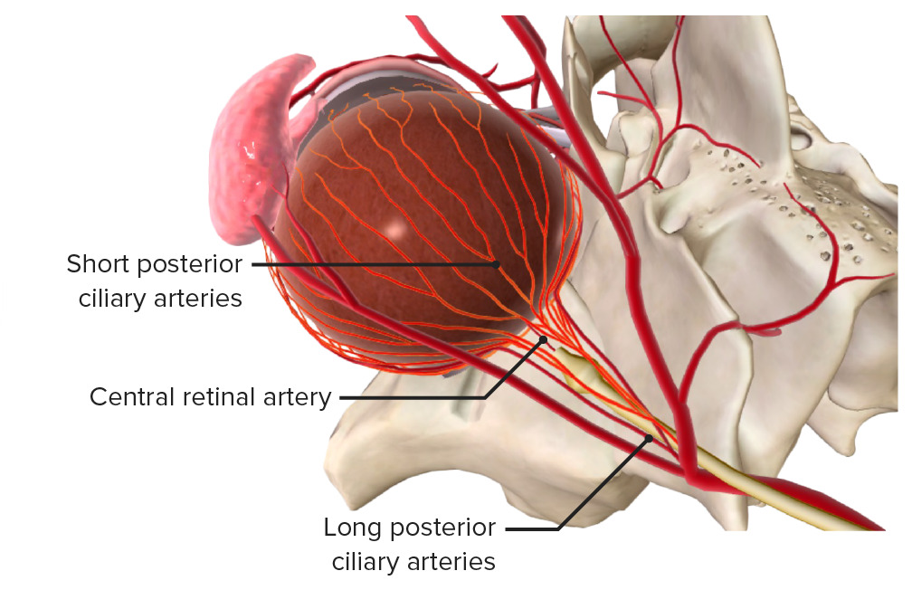
Vista superior: A artéria carótida interna ramifica-se na artéria oftálmica, que se subdivide na artéria retiniana central e nas artérias ciliares posteriores.
Imagem por BioDigital, editado por Lecturio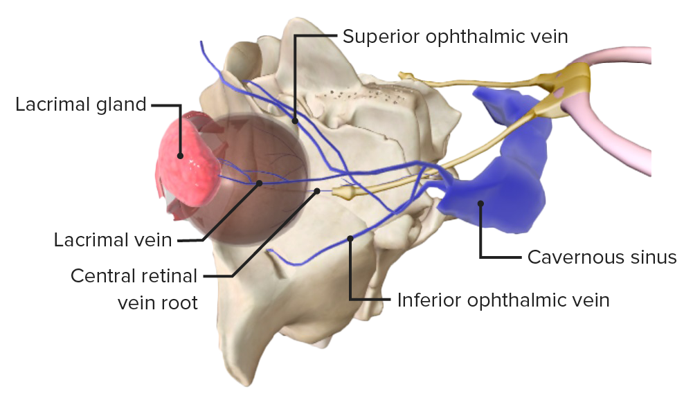
A veia oftálmica superior e o seio cavernoso, para onde drena a veia retiniana central.
Imagem por BioDigital, editado por LecturioAs causas exatas são desconhecidas, porém, está associada com as seguintes condições:
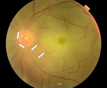
Imagem que mostra uma fundoscopia a um olho esquerdo com OACR; pode-se verificar atenuação das artérias da retina (setas) e palidez do disco ótico
Imagem: “Color fundus photography of the left eye” por Division of Cardiology, Department of Internal Medicine, Pohang St. Mary’s Hospital, Pohang, Republic of Korea. Licença: CC BY 4.0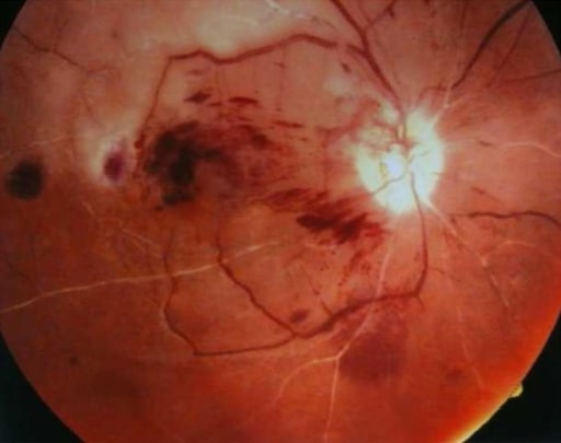
Fundoscopia do olho direito com uma oclusão combinada da artéria central da retina e da veia central da retina: pode ser observado um fundo ocular do tipo “sangue” e “trovão” combinado com uma mácula vermelho-cereja (caso de doente com lúpus eritematoso sistémico).
Imagem: “Chorea and retinal vessel occlusion” por Department of Neurology, Shariati Hospital, Tehran University of Medical Sciences AND Iranian Center of Neurological Research, Tehran, Iran. Licença: CC BY 2.0Oclusão da artéria central da retina Retina The ten-layered nervous tissue membrane of the eye. It is continuous with the optic nerve and receives images of external objects and transmits visual impulses to the brain. Its outer surface is in contact with the choroid and the inner surface with the vitreous body. The outermost layer is pigmented, whereas the inner nine layers are transparent. Eye: Anatomy
Oclusão da veia central da retina Retina The ten-layered nervous tissue membrane of the eye. It is continuous with the optic nerve and receives images of external objects and transmits visual impulses to the brain. Its outer surface is in contact with the choroid and the inner surface with the vitreous body. The outermost layer is pigmented, whereas the inner nine layers are transparent. Eye: Anatomy
O diagnóstico de ambas as situações é geralmente clínico, embora possam ser necessários exames adicionais.
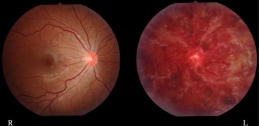
Imagem de uma hemorragia retiniana esquerda (OVCR) numa doente com 15 anos (com história de lúpus sistémico e síndrome antifosfolipídico)
Imagem: “Finding of eye fundus” por Department of Pediatrics and Child Neurology, Oita University Faculty of Medicine, Hasama, Yufu, Oita 879-5593, Japan. Licença: CC BY 2.0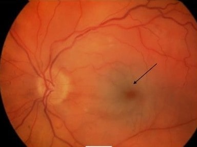
Olho esquerdo com mácula vermelho-cereja com palidez de retina típica de OACR (seta)
Imagem: “Acute central retinal artery occlusion” por James Paget University Hospital NHS Foundation Trust, Lowestoft Road, Gorleston, Great Yarmouth NR31 6LA, Norfolk, UK. Licença: CC BY 2.0