


O cérebro e a medula espinhal são envolvidos por 3 camadas sobrepostas de tecido conjuntivo chamadas meninges Meninges The brain and the spinal cord are enveloped by 3 overlapping layers of connective tissue called the meninges. The layers are, from the most external layer to the most internal layer, the dura mater, arachnoid mater, and pia mater. Between these layers are 3 potential spaces called the epidural, subdural, and subarachnoid spaces. Meninges: Anatomy. As camadas são, da camada mais MAIS Androgen Insensitivity Syndrome externa à camada mais MAIS Androgen Insensitivity Syndrome interna, a dura-máter, aracnoide-máter e pia-máter. Entre estas camadas existem 3 espaços potenciais chamados espaço epidural, subdural e subaracnoide. A função das meninges Meninges The brain and the spinal cord are enveloped by 3 overlapping layers of connective tissue called the meninges. The layers are, from the most external layer to the most internal layer, the dura mater, arachnoid mater, and pia mater. Between these layers are 3 potential spaces called the epidural, subdural, and subarachnoid spaces. Meninges: Anatomy é proteger o conteúdo do cérebro e da medula espinhal. A infeção do SNC apresenta-se com inflamação das meninges Meninges The brain and the spinal cord are enveloped by 3 overlapping layers of connective tissue called the meninges. The layers are, from the most external layer to the most internal layer, the dura mater, arachnoid mater, and pia mater. Between these layers are 3 potential spaces called the epidural, subdural, and subarachnoid spaces. Meninges: Anatomy, e a etiologia pode ser estudada a partir do exame do LCR, que está contido no espaço subaracnoide.
Last updated: Dec 15, 2025
As meninges Meninges The brain and the spinal cord are enveloped by 3 overlapping layers of connective tissue called the meninges. The layers are, from the most external layer to the most internal layer, the dura mater, arachnoid mater, and pia mater. Between these layers are 3 potential spaces called the epidural, subdural, and subarachnoid spaces. Meninges: Anatomy são camadas de tecido conjuntivo que protegem o cérebro e a medula espinhal.
As meninges Meninges The brain and the spinal cord are enveloped by 3 overlapping layers of connective tissue called the meninges. The layers are, from the most external layer to the most internal layer, the dura mater, arachnoid mater, and pia mater. Between these layers are 3 potential spaces called the epidural, subdural, and subarachnoid spaces. Meninges: Anatomy consistem em 3 camadas de tecido conjuntivo, com espaços potenciais entre elas:
| Camada | Origem | Características |
|---|---|---|
| Espaço epidural | NA |
|
| Dura-máter | Mesoderme |
|
| Espaço subdural | NA |
|
| Aracnoide ( leptomeninges Leptomeninges Meninges: Anatomy) | Crista neural |
|
| Espaço subaracnoide | Plexo Coroide |
|
| Pia mater Pia mater The innermost layer of the three meninges covering the brain and spinal cord. It is the fine vascular membrane that lies under the arachnoid and the dura mater. Meninges: Anatomy ( leptomeninges Leptomeninges Meninges: Anatomy) | Crista neural |
|
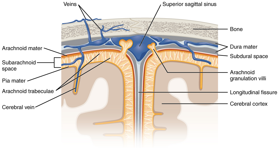
Camadas das meninges e as suas relações abaixo do crânio
Imagem: “Diagram of section of top of brain showing the meninges and subarachnoid space” por OpenStax. Licença: CC BY 4.0A dura-máter é a camada mais MAIS Androgen Insensitivity Syndrome espessa das meninges Meninges The brain and the spinal cord are enveloped by 3 overlapping layers of connective tissue called the meninges. The layers are, from the most external layer to the most internal layer, the dura mater, arachnoid mater, and pia mater. Between these layers are 3 potential spaces called the epidural, subdural, and subarachnoid spaces. Meninges: Anatomy e fornece estrutura ao cérebro.
As pregas durais e os seios durais vêm da dura-máter.
| Estrutura | Características |
|---|---|
| Foice cerebral | Separa os hemisférios cerebrais direito e esquerdo |
| Foice do cerebelo | Separa os hemisférios cerebelares direito e esquerdo |
| Tenda do cerebelo | Tenda, ou teto, sobre o cerebelo |
| Diafragma selar | Teto sobre a glândula pituitária |
| Seios durais | As 2 camadas de dura-máter atravessam juntas na maior parte do crânio. No local onde se separam, o espaço entre elas é denominado de seio venoso dural. Estes seios drenam sangue e LCR do cérebro e desembocam na veia jugular interna. |
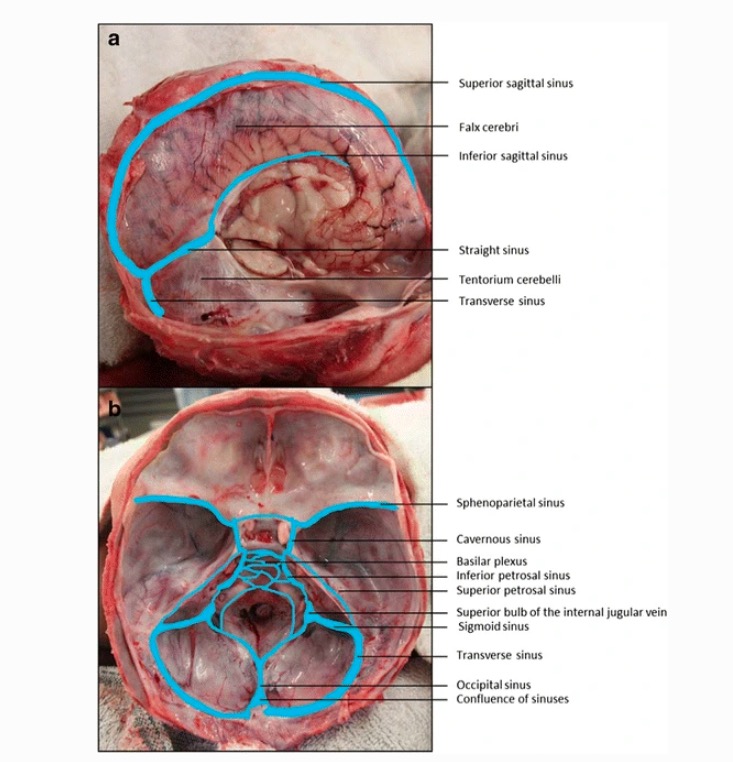
Seios venosos durais
Imagem: “Dural venous sinuses” por Jmarchn. Licença: CC BY-SA 3.0, editado por Emma C. Cheshire et al. (2017).Distúrbios neoplásicos:
Distúrbios infeciosos:
Distúrbios traumáticos: