Esofagite

A esofagite é a inflamação ou irritação do esófago. Os principais tipos de esofagite são a induzida por fármacos, a infeciosa, a eosinofílica, a corrosiva e a de refluxo ácido. Os doentes apresentam-se tipicamente com odinofagia, disfagia e dor torácica retroesternal. O diagnóstico é feito por endoscopia com biópsia. São realizados exames laboratoriais e de imagem dependendo do grau de lesão e envolvimento de outros sistemas de órgãos. O tratamento da esofagite depende da etiologia subjacente e inclui alterações dietéticas, evicção de agentes agressores, antibioterapia ou utilização de um inibidor da bomba de protões. Nos casos graves, como nas lesões corrosivas, pode ser necessário realizar uma cirurgia. Se não for tratada, a esofagite pode levar a complicações como estenoses, metaplasia do esófago e desenvolvimento de neoplasia maligna.

Última atualização: Jan 16, 2024

Responsibilidade editorial: Stanley Oiseth, Lindsay Jones, Evelin Maza

Descrição Geral

  • Esophagitis: inflammation of or injury to the esophageal mucosa
  • Major etiologies:
    • Medication-induced/pill-induced esophagitis
    • Infectious esophagitis
    • Eosinophilic esophagitis
    • Corrosive esophagitis
    • Reflux esophagitis
  • Diagnosis is done by upper endoscopy; findings vary per etiology.

Mnemónica

Para lembrar as causas mais comuns de esofagite: “PIECE

  • Pill-induced esophagitis
  • Infectious esophagitis
  • Eosinophilic esophagitis
  • Corrosive esophagitis
  • Etc. (reflux esophagitis)

Vídeos recomendados

Medication-induced Esophagitis

Etiologia

  • Antibiotics: tetracyclines, doxycycline, clindamycin
  • Anti-inflammatories: Nonsteroidal anti-inflammatory drugs (NSAIDs), aspirin
  • Bisphosphonates: alendronate, risedronate
  • Others: potassium chloride, quinidine, iron supplements, ascorbic acid

Fisiopatologia

  • Prolonged, direct contact between the medication(s) and the mucosa causes irritation, erosions, and ulcerations
  • Frequently affects site(s) of anatomical narrowing (mid-esophagus, near level of the aortic arch)
  • Mechanisms:
    • Disruption of the normal protective mucosal barrier: aspirin, NSAIDs
    • Local caustic damage (e.g., doxycycline is acidic once dissolved)
    • Hyperosmotic solutions contribute to tissue and vascular injury (e.g., potassium chloride).

Fatores de risco

  • Occurs more often in elderly patients
  • Position of patient (supine > upright)
  • Size of medication (delayed transit with large tablets)
  • Amount of fluid ingested with medication (intake of at least > 100 mL advised)

Apresentação clínica

  • Heartburn
  • Retrosternal pain
  • Odynophagia, dysphagia

Diagnóstico

  • Clinical diagnosis based on history
  • Upper endoscopy:
    • Perform if with severe or persistent symptoms (despite discontinuation of the offending medication) 
    • Findings: discrete ulcer, with normal bordering mucosa
Esofagite induzida por cloxacilina

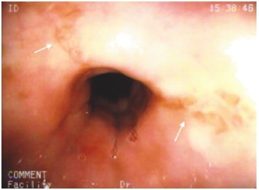
Erosões em estrela no esófago médio (setas brancas) num doente com esofagite induzida por cloxacilina

Imagem: “Cloxacillin: A New Cause of Pill-Induced Esophagitis” de Zezos P, Harel Z, Saibil F. Licença: CC BY 4.0

Prevenção e tratamento

  • Avoid the medication. 
  • Resolution expected in < 2 weeks after discontinuation.
  • If medication is necessary, take the medication with enough water and maintain an upright position for at least 30 minutes. 

Infectious Esophagitis: Cytomegalovirus (CMV)

Etiologia

  • Past CMV infection + CD4 cell count < 50 cells/microL → ↑ risk of reactivation → infection of organs such as the esophagus
  • Gastrointestinal (GI) disease from CMV is not frequent but is a serious complication of AIDS (acquired immunodeficiency syndrome).

Fatores de risco

  • Patients with AIDS (occurrence decreased since availability of antiretroviral therapy)
  • Solid-organ and bone marrow transplant recipients
  • Patients receiving immunosuppressive therapy and chemotherapy

Apresentação clínica

  • Odynophagia with dysphagia
  • Nausea, fever, substernal burning pain
  • Can have concurrent infection in retina, lung, liver, colon

Diagnóstico

O diagnóstico é feito por endoscopia digestiva alta.

  • Linear or longitudinal deep ulcerations, erosions
  • Biopsy: cytomegalic cells (with intracytoplasmic or intranuclear inclusions)
Esofagite por citomegalovírus

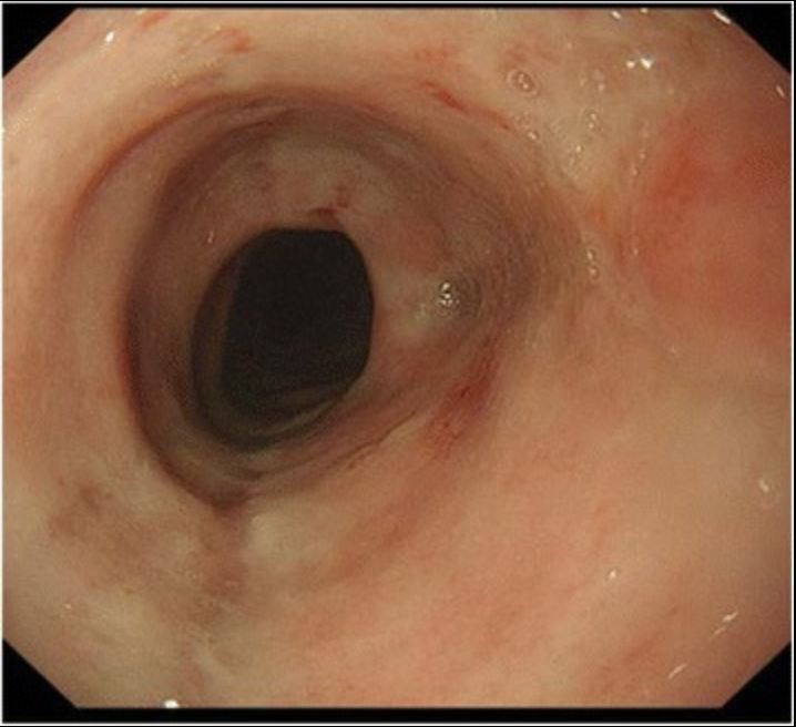
Achados na endoscopia digestiva alta de um doente com esofagite por citomegalovírus. É possível ver uma úlcera com uma base branca no esófago.

Imagem: “Cytomegalovirus esophagitis developing during chemoradiotherapy for esophageal cancer” do Journal of Medical Case Reports. Licença: CC BY 4.0

Tratamento

  • Ganciclovir or foscarnet
  • With AIDS: antiretroviral therapy

Infectious Esophagitis: Herpes Simplex Virus (HSV)

Etiologia

  • Majority are HSV type 1.
  • Can be primary infection or reactivation of HSV

Fatores de risco

  • AIDS
  • Organ or bone marrow transplant recipients
  • Patients on immunosuppressive therapy

Apresentação clínica

  • Odynophagia with dysphagia
  • Retrosternal chest pain, +/- fever, concurrent oropharyngeal ulcers

Diagnóstico

O diagnóstico é feito por endoscopia digestiva alta.

  • Early lesion: vesicles (rarely seen)
  • Punched-out ulcers, +/- plaques
  • Biopsy: HSV-infected cells with Cowdry type A inclusion bodies
  • Viral culture if no treatment response
Esofagite herpética

Endoscopia digestiva alta de um doente, que mostra numerosas lesões na superfície esofágica, de coloração amarelo-esbranquiçada, pleomórficas e pequenas placas circulares isoladas com erosões centrais.

Imagem: “Herpetic esophagitis in immunocompetent medical student” de Marinho AV, Bonfim VM, de Alencar LR, Pinto SA, de Araújo Filho JA. Licença: CC BY 3.0

Tratamento

  • Acyclovir
  • With HIV/AIDS: antiretroviral therapy

Infectious Esophagitis: Candidiasis

Etiologia

  • Candida albicans: most frequently occurring
  • Candida glabrata, Candida krusei: less common

Fatores de risco

  • Human immunodeficiency virus (HIV) with CD4 cell count < 100 cells/μL
  • Hematologic malignancy
  • Hematopoietic cell transplant recipient
  • Patients under cytotoxic chemotherapy
  • Possible in patients on chronic inhaled corticosteroids

Apresentação clínica

  • Odynophagia with dysphagia
  • May or may not have oral thrush

Diagnóstico

O diagnóstico é feito por endoscopia digestiva alta.

  • White pseudomembrane or plaque lesions
  • Biopsy: yeasts and hyphae in mucosal cells
  • Culture shows Candida
Desconforto epigástrico causado por candidíase esofágica

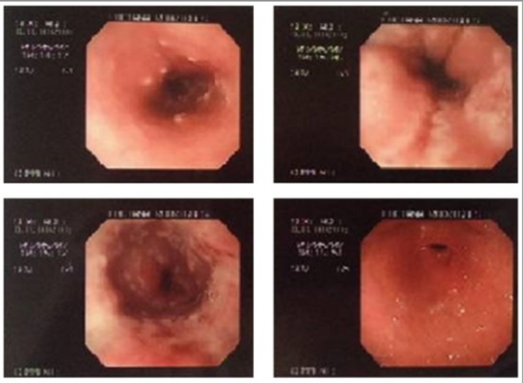
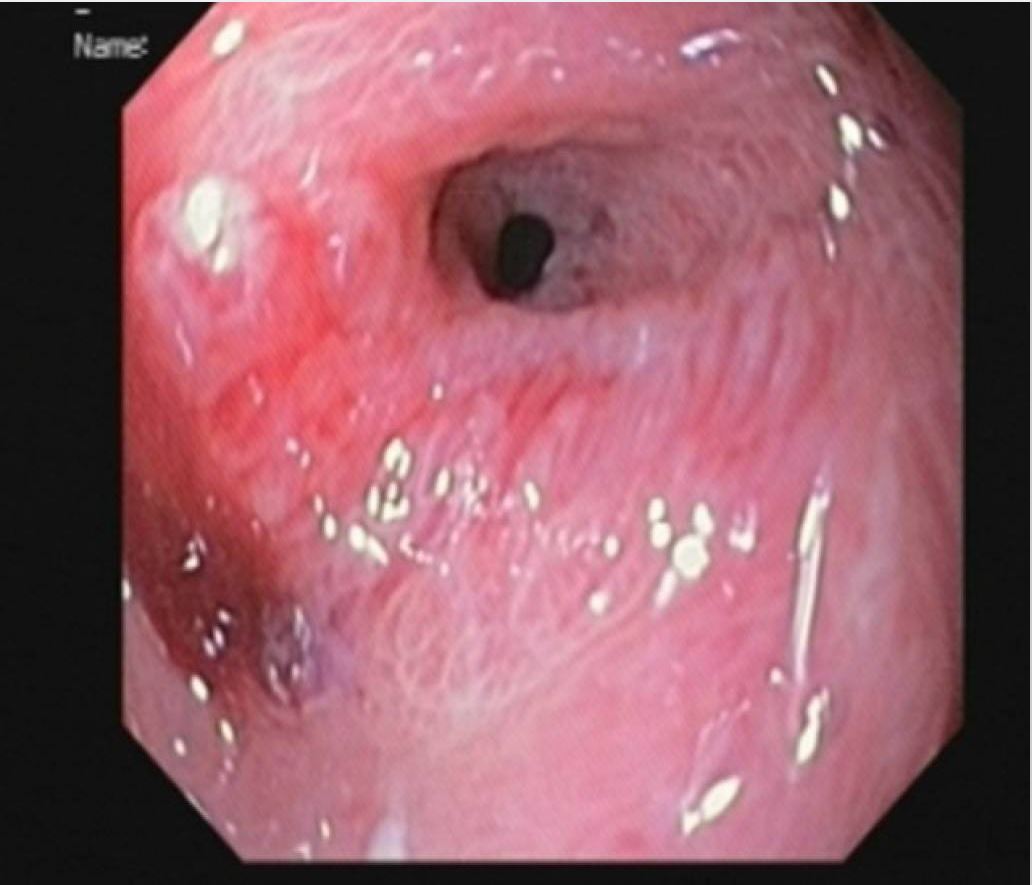
A endoscopia digestiva alta mostra lesões brancas difusas no esófago, características de esofagite por Candida .

Imagem: “Epigastric Distress Caused by Esophageal Candidiasis” de Chen KH, Weng MT, Chou YH, Lu YF, Hsieh CH. Licença: CC BY 4.0

Tratamento

  • Candida: oral fluconazole (nystatin for concurrent oral thrush)
  • With HIV/AIDS: antiretroviral therapy

Eosinophilic Esophagitis

Etiologia

  • Allergic disorder associated with:
    • Antigen sensitization through foods or aeroallergens
    • Eosinophilia and esophageal dysfunction
  • Note: Eosinophils are not normally found in the esophagus.

Fatores de risco

  • Asthma
  • Atopy
  • Allergies

Epidemiologia

  • Men > women
  • Common age of presentation: 20–30 years
  • Caucasians are more affected.

Fisiopatologia

  • Eosinophils are recruited on exposure to inhaled or congested allergens.
  • Eosinophilic infiltration occurs → eosinophils release interleukins → inflammatory response

Apresentação clínica

  • Dysphagia (to solid foods)
  • Food impaction
  • Odynophagia

Diagnóstico

O diagnóstico é feito por endoscopia digestiva alta.

  • Mucosal rings in the esophagus
  • Biopsy showing eosinophils (> 15/HPF (high-power field)) 
  • Other features:
    • Esophageal eosinophilia persists even with proton pump inhibitor (PPI) intake.
    • Normal pH monitoring
Esofagite eosinofílica

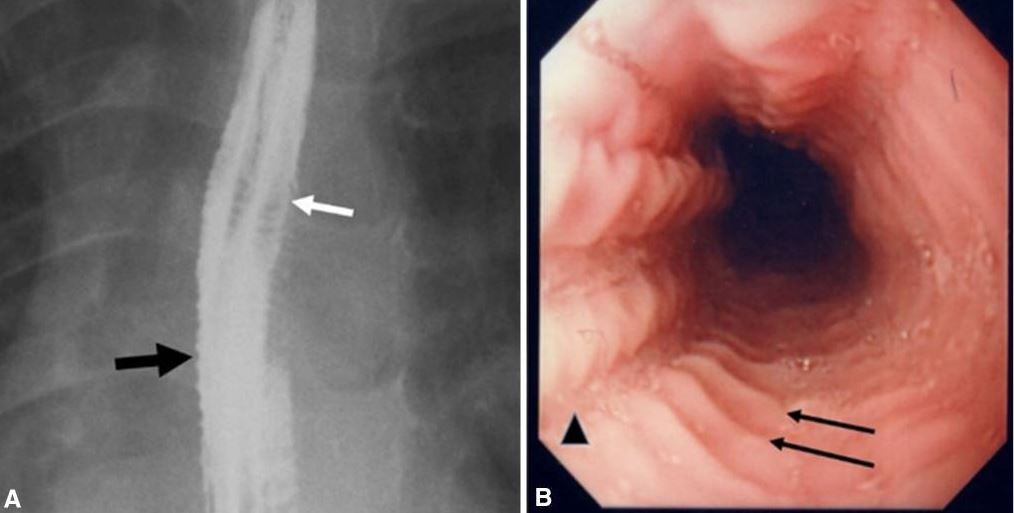
Images of eosinophilic esophagitis:
A. Esophagram in a patient with history of recurrent food impactions and dysphagia shows multiple esophageal rings (white arrow), giving the appearance of a corrugated or ringed esophagus, and mucosal irregularity (black arrow).
B. Endoscopy shows multiple transverse rings (arrows) and mucosal furrowing (arrowhead).

Image: “Eosinophilic esophagitis” by Al-Hussaini A, AboZeid A, Hai A. License: CC BY 4.0

Tratamento

  • Referral to an allergist for guidance (dietary therapy)
  • Avoid known food allergens.
  • Medication therapy:
    • Topical glucocorticoids (e.g., swallowed fluticasone)
    • Systemic glucocorticoids if with significant dysphagia, weight loss, dehydration
    • PPI for reflux symptoms

Corrosive Esophagitis

Etiologia

  • Alkali:
    • Drain cleaners, household cleaning products, batteries, bleaches
    • Viscous, tasteless, colorless
  • Acids:
    • Battery fluid, toilet bowl cleaners, metal-cleaning liquids, anti-rust solutions
    • Unpleasant taste, malodorous

Fatores de risco

  • Intentional (suicidality, underlying psychiatric illness)
  • Accidental (usually in children)

Fisiopatologia

  • Alkali-induced injury:
    • Rapid damage, affecting esophagus more than the stomach
    • Large amounts, however, result in gastric injury.
    • Process: liquefactive necrosis in the esophagus
  • Acid-induced injury:
    • Pain on contact with the oropharynx limits the amount ingested.
    • More oropharyngeal and airway damage than with alkali solutions
    • Acid passes down faster, causing more stomach damage.
    • Process: superficial coagulation necrosis in the esophagus

Complicações das lesões graves

  • Perforation
  • Bleeding
  • Mediastinitis
  • Obstruction
  • Strictures
  • Fistulas
  • Laryngeal and tracheobronchial damage from aspiration
  • Increased risk for squamous cell cancer (SCC) of the esophagus

Apresentação clínica

  • Oropharyngeal, retrosternal, or epigastric pain
  • Odynophagia and/or dysphagia
  • Hypersalivation
  • Stridor or wheezing from burning of the larynx
  • Dyspnea
  • Nausea/vomiting
  • Severe retrosternal pain (perforation and mediastinitis)
  • Abdominal tenderness and rigidity (perforation and peritonitis)

Diagnóstico

  • History: Note type and amount of ingested agent.
  • X-rays:
    • Chest: check for pneumomediastinum, aspiration pneumonia, foreign body (e.g., battery)
    • Abdominal: check for pneumoperitoneum, foreign body 
  • Computed tomography (CT) scan: checks depth of necrosis and helps assess need for emergency surgery
  • Upper endoscopy:
    • Within 24 hours if without contraindications 
    • Contraindicated in hemodynamic instability, gastrointestinal perforation
Estenose pilórica esofagite

Achados na endoscopia digestiva alta de uma criança que ingeriu uma substância alcalina: estenose pilórica desenvolvida 2 meses após a lesão.

Imagem: “Pyloric stenosis after 2 months” de Dehghani SM, Aldaghi M, Javaherizadeh H. Licença: CC BY 3.0

Classificação da lesão

  • 1st-degree injury: superficial mucosa affected; erythema, edema, hemorrhage, with healing expected
  • 2nd-degree injury: ulcers, exudates affect up to submucosal layer; scarring and strictures possible
  • 3rd-degree injury: transmural in-depth, with deep ulcers and perforation of the wall

Tratamento

  • Airway protection; assess need for intubation
  • Fluid resuscitation
  • NPO (nothing by mouth)
  • No nasogastric insertion, no emetics
  • PPIs
  • Antibiotics for suspected perforation
  • Evaluate for surgical indications
  • Surveillance: upper endoscopy 15–20 years later to screen for SCC

Esofagite de Refluxo

A esofagite de refluxo é também conhecida como doença de refluxo gastroesofágico (DRGE).

Etiologia e Fisiopatologia

  • Retrograde flow of stomach acid into the esophagus due to transient relaxation of the lower esophageal sphincter (LES) 
  • Most episodes of relaxation are triggered by gastric distention.
  • Some have incompetent LES.
  • Toxic substances (gastric acid, pepsin, bile salts) cause damage to the distal esophageal mucosa.

Fatores de risco

  • Smoking
  • Alcohol consumption
  • Obesity
  • Hiatal hernia
  • Stress
  • Scleroderma
  • Zollinger-Ellison syndrome causing increased acid secretion

Apresentação clínica

  • Retrosternal “heartburn” or pyrosis
    • Worsens when postprandial or lying down
    • May cause awakening from sleep
    • May be exacerbated by emotional stress
  • Regurgitation
  • Hoarseness
  • “Brackish” taste in the mouth
  • Globus sensation
  • Chronic, non-productive cough

Complicações

  • Erosive esophagitis
  • Barrett’s esophagus (at risk for adenocarcinoma)
  • Esophageal stricture

Diagnóstico

  • Clinical (classic signs of heartburn and regurgitation)
  • Confirmed with esophageal pH monitoring if:
    • With unsatisfactory treatment
    • With atypical symptoms
    • Antireflux surgery being considered
  • Upper endoscopy is performed if with:
    • Dysphagia
    • Unexplained weight loss
    • Anemia or evidence of gastrointestinal bleeding
    • Refractory symptoms
    • New-onset GERD > 60 years
    • Odynophagia
    • High risk for Barrett’s esophagus
    • 1st-degree relative with gastrointestinal cancer
Esofagite de refluxo

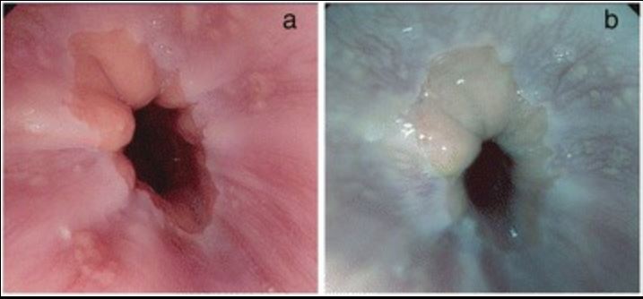
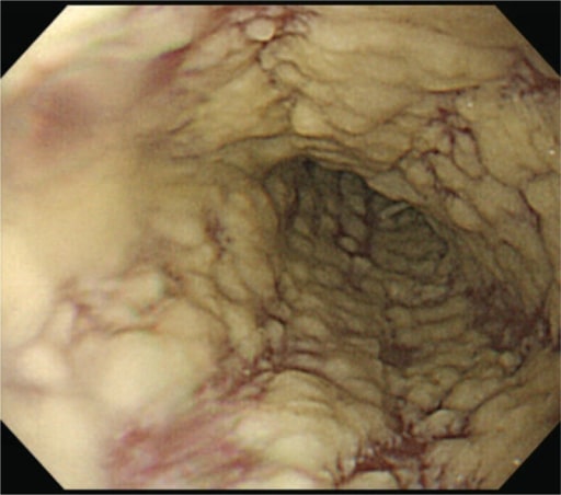
Esofagite de refluxo na endoscopia digestiva alta:
A: imagem de alta definição das alterações esofágicas na DRGE
B: imagem de DRGE com melhoria da tonalidade

Imagem: “Reflux esophagitis” de Netinatsunton N, Sottisuporn J, Attasaranya S, Witeerungrot T, Chamroonkul N, Jongboonyanuparp T, Geater A, Ovartlarnporn B. Licença: CC BY 4.0

Tratamento

  • Lifestyle modification:
    • Weight loss 
    • Elevation of the head of the bed
    • Elimination of dietary triggers
    • Smoking/alcohol cessation
  • Medication therapy: PPIs
  • Surgical options include fundoplication, which is indicated for refractory symptoms.

Referências

  1. Azer, S., Reddivari, A. (2020). Reflux esophagitis. Retrieved Nov 9, 2020, from https://www.ncbi.nlm.nih.gov/books/NBK554462
  2. Bonnis, P., Kotton, C., Hirsch, M., Mitty, J. (2020). Herpes simplex virus infection of the esophagus. UpToDate. Retrieved Nov 8, 2020, from https://www.uptodate.com/contents/herpes-simplex-virus-infection-of-the-esophagus
  3. Castell, D., Lamont, J., Grover, S. (2020). Medication-induced esophagitis. UpToDate. Retrieved 8 Nov 2020, from https://www.uptodate.com/contents/medication-induced-esophagitis
  4. De Lusong, M., Timbol, A., Tuazon, D. (2017). Management of esophageal caustic injury. World Journal of Gastrointestinal Pharmacology and Therapeutics 6; 8(2): 90–98.
  5. Greenberger, N.J. (2016). Eosinophilic esophagitis. Greenberger N.J., Blumberg R.S., Burakoff, R. (Eds.) Current Diagnosis & Treatment: Gastroenterology, Hepatology, & Endoscopy, 3rd ed. McGraw-Hill.
  6. Jacobson, M., Bartlett, J., Mitty, J. (2019). AIDS-related cytomegalovirus gastrointestinal disease. UpToDate. Retrieved Nov 8, 2020 from https://www.uptodate.com/contents/aids-related-cytomegalovirus-gastrointestinal-disease
  7. Kardon, E., Vearrier, D. (2018). Caustic ingestions. Medscape. Retrieved 8 Nov 2020, from https://emedicine.medscape.com/article/813772-overview
  8. McQuaid K.R. (2021). Caustic esophageal injury. Papadakis M.A., McPhee S.J., Rabow M.W. (Eds.) Current Medical Diagnosis & Treatment 2021. McGraw-Hill.
  9. McQuaid K.R. (2021). Gastroesophageal reflux disease. Papadakis, M.A., McPhee S.J., Rabow M.W. (Eds.) Current Medical Diagnosis & Treatment 2021. McGraw-Hill.
  10. Triadafilopoulos, G., Saltzman, J., Grover, S., Chen, W. (2019). Caustic esophageal injury in adults. UpToDate. Retrieved Nov 8, 2020 from https://www.uptodate.com/contents/caustic-esophageal-injury-in-adults

Aprende mais com a Lecturio:

Complementa o teu estudo da faculdade com o companheiro de estudo tudo-em-um da Lecturio, através de métodos de ensino baseados em evidência.

Estuda onde quiseres

A Lecturio Medical complementa o teu estudo através de métodos de ensino baseados em evidência, vídeos de palestras, perguntas e muito mais – tudo combinado num só lugar e fácil de usar.

User Reviews

Details