La hipertensión tiene muchos efectos adversos en EN Erythema nodosum is an immune-mediated panniculitis (inflammation of the subcutaneous fat) caused by a type IV (delayed-type) hypersensitivity reaction. It commonly manifests in young women as tender, erythematous nodules on the shins. Erythema Nodosum el ojo, de los LOS Neisseria cuales la retinopatía es la presentación más común. La retinopatía hipertensiva consiste en EN Erythema nodosum is an immune-mediated panniculitis (inflammation of the subcutaneous fat) caused by a type IV (delayed-type) hypersensitivity reaction. It commonly manifests in young women as tender, erythematous nodules on the shins. Erythema Nodosum alteraciones vasculares de la retina Retina The ten-layered nervous tissue membrane of the eye. It is continuous with the optic nerve and receives images of external objects and transmits visual impulses to the brain. Its outer surface is in contact with the choroid and the inner surface with the vitreous body. The outermost layer is pigmented, whereas the inner nine layers are transparent. Eye: Anatomy que se desarrollan como efecto directo de una presión arterial elevada. En EN Erythema nodosum is an immune-mediated panniculitis (inflammation of the subcutaneous fat) caused by a type IV (delayed-type) hypersensitivity reaction. It commonly manifests in young women as tender, erythematous nodules on the shins. Erythema Nodosum los LOS Neisseria aumentos agudos de la presión arterial, la autorregulación da lugar a una contracción de las arteriolas de la retina Retina The ten-layered nervous tissue membrane of the eye. It is continuous with the optic nerve and receives images of external objects and transmits visual impulses to the brain. Its outer surface is in contact with the choroid and the inner surface with the vitreous body. The outermost layer is pigmented, whereas the inner nine layers are transparent. Eye: Anatomy. En EN Erythema nodosum is an immune-mediated panniculitis (inflammation of the subcutaneous fat) caused by a type IV (delayed-type) hypersensitivity reaction. It commonly manifests in young women as tender, erythematous nodules on the shins. Erythema Nodosum la hipertensión crónica, los LOS Neisseria cambios estructurales consistentes con la arteriosclerosis afectan la vasculatura de la retina Retina The ten-layered nervous tissue membrane of the eye. It is continuous with the optic nerve and receives images of external objects and transmits visual impulses to the brain. Its outer surface is in contact with the choroid and the inner surface with the vitreous body. The outermost layer is pigmented, whereas the inner nine layers are transparent. Eye: Anatomy. Se produce un daño en EN Erythema nodosum is an immune-mediated panniculitis (inflammation of the subcutaneous fat) caused by a type IV (delayed-type) hypersensitivity reaction. It commonly manifests in young women as tender, erythematous nodules on the shins. Erythema Nodosum la pared endotelial y aparecen diversos signos como hemorragias, manchas algodonosas y exudados. En EN Erythema nodosum is an immune-mediated panniculitis (inflammation of the subcutaneous fat) caused by a type IV (delayed-type) hypersensitivity reaction. It commonly manifests in young women as tender, erythematous nodules on the shins. Erythema Nodosum los LOS Neisseria casos graves de hipertensión no controlada, se observa un papiledema. El tratamiento se centra en EN Erythema nodosum is an immune-mediated panniculitis (inflammation of the subcutaneous fat) caused by a type IV (delayed-type) hypersensitivity reaction. It commonly manifests in young women as tender, erythematous nodules on the shins. Erythema Nodosum el control de la hipertensión. Los LOS Neisseria pacientes con retinopatía hipertensiva grave tienen un mayor riesgo de sufrir una enfermedad arterial coronaria y un accidente cerebrovascular; por lo tanto, la detección y el tratamiento de la hipertensión subyacente son importantes.
Last updated: Dec 15, 2025
Retinopatía hipertensiva:
Hipertensión:
| Categoría de presión arterial | Presión arterial sistólica | Presión arterial diastólica | |
|---|---|---|---|
| Presión arterial elevada | 120-129 mm Hg | Y | <80 mm Hg |
| Hipertensión en EN Erythema nodosum is an immune-mediated panniculitis (inflammation of the subcutaneous fat) caused by a type IV (delayed-type) hypersensitivity reaction. It commonly manifests in young women as tender, erythematous nodules on the shins. Erythema Nodosum fase 1 | 130-139 mm Hg | O | 80-89 mm Hg |
| Hipertensión en EN Erythema nodosum is an immune-mediated panniculitis (inflammation of the subcutaneous fat) caused by a type IV (delayed-type) hypersensitivity reaction. It commonly manifests in young women as tender, erythematous nodules on the shins. Erythema Nodosum fase 2 | ≥ 140 mm Hg | O | ≥ 90 mm Hg |
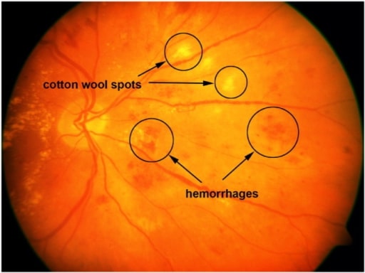
Imagen de la retina con manchas algodonosas y hemorragias.
Imagen: “A sample retinal image with cotton-wool spots and hemorrhages” por the United States National Library of Medicine. Licencia: CC BY 4.0.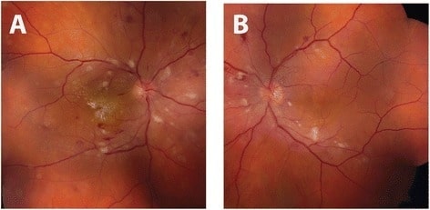
Fotografía del fondo de ojo de una retinopatía hipertensiva: Ojo derecho (A) y ojo izquierdo (B) demuestran una vasoconstricción arteriolar bilateral y manchas algodonosas con hemorragias intrarretinianas puntiformes y en forma de llamas. En el ojo derecho también se aprecian exudados intrarretinianos en forma de estrella macular.
Imagen: “Initial fundus exam” por the Department of Ophthalmology and Visual Sciences, University of Michigan, 500 S State St, Ann Arbor, MI 48109 USA. Licencia: CC BY 4.0.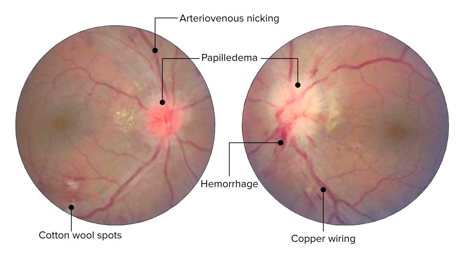
Cambios de la retina en la retinopatía hipertensiva aguda. La imagen muestra muescas arteriovenosas, cambios arteriales en hilo de cobre, hemorragias y manchas algodonosas. Edema del disco óptico bilateral presente (el izquierdo más que el derecho).
Imagen: “Central retinal vein occlusion” por Doheny Eye Institute, University of Southern California Los Angeles, CA 90033 USA. Licencia: CC BY 3.0. Editado por Lecturio.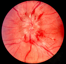
Foto que muestra el papiledema (disco óptico inflamado con márgenes de disco borrosos).
Imagen: “Papilledema” por Jonathan Trobe, MD. Licencia: CC BY 3.0.| Grado I | Vasoconstricción leve o moderada de las arteriolas de la retina Retina The ten-layered nervous tissue membrane of the eye. It is continuous with the optic nerve and receives images of external objects and transmits visual impulses to the brain. Its outer surface is in contact with the choroid and the inner surface with the vitreous body. The outermost layer is pigmented, whereas the inner nine layers are transparent. Eye: Anatomy, con una relación arteriovenosa de ≥ 1:2 |
|---|---|
| Grado II | Vasoconstricción moderada a grave de las arteriolas de la retina Retina The ten-layered nervous tissue membrane of the eye. It is continuous with the optic nerve and receives images of external objects and transmits visual impulses to the brain. Its outer surface is in contact with the choroid and the inner surface with the vitreous body. The outermost layer is pigmented, whereas the inner nine layers are transparent. Eye: Anatomy con una relación arteriovenosa <1:2 o muesca arteriovenosa |
| Grado III | Exudados blandos o hemorragias en EN Erythema nodosum is an immune-mediated panniculitis (inflammation of the subcutaneous fat) caused by a type IV (delayed-type) hypersensitivity reaction. It commonly manifests in young women as tender, erythematous nodules on the shins. Erythema Nodosum forma de llama |
| Grado IV | Edema Edema Edema is a condition in which excess serous fluid accumulates in the body cavity or interstitial space of connective tissues. Edema is a symptom observed in several medical conditions. It can be categorized into 2 types, namely, peripheral (in the extremities) and internal (in an organ or body cavity). Edema óptico bilateral |
| Grado de retinopatía | Hallazgos en EN Erythema nodosum is an immune-mediated panniculitis (inflammation of the subcutaneous fat) caused by a type IV (delayed-type) hypersensitivity reaction. It commonly manifests in young women as tender, erythematous nodules on the shins. Erythema Nodosum la retina Retina The ten-layered nervous tissue membrane of the eye. It is continuous with the optic nerve and receives images of external objects and transmits visual impulses to the brain. Its outer surface is in contact with the choroid and the inner surface with the vitreous body. The outermost layer is pigmented, whereas the inner nine layers are transparent. Eye: Anatomy | Riesgos sistémicos |
|---|---|---|
| Ninguno | No hay signos detectables | Ninguno |
| Leve |
|
Asociación moderada con el riesgo de accidente cerebrovascular clínico, accidente cerebrovascular subclínico, enfermedad coronaria y mortalidad |
| Moderado |
|
Fuerte asociación con el riesgo de accidente cerebrovascular clínico, accidente cerebrovascular subclínico, deterioro cognitivo, enfermedad coronaria y mortalidad |
| Maligno | Signos de retinopatía moderada más inflamación del disco óptico | Fuerte asociación con la mortalidad |
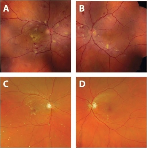
Mejora del aspecto del fondo de ojo tras el tratamiento de la hipertensión.
Fotografías del fondo de ojo tomadas en el momento de la presentación (A, B): Se observan vasoconstricciones arteriolares, hemorragias retinianas, manchas algodonosas y exudados duros (a la derecha).
Cuatro meses después del diagnóstico y tratamiento de la hipertensión sistémica (C, D): La normalización de la presión arterial dio lugar a la resolución de las hemorragias retinianas y las manchas algodonosas.
Hay una mejora de intervalo en los exudados duros en el ojo derecho con una estrella macular residual.