La región glútea se encuentra en EN Erythema nodosum is an immune-mediated panniculitis (inflammation of the subcutaneous fat) caused by a type IV (delayed-type) hypersensitivity reaction. It commonly manifests in young women as tender, erythematous nodules on the shins. Erythema Nodosum la parte posterior de la cintura pélvica y se extiende distalmente hacia la parte superior de la pierna como al AL Amyloidosis muslo posterior. La región glútea está formada por los LOS Neisseria músculos glúteos y varias arterias, venas y nervios clínicamente importantes. Los LOS Neisseria músculos de la región glútea ayudan a mover la articulación de la cadera durante la caminata, la carrera, la bipedestación y la sedestación y están especializados en EN Erythema nodosum is an immune-mediated panniculitis (inflammation of the subcutaneous fat) caused by a type IV (delayed-type) hypersensitivity reaction. It commonly manifests in young women as tender, erythematous nodules on the shins. Erythema Nodosum soportar el peso y mantener el equilibrio horizontal de la pelvis Pelvis The pelvis consists of the bony pelvic girdle, the muscular and ligamentous pelvic floor, and the pelvic cavity, which contains viscera, vessels, and multiple nerves and muscles. The pelvic girdle, composed of 2 "hip" bones and the sacrum, is a ring-like bony structure of the axial skeleton that links the vertebral column with the lower extremities. Pelvis: Anatomy.
Last updated: Dec 15, 2025
La región glútea es la zona posterior a la cintura pélvica entre la cresta ilíaca y el pliegue glúteo. La región comprende lo siguiente:
Grupos musculares:
Nervios:
Vasos:
Forámenes:
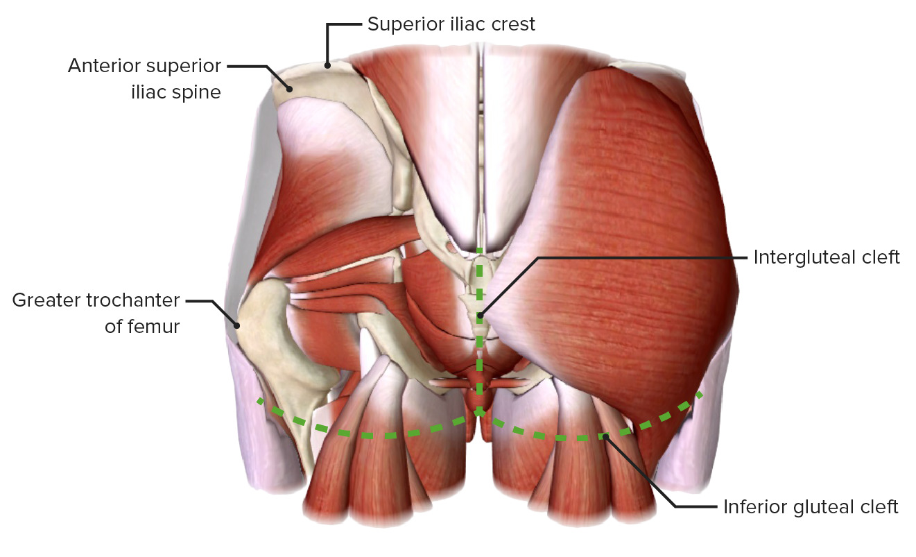
Límites de la región glútea
Imagen por BioDigital, editado por Lecturio.Los LOS Neisseria músculos de los LOS Neisseria glúteos pueden dividirse en EN Erythema nodosum is an immune-mediated panniculitis (inflammation of the subcutaneous fat) caused by a type IV (delayed-type) hypersensitivity reaction. It commonly manifests in young women as tender, erythematous nodules on the shins. Erythema Nodosum 2 grupos que son responsables de los LOS Neisseria principales movimientos de la articulación de la cadera:
| Músculo | Origen | Inserción | Inervación | Función |
|---|---|---|---|---|
| Glúteo mayor | Ilión posterior a la línea glútea posterior, sacro y cóccix posteriores, y ligamento sacrotuberoso | Tracto iliotibial (75%) y tuberosidad glútea (25%) | Nervio glúteo inferior ( S1 S1 Heart Sounds, S2 S2 Heart Sounds) |
|
| Glúteo medio | Ilio externo entre las líneas glúteas anterior y posterior | Trocánter mayor del fémur | Nervio glúteo superior (L4, L5, S1 S1 Heart Sounds) |
|
| Glúteo menor | Ilio externo entre las líneas glúteas anterior e inferior | Trocánter mayor del fémur | ||
| Tensor de la fascia lata Fascia lata Femoral Region and Hernias: Anatomy | Espina ilíaca anterosuperior | Tracto iliotibial hasta el cóndilo lateral de la tibia Tibia The second longest bone of the skeleton. It is located on the medial side of the lower leg, articulating with the fibula laterally, the talus distally, and the femur proximally. Knee Joint: Anatomy |
|

Los 3 músculos glúteos: glúteo mayor, glúteo medio y glúteo menor
Imagen por BioDigital, editado por Lecturio.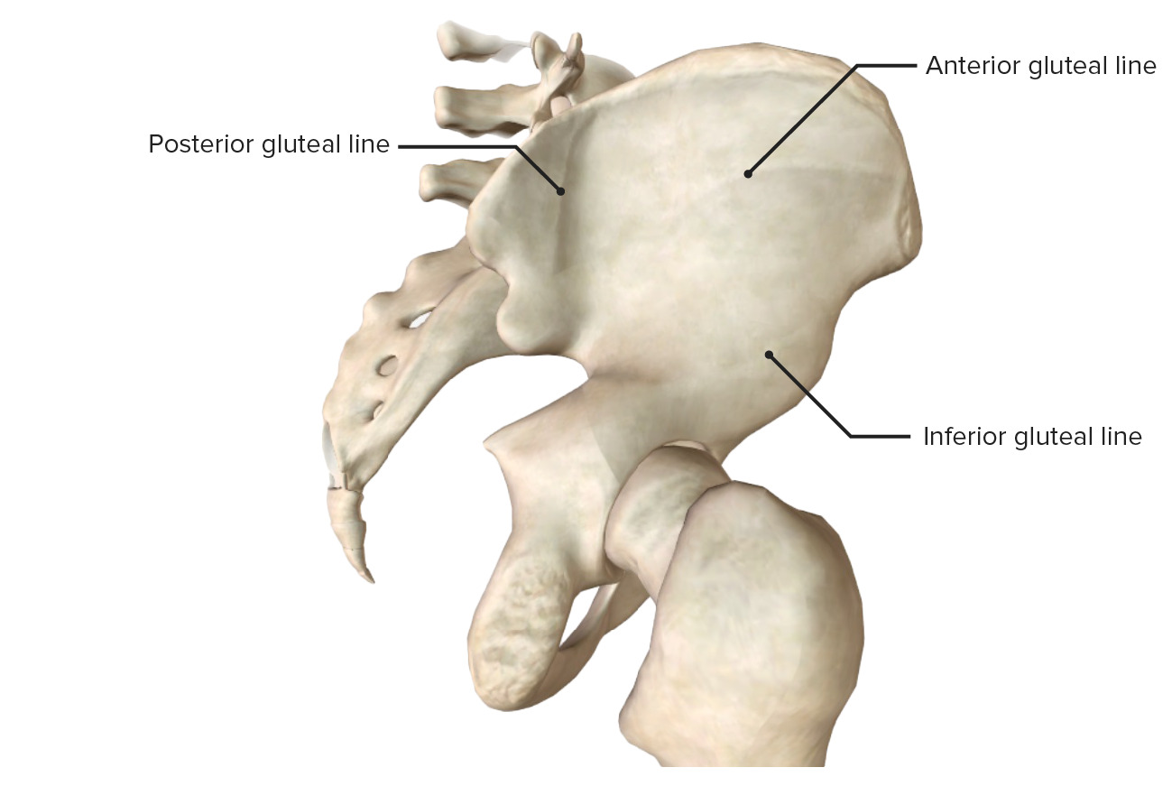
Las líneas glúteas del ilion, creadas por la inserción de los músculos glúteos
Imagen por BioDigital, editado por Lecturio.
Músculo glúteo medio: mostrando su origen e inserción en vistas posterior y lateral
Imagen por BioDigital, editado por Lecturio.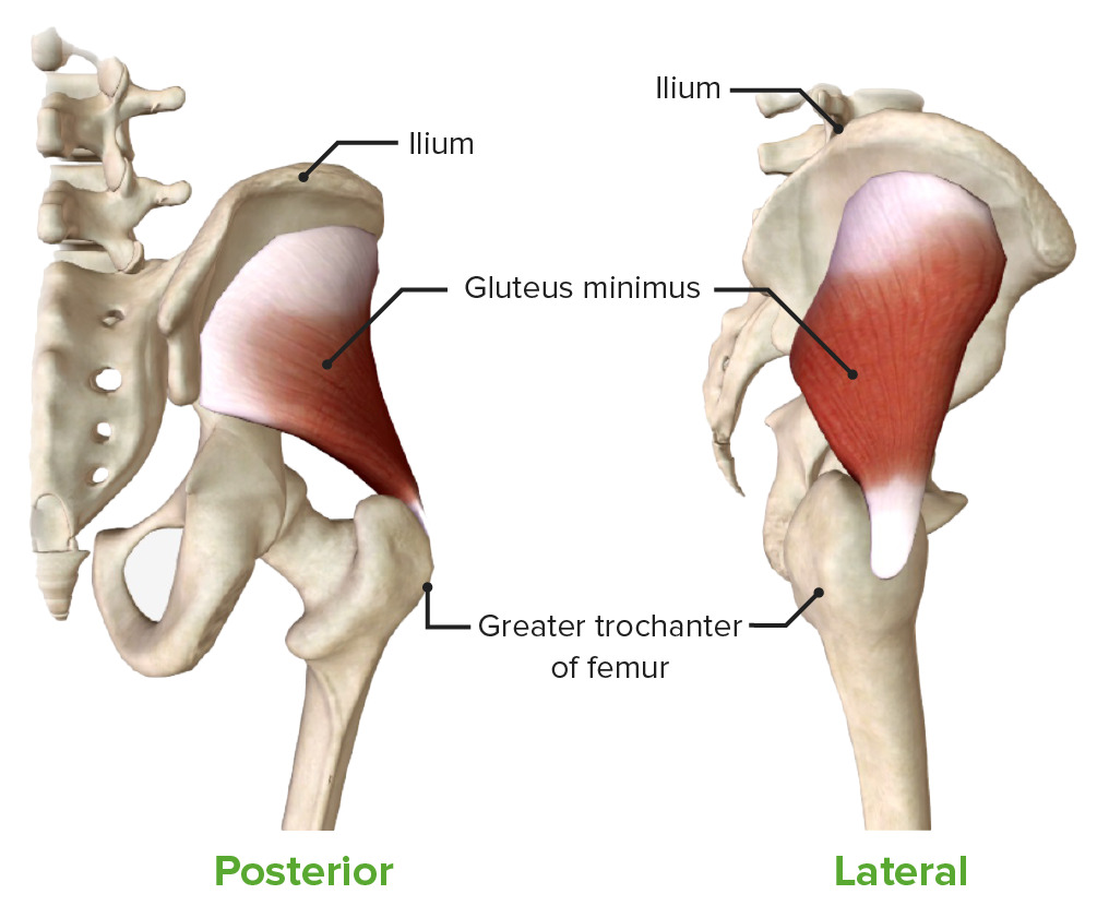
Vistas lateral y posterior del músculo glúteo medio
Imagen por BioDigital, editado por Lecturio.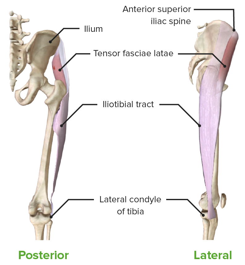
Músculo tensor de la fascia lata: mostrando su origen e inserción en vistas posterior y lateral
Imagen por BioDigital, editado por Lecturio.| Músculo | Origen | Inserción | Inervación | Función |
|---|---|---|---|---|
| Piriforme |
|
Trocánter mayor (superficie superior) | Ramas anteriores de S1 S1 Heart Sounds |
|
| Gemelos de la cadera |
|
Trocánter mayor (superficie medial) | ||
| Obturador interno |
|
Trocánter mayor (superficie medial) | ||
| Cuadrado femoral |
|
Cresta intertrocantérica | Nervio al AL Amyloidosis cuadrado femoral (L5, S1 S1 Heart Sounds) |
|
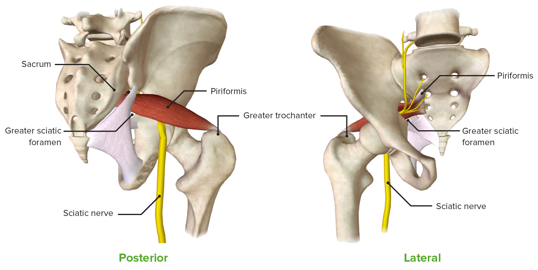
El músculo piriforme: mostrando su origen e inserción en vistas anterior y posterior, junto con su estrecha relación espacial con el nervio ciático
Imagen por BioDigital, editado por Lecturio.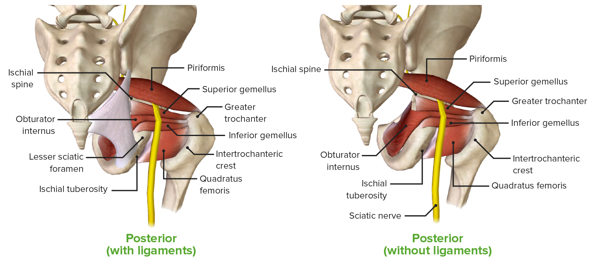
La región glútea, mostrando los músculos glúteos profundos: piriforme, obturador interno, gemelo superior, gemelo inferior y cuadrado femoral.
Imagen por BioDigital, editado por Lecturio.Los LOS Neisseria forámenes superior e inferior están formados por los LOS Neisseria siguientes ligamentos insertados en EN Erythema nodosum is an immune-mediated panniculitis (inflammation of the subcutaneous fat) caused by a type IV (delayed-type) hypersensitivity reaction. It commonly manifests in young women as tender, erythematous nodules on the shins. Erythema Nodosum la pelvis Pelvis The pelvis consists of the bony pelvic girdle, the muscular and ligamentous pelvic floor, and the pelvic cavity, which contains viscera, vessels, and multiple nerves and muscles. The pelvic girdle, composed of 2 “hip” bones and the sacrum, is a ring-like bony structure of the axial skeleton that links the vertebral column with the lower extremities. Pelvis: Anatomy ósea:
Los LOS Neisseria ligamentos sacroespinoso y sacrotuberoso crean los LOS Neisseria siguientes forámenes o pasajes:
| Foramen ciático mayor | Foramen ciático menor | |
|---|---|---|
| Límites |
|
|
| Contenido |
|
|
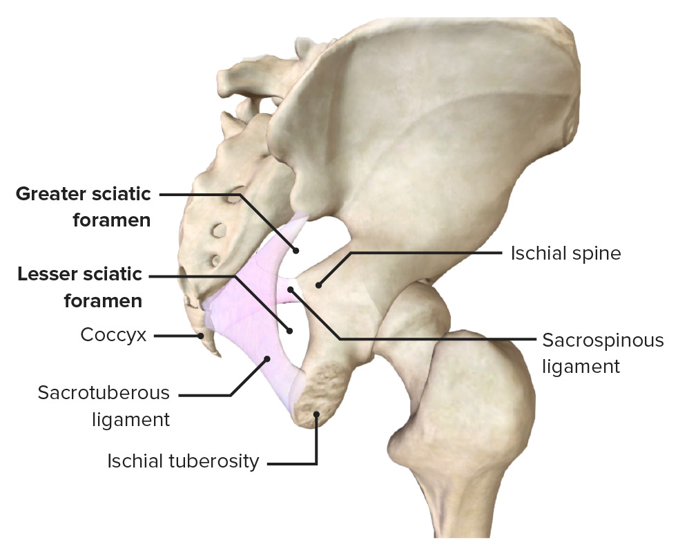
Los agujeros ciáticos mayor y menor se crean por los espacios entre los ligamentos sacroespinoso y sacrotuberoso.
Imagen por BioDigital, editado por Lecturio.Dos ramas de las arterias ilíacas internas:

Los vasos glúteos que emergen de los forámenes suprapiriforme e infrapiriforme
Imagen por BioDigital, editado por Lecturio.| Nervio | Origen | Músculos que inerva |
|---|---|---|
| Ciático | Divisiones anteriores y posteriores de las raíces nerviosas L4-S3 | |
| Glúteo superior | L4-S1 (plexo sacro) |
|
| Glúteo inferior | L5-S2 (plexo sacro) |
|
| Cutáneo femoral posterior | S1-S3 (plexo sacro) |
|
| Pudendo | S2-S4 (plexo pudendo) |
|
| Plexo sacro | L4-S4 (ramas directas) |
|
La capa profunda de la región glútea, mostrando los nervios de la región glútea
Imagen por BioDigital, editado por Lecturio.Las siguientes son clínicamente relevantes para la región glútea: