La pelvisPelvisThe pelvis consists of the bony pelvic girdle, the muscular and ligamentous pelvic floor, and the pelvic cavity, which contains viscera, vessels, and multiple nerves and muscles. The pelvic girdle, composed of 2 "hip" bones and the sacrum, is a ring-like bony structure of the axial skeleton that links the vertebral column with the lower extremities.Pelvis: Anatomy está formada por la cintura pélvica ósea, el piso pélvico muscular y ligamentoso y la cavidad pélvica, que contiene vísceras, vasos y múltiples nervios y músculos. La cintura pélvica, compuesta por 2 huesos de la “cadera” y el sacro, es una estructura ósea enENErythema nodosum is an immune-mediated panniculitis (inflammation of the subcutaneous fat) caused by a type IV (delayed-type) hypersensitivity reaction. It commonly manifests in young women as tender, erythematous nodules on the shins.Erythema Nodosum forma de anillo del esqueleto axialAxialComputed Tomography (CT) que une la columna vertebral con las extremidades inferiores. Las articulaciones del anillo pélvico incluyen la sínfisis púbica enENErythema nodosum is an immune-mediated panniculitis (inflammation of the subcutaneous fat) caused by a type IV (delayed-type) hypersensitivity reaction. It commonly manifests in young women as tender, erythematous nodules on the shins.Erythema Nodosum la parte anterior y las articulaciones sacroilíacas enENErythema nodosum is an immune-mediated panniculitis (inflammation of the subcutaneous fat) caused by a type IV (delayed-type) hypersensitivity reaction. It commonly manifests in young women as tender, erythematous nodules on the shins.Erythema Nodosum la parte posterior. LosLOSNeisseria huesos de la cadera están formados por 3 huesos fusionados: el pubis, el isquion y el ilion. La cavidad pélvica alberga varias estructuras gastrointestinales, urinarias y reproductivas, que son sostenidas por losLOSNeisseria músculos y el tejido conectivo del piso pélvico. La pelvisPelvisThe pelvis consists of the bony pelvic girdle, the muscular and ligamentous pelvic floor, and the pelvic cavity, which contains viscera, vessels, and multiple nerves and muscles. The pelvic girdle, composed of 2 "hip" bones and the sacrum, is a ring-like bony structure of the axial skeleton that links the vertebral column with the lower extremities.Pelvis: Anatomy femenina, que se adapta alALAmyloidosis parto, es generalmente más ancha y más grande que la pelvisPelvisThe pelvis consists of the bony pelvic girdle, the muscular and ligamentous pelvic floor, and the pelvic cavity, which contains viscera, vessels, and multiple nerves and muscles. The pelvic girdle, composed of 2 "hip" bones and the sacrum, is a ring-like bony structure of the axial skeleton that links the vertebral column with the lower extremities.Pelvis: Anatomy masculina.
La pelvisPelvisThe pelvis consists of the bony pelvic girdle, the muscular and ligamentous pelvic floor, and the pelvic cavity, which contains viscera, vessels, and multiple nerves and muscles. The pelvic girdle, composed of 2 “hip” bones and the sacrum, is a ring-like bony structure of the axial skeleton that links the vertebral column with the lower extremities.Pelvis: Anatomy es una estructura enENErythema nodosum is an immune-mediated panniculitis (inflammation of the subcutaneous fat) caused by a type IV (delayed-type) hypersensitivity reaction. It commonly manifests in young women as tender, erythematous nodules on the shins.Erythema Nodosum forma de anillo que rodea y protege la cavidad pélvica. La pelvisPelvisThe pelvis consists of the bony pelvic girdle, the muscular and ligamentous pelvic floor, and the pelvic cavity, which contains viscera, vessels, and multiple nerves and muscles. The pelvic girdle, composed of 2 “hip” bones and the sacrum, is a ring-like bony structure of the axial skeleton that links the vertebral column with the lower extremities.Pelvis: Anatomy está compuesta por losLOSNeisseria siguientes huesos:
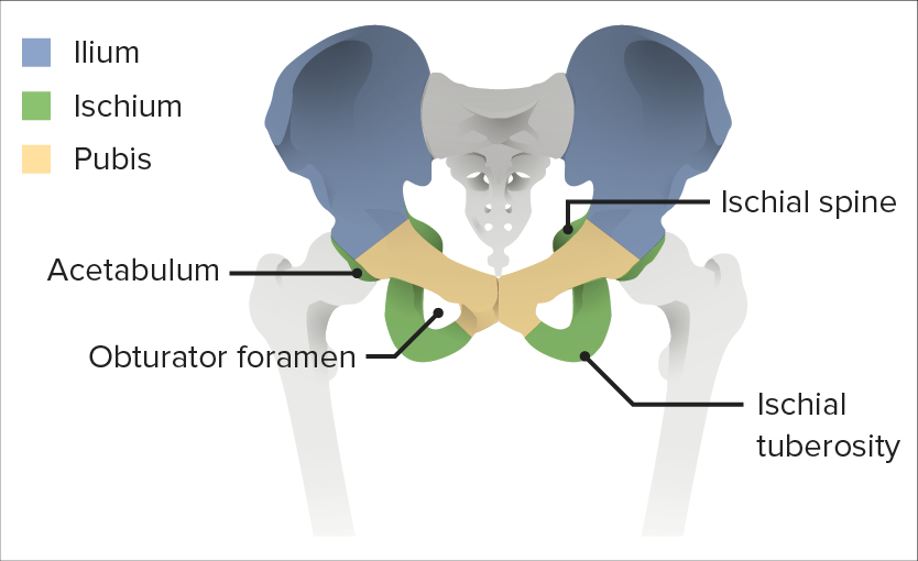
2 “huesos de la cadera”, que constan de 3 huesos cada uno; estos 3 huesos están separados alALAmyloidosis nacer, unidos por cartílago hialino y se fusionan por completo alALAmyloidosis final de la adolescencia:
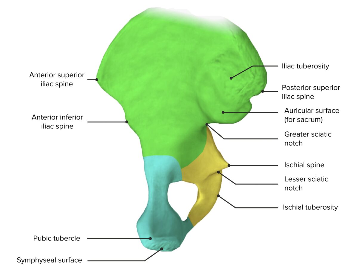
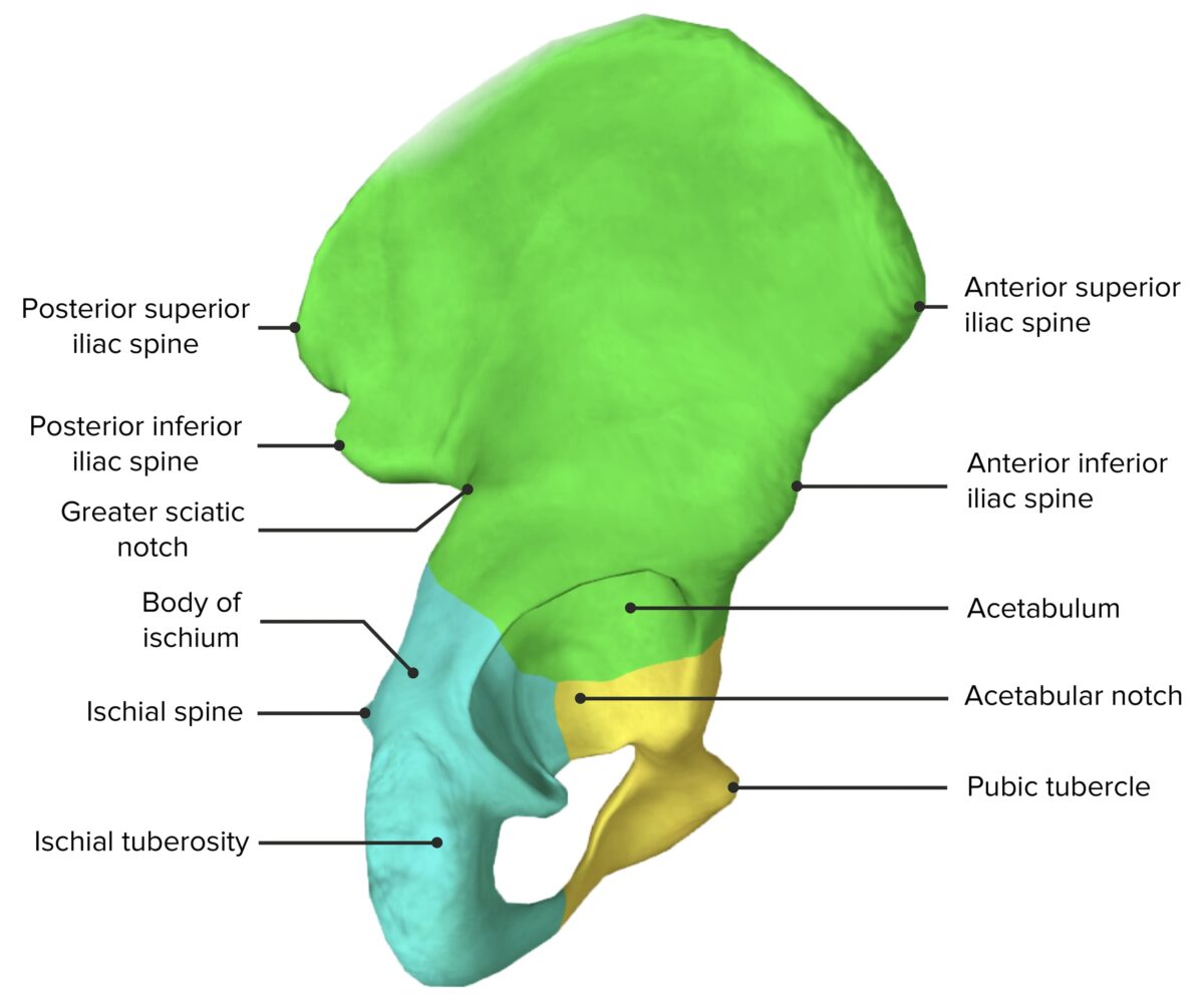
La espina iliaca anterosuperior, espina iliaca anteroinferior, cresta iliaca y la espina iliaca posterosuperior son puntos de referencia del examen físico anatómico.
Isquion:
Cara inferior de la pelvisPelvisThe pelvis consists of the bony pelvic girdle, the muscular and ligamentous pelvic floor, and the pelvic cavity, which contains viscera, vessels, and multiple nerves and muscles. The pelvic girdle, composed of 2 “hip” bones and the sacrum, is a ring-like bony structure of the axial skeleton that links the vertebral column with the lower extremities.Pelvis: Anatomy y cara posteroinferior del acetábulo
La proyección ósea inferior es la tuberosidad isquiática, que es el lugar de inserción de losLOSNeisseria isquiotibiales proximales.
Pubis:
Porción anteromedial de la pelvisPelvisThe pelvis consists of the bony pelvic girdle, the muscular and ligamentous pelvic floor, and the pelvic cavity, which contains viscera, vessels, and multiple nerves and muscles. The pelvic girdle, composed of 2 “hip” bones and the sacrum, is a ring-like bony structure of the axial skeleton that links the vertebral column with the lower extremities.Pelvis: Anatomy
Se unen enENErythema nodosum is an immune-mediated panniculitis (inflammation of the subcutaneous fat) caused by a type IV (delayed-type) hypersensitivity reaction. It commonly manifests in young women as tender, erythematous nodules on the shins.Erythema Nodosum la sínfisis púbica enENErythema nodosum is an immune-mediated panniculitis (inflammation of the subcutaneous fat) caused by a type IV (delayed-type) hypersensitivity reaction. It commonly manifests in young women as tender, erythematous nodules on the shins.Erythema Nodosum dirección anteromedial
Acetábulo:
Formado a partir de partes del ilion, isquion y pubis
Encaje enENErythema nodosum is an immune-mediated panniculitis (inflammation of the subcutaneous fat) caused by a type IV (delayed-type) hypersensitivity reaction. It commonly manifests in young women as tender, erythematous nodules on the shins.Erythema Nodosum forma de copa de la articulación de la cadera (forma la articulación acetabulofemoral con la cabeza del fémur)
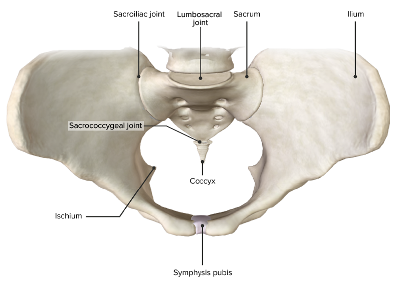
Sacro:
Hueso enENErythema nodosum is an immune-mediated panniculitis (inflammation of the subcutaneous fat) caused by a type IV (delayed-type) hypersensitivity reaction. It commonly manifests in young women as tender, erythematous nodules on the shins.Erythema Nodosum forma de mariposa formado por la fusión de las 5 vértebras sacras
Se articula con losLOSNeisseria 2 huesos de la cadera enENErythema nodosum is an immune-mediated panniculitis (inflammation of the subcutaneous fat) caused by a type IV (delayed-type) hypersensitivity reaction. It commonly manifests in young women as tender, erythematous nodules on the shins.Erythema Nodosum la parte posterior
Cada lado se articula con el ilion a través de las articulaciones sacroilíacas.
Lugar de inserción para varios músculos, tendones y ligamentos de la pelvisPelvisThe pelvis consists of the bony pelvic girdle, the muscular and ligamentous pelvic floor, and the pelvic cavity, which contains viscera, vessels, and multiple nerves and muscles. The pelvic girdle, composed of 2 “hip” bones and the sacrum, is a ring-like bony structure of the axial skeleton that links the vertebral column with the lower extremities.Pelvis: Anatomy
Vista medial del hueso de la cadera, con sus componentes: ilion (verde), isquion (amarillo) y pubis (azul)
Hay 4 articulaciones primarias dentro de la pelvisPelvisThe pelvis consists of the bony pelvic girdle, the muscular and ligamentous pelvic floor, and the pelvic cavity, which contains viscera, vessels, and multiple nerves and muscles. The pelvic girdle, composed of 2 “hip” bones and the sacrum, is a ring-like bony structure of the axial skeleton that links the vertebral column with the lower extremities.Pelvis: Anatomy:
Sacroilíacas:
Articulaciones sinoviales
Compuestas por las superficies articulares del sacro y el ilion
Transmiten peso desde la columna vertebral a losLOSNeisseria huesos de la cadera y las extremidades inferiores
Sacrococcígea:
Articulación cartilaginosa
Compuesta por la base del cóccix y la superficie ovalada enENErythema nodosum is an immune-mediated panniculitis (inflammation of the subcutaneous fat) caused by a type IV (delayed-type) hypersensitivity reaction. It commonly manifests in young women as tender, erythematous nodules on the shins.Erythema Nodosum el vértice del sacro
Sínfisis púbica:
Articulación cartilaginosa
Compuesta por las ramas superiores izquierda y derecha de losLOSNeisseria huesos púbicos
Articulación lumbosacra:
Localizada entre la columna lumbar (L5) y el sacro
Estabilizada por losLOSNeisseria ligamentos iliolumbares
Vista superior de la cintura pélvica, mostrando las 4 articulaciones primarias de la pelvis
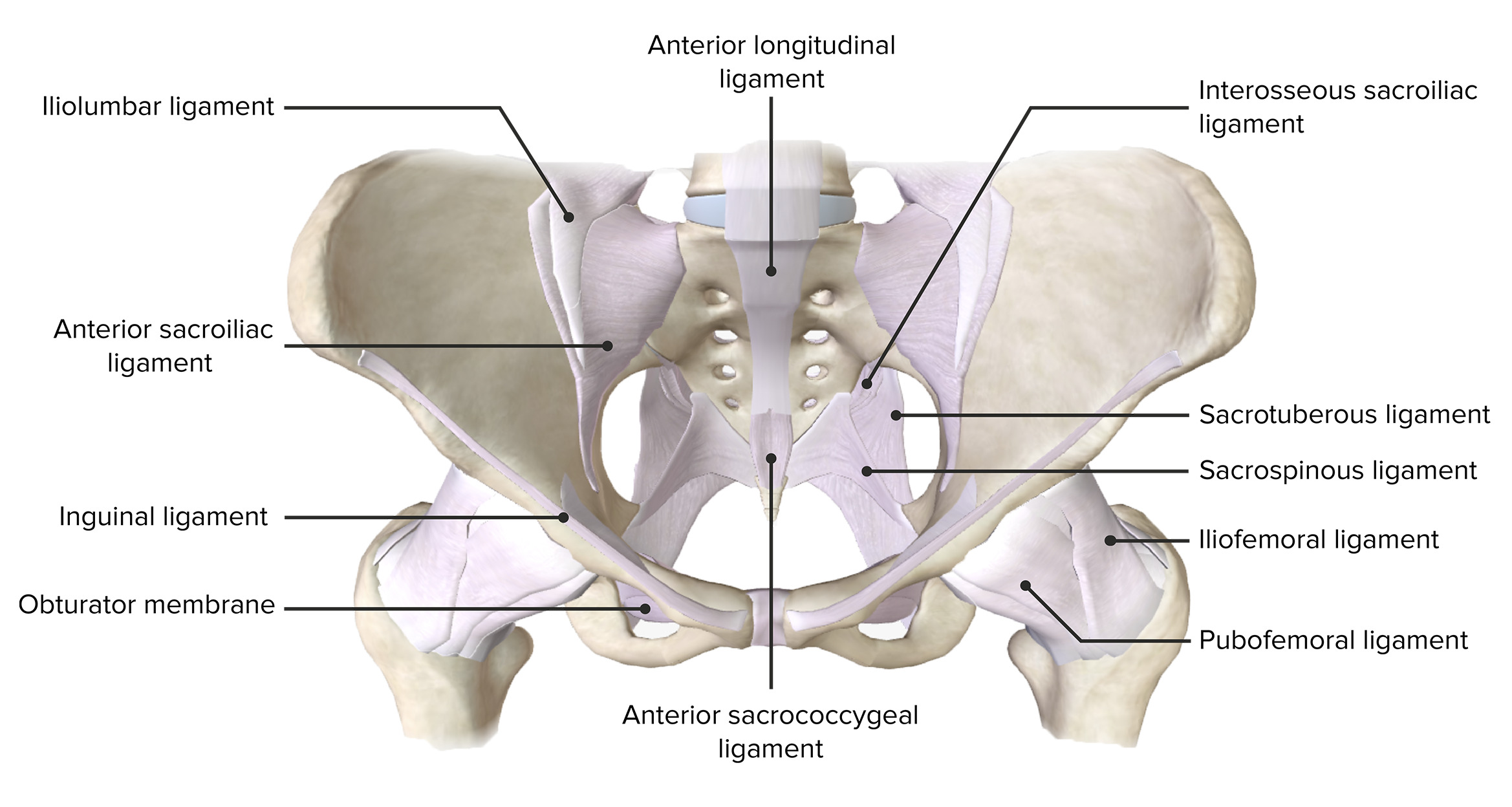
La pelvisPelvisThe pelvis consists of the bony pelvic girdle, the muscular and ligamentous pelvic floor, and the pelvic cavity, which contains viscera, vessels, and multiple nerves and muscles. The pelvic girdle, composed of 2 “hip” bones and the sacrum, is a ring-like bony structure of the axial skeleton that links the vertebral column with the lower extremities.Pelvis: Anatomy ósea está estabilizada principalmente por losLOSNeisseria siguientes ligamentos:
Ligamento sacroilíaco anterior y posterior: soporta la articulación sacroilíaca
La cavidad pélvica está delimitada por losLOSNeisseria huesos de la cintura pélvica y contiene principalmente órganos reproductores, órganos urinarios y el recto. Como la cavidad peritoneal descansa enENErythema nodosum is an immune-mediated panniculitis (inflammation of the subcutaneous fat) caused by a type IV (delayed-type) hypersensitivity reaction. It commonly manifests in young women as tender, erythematous nodules on the shins.Erythema Nodosum la cavidad pélvica, losLOSNeisseria espacios de esta región también se consideran parte del espacio de la cavidad pélvica. Mediante el uso de ligamentos y puntos de referencia óseos, la pelvisPelvisThe pelvis consists of the bony pelvic girdle, the muscular and ligamentous pelvic floor, and the pelvic cavity, which contains viscera, vessels, and multiple nerves and muscles. The pelvic girdle, composed of 2 “hip” bones and the sacrum, is a ring-like bony structure of the axial skeleton that links the vertebral column with the lower extremities.Pelvis: Anatomy se puede dividir enENErythema nodosum is an immune-mediated panniculitis (inflammation of the subcutaneous fat) caused by a type IV (delayed-type) hypersensitivity reaction. It commonly manifests in young women as tender, erythematous nodules on the shins.Erythema Nodosum varias aberturas, agujeros y espacios.
Aberturas de la pelvisPelvisThe pelvis consists of the bony pelvic girdle, the muscular and ligamentous pelvic floor, and the pelvic cavity, which contains viscera, vessels, and multiple nerves and muscles. The pelvic girdle, composed of 2 “hip” bones and the sacrum, is a ring-like bony structure of the axial skeleton that links the vertebral column with the lower extremities.Pelvis: Anatomy
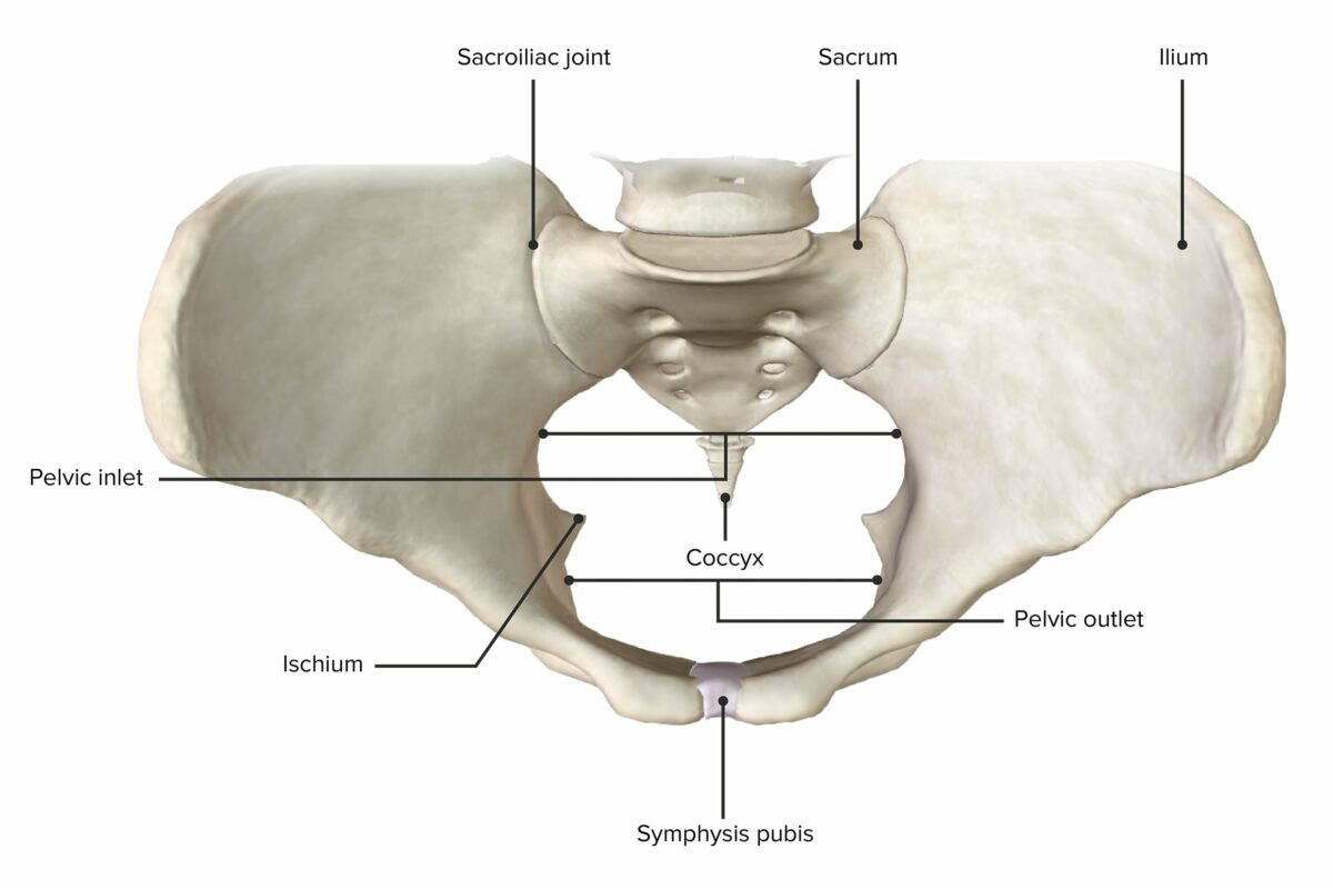
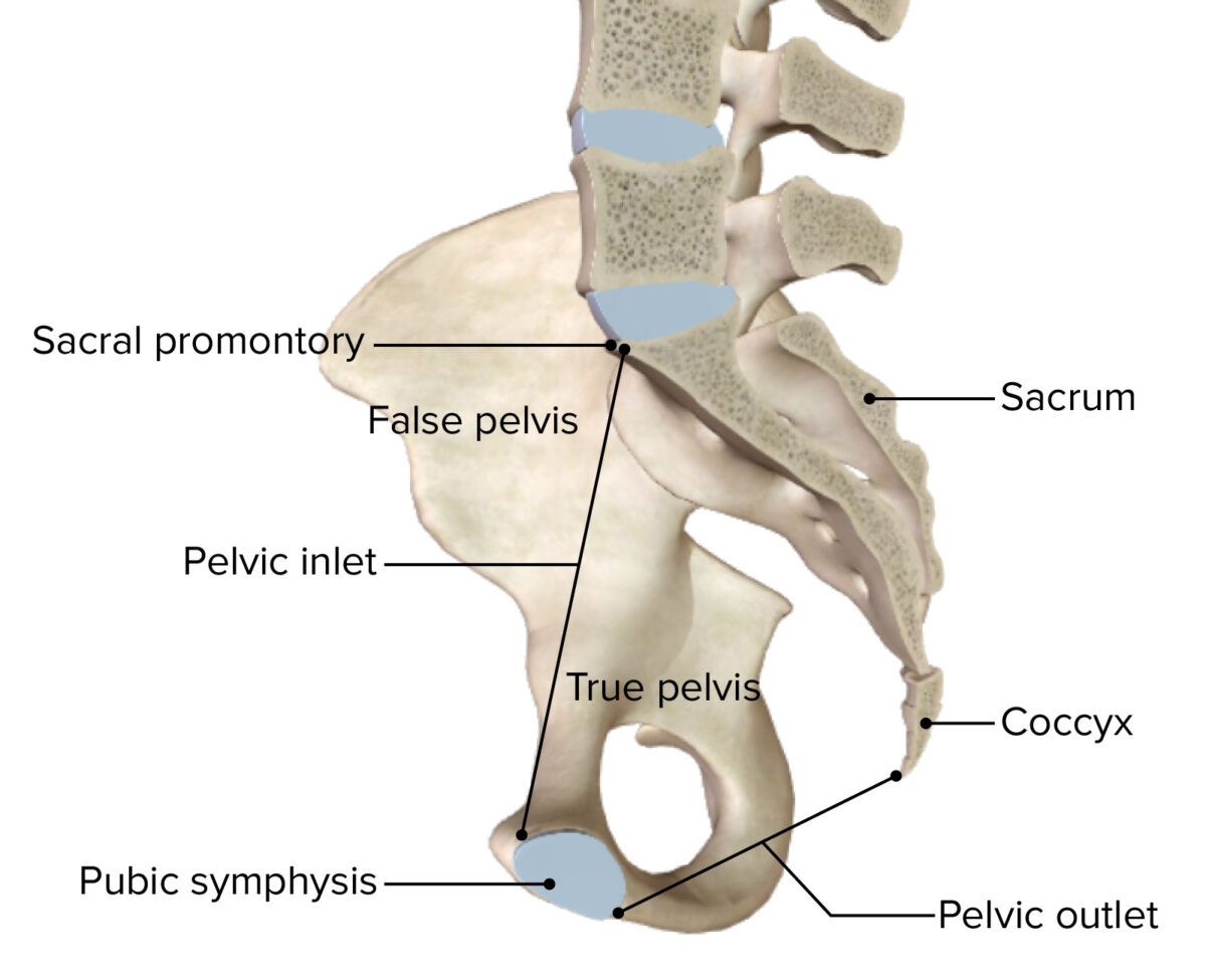
Entrada pélvica:
Abertura superior de la pelvisPelvisThe pelvis consists of the bony pelvic girdle, the muscular and ligamentous pelvic floor, and the pelvic cavity, which contains viscera, vessels, and multiple nerves and muscles. The pelvic girdle, composed of 2 “hip” bones and the sacrum, is a ring-like bony structure of the axial skeleton that links the vertebral column with the lower extremities.Pelvis: Anatomy
Límite entre las cavidades pélvica y abdominal
Salida pélvica:
Abertura inferior de la pelvisPelvisThe pelvis consists of the bony pelvic girdle, the muscular and ligamentous pelvic floor, and the pelvic cavity, which contains viscera, vessels, and multiple nerves and muscles. The pelvic girdle, composed of 2 “hip” bones and the sacrum, is a ring-like bony structure of the axial skeleton that links the vertebral column with the lower extremities.Pelvis: Anatomy
Circunferencia inferior de la pelvisPelvisThe pelvis consists of the bony pelvic girdle, the muscular and ligamentous pelvic floor, and the pelvic cavity, which contains viscera, vessels, and multiple nerves and muscles. The pelvic girdle, composed of 2 “hip” bones and the sacrum, is a ring-like bony structure of the axial skeleton that links the vertebral column with the lower extremities.Pelvis: Anatomy menor
Tabla: Aberturas de la pelvisPelvisThe pelvis consists of the bony pelvic girdle, the muscular and ligamentous pelvic floor, and the pelvic cavity, which contains viscera, vessels, and multiple nerves and muscles. The pelvic girdle, composed of 2 “hip” bones and the sacrum, is a ring-like bony structure of the axial skeleton that links the vertebral column with the lower extremities.Pelvis: Anatomy
Localización
Límites
Contenido
Entrada pélvica
Abertura pélvica superior
Anterior: línea pectínea, cresta púbica y margen superior de la sínfisis púbica
Posterior: promontorio sacro, borde anterior del ala sacra
Lateral: línea arqueada
Uréter
Cordón espermático
Ligamento redondo del útero
Ligamento suspensorio del ovario
Vasos medios del sacro
Vasos gonadales
Vasos iliolumbares
Tronco lumbosacro
Tronco simpático
Nervio obturador
Salida pélvica
Abertura pélvica inferior
Anterior: sínfisis púbica, rama isquiopúbica y ligamento púbico arqueado
Posterior: sacro y cóccix
Lateral: tuberosidades isquiáticas y ligamentos sacrotuberosos
Inferior: diafragma pélvico y urogenital
Partes terminales de losLOSNeisseria sistemas excretor, reproductivo y digestivo
Vista superior de la cintura pélvica, que muestra el diámetro horizontal más ancho de la entrada pélvica y la salida pélvica.
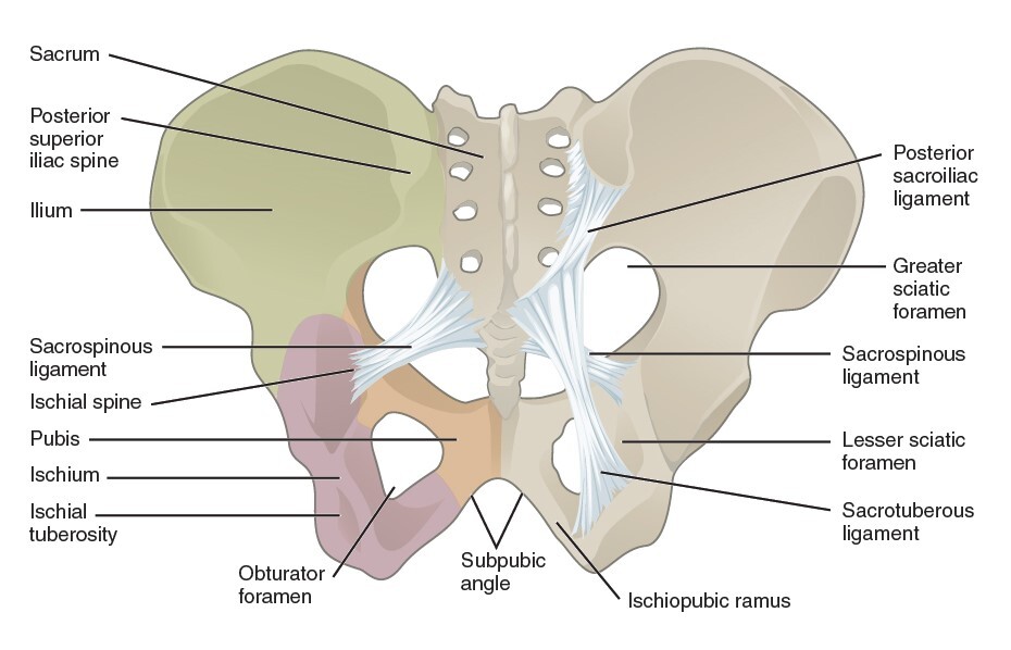
Agujeros de la pelvisPelvisThe pelvis consists of the bony pelvic girdle, the muscular and ligamentous pelvic floor, and the pelvic cavity, which contains viscera, vessels, and multiple nerves and muscles. The pelvic girdle, composed of 2 “hip” bones and the sacrum, is a ring-like bony structure of the axial skeleton that links the vertebral column with the lower extremities.Pelvis: Anatomy
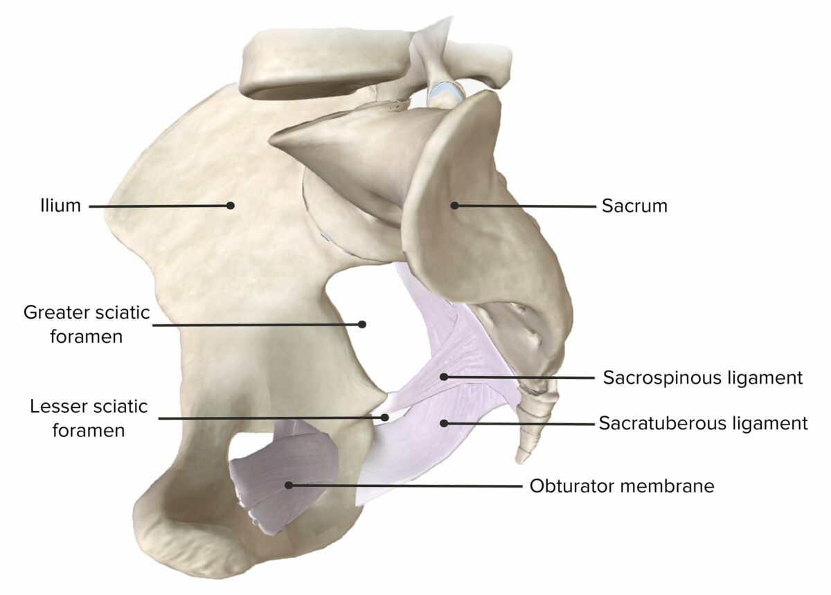
Agujero ciático mayor:
Conecta la pelvisPelvisThe pelvis consists of the bony pelvic girdle, the muscular and ligamentous pelvic floor, and the pelvic cavity, which contains viscera, vessels, and multiple nerves and muscles. The pelvic girdle, composed of 2 “hip” bones and the sacrum, is a ring-like bony structure of the axial skeleton that links the vertebral column with the lower extremities.Pelvis: Anatomy con la región de losLOSNeisseria glúteos
Separado del foramen ciático menor por el ligamento sacroespinoso
Agujero ciático menor: conecta la pelvisPelvisThe pelvis consists of the bony pelvic girdle, the muscular and ligamentous pelvic floor, and the pelvic cavity, which contains viscera, vessels, and multiple nerves and muscles. The pelvic girdle, composed of 2 “hip” bones and the sacrum, is a ring-like bony structure of the axial skeleton that links the vertebral column with the lower extremities.Pelvis: Anatomy con la parte posterior del muslo
Agujero obturador:
Conecta la pelvisPelvisThe pelvis consists of the bony pelvic girdle, the muscular and ligamentous pelvic floor, and the pelvic cavity, which contains viscera, vessels, and multiple nerves and muscles. The pelvic girdle, composed of 2 “hip” bones and the sacrum, is a ring-like bony structure of the axial skeleton that links the vertebral column with the lower extremities.Pelvis: AnatomyalALAmyloidosis muslo
Casi completamente cubierto por la membrana obturadora
Tabla: Agujeros de la pelvisPelvisThe pelvis consists of the bony pelvic girdle, the muscular and ligamentous pelvic floor, and the pelvic cavity, which contains viscera, vessels, and multiple nerves and muscles. The pelvic girdle, composed of 2 “hip” bones and the sacrum, is a ring-like bony structure of the axial skeleton that links the vertebral column with the lower extremities.Pelvis: Anatomy
Estructura
Límites
Contenido
Agujero ciático mayor
Formado por:
Ligamento sacroespinoso (se extiende desde el borde lateral del sacro y el cóccix hasta la espina isquiática)
Ligamento sacrotuberoso (se extiende desde el borde lateral del sacro y el cóccix hasta la tuberosidad isquiática)
Inferior: ligamento sacroespinoso y espina isquiática
Superior: ligamento sacroilíaco anterior
Anterolateral: escotadura ciática mayor del ilion
Posteromedial: ligamento sacrotuberoso
Músculo piriforme
Agujero suprapiriforme: vasos y nervios glúteos superiores
Agujero infrapiriforme:
Nervio pudendo
Vasos pudendos internos
Nervio ciático
Nervio y vasos glúteos inferiores
Nervio del músculo obturador interno
Nervio del músculo cuadrado femoral
Nervio cutáneo femoral posterior
Agujero ciático menor
Superior: ligamento sacroespinoso y espina isquiática
Anterior: tuberosidad isquiática
Posterior: ligamento sacrotuberoso
Nervio pudendo
Vasos pudendos internos
Tendón y nervio del músculo obturador interno
Agujero obturador
Formado por losLOSNeisseria huesos del isquion y el pubis.
Delimitado por un margen delgado (surco del obturador) alALAmyloidosis que se une la membrana del obturador, creando el canal obturador
Tubérculo obturador anterior (pubis)
Tubérculo obturador posterior (isquion)
Arteria obturatriz
Vena obturatriz
Nervio obturatriz
Ligamentos de la pelvis:
Vista anterior de la pelvis, con el agujero ciático mayor y menor y el agujero obturador
Imagen: “Ligaments of the pelvis” por OpenStax College. Licencia: CC BY 4.0
Vista lateral de la hemipelvis, con el agujero ciático mayor y menor.
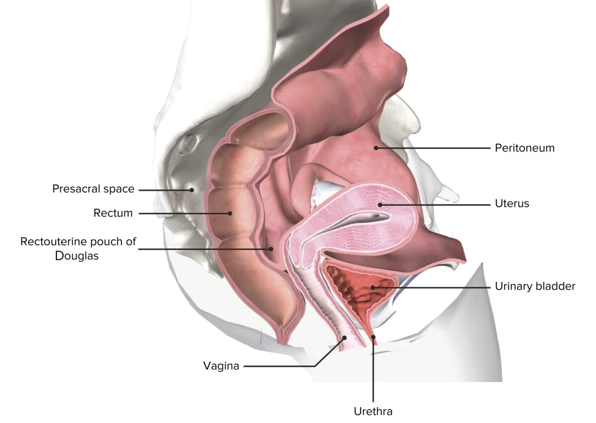
Espacios peritoneales de la pelvisPelvisThe pelvis consists of the bony pelvic girdle, the muscular and ligamentous pelvic floor, and the pelvic cavity, which contains viscera, vessels, and multiple nerves and muscles. The pelvic girdle, composed of 2 “hip” bones and the sacrum, is a ring-like bony structure of the axial skeleton that links the vertebral column with the lower extremities.Pelvis: Anatomy
La pelvisPelvisThe pelvis consists of the bony pelvic girdle, the muscular and ligamentous pelvic floor, and the pelvic cavity, which contains viscera, vessels, and multiple nerves and muscles. The pelvic girdle, composed of 2 “hip” bones and the sacrum, is a ring-like bony structure of the axial skeleton that links the vertebral column with the lower extremities.Pelvis: Anatomy tiene múltiples espacios relacionados con el extremo caudal de la cavidad peritoneal y losLOSNeisseria pliegues peritoneales que se encuentran dentro de las vísceras:
Bolsa rectovesical:
Existe solo enENErythema nodosum is an immune-mediated panniculitis (inflammation of the subcutaneous fat) caused by a type IV (delayed-type) hypersensitivity reaction. It commonly manifests in young women as tender, erythematous nodules on the shins.Erythema Nodosum hombres
Localizada entre la parte superoposterior de la vejiga urinaria y la parte superomedial del recto
Bolsa rectouterina de Douglas:
Existe solo enENErythema nodosum is an immune-mediated panniculitis (inflammation of the subcutaneous fat) caused by a type IV (delayed-type) hypersensitivity reaction. It commonly manifests in young women as tender, erythematous nodules on the shins.Erythema Nodosum mujeres
Localizada entre la parte superoposterior del útero y la parte superomedial del recto
Fosa pararrectal:
Se conecta enENErythema nodosum is an immune-mediated panniculitis (inflammation of the subcutaneous fat) caused by a type IV (delayed-type) hypersensitivity reaction. It commonly manifests in young women as tender, erythematous nodules on the shins.Erythema Nodosum ambos lados con la bolsa rectovesical/rectouterina
Existe solo enENErythema nodosum is an immune-mediated panniculitis (inflammation of the subcutaneous fat) caused by a type IV (delayed-type) hypersensitivity reaction. It commonly manifests in young women as tender, erythematous nodules on the shins.Erythema Nodosum mujeres
Localizada entre la superficie posterior de la vejiga urinaria y la superficie anterior del útero
Espacio rectovesical:
Existe solo enENErythema nodosum is an immune-mediated panniculitis (inflammation of the subcutaneous fat) caused by a type IV (delayed-type) hypersensitivity reaction. It commonly manifests in young women as tender, erythematous nodules on the shins.Erythema Nodosum hombres
Localizado posterior a la porción inferior de la vejiga urinaria y la próstata, inferior a la bolsa rectovesical
Fosa isquiorrectal: de forma triangular, a ambos lados del recto inferior y del ano
Espacio retropúbico de Retzius: localizado entre la superficie posterior del pubis y la superficie anterior de la vejiga/próstata enENErythema nodosum is an immune-mediated panniculitis (inflammation of the subcutaneous fat) caused by a type IV (delayed-type) hypersensitivity reaction. It commonly manifests in young women as tender, erythematous nodules on the shins.Erythema NodosumlosLOSNeisseria hombres
Pelvis femenina seccionada que representa el útero in situ
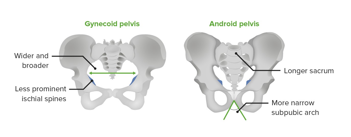
Variaciones anatómicas de la pelvisPelvisThe pelvis consists of the bony pelvic girdle, the muscular and ligamentous pelvic floor, and the pelvic cavity, which contains viscera, vessels, and multiple nerves and muscles. The pelvic girdle, composed of 2 “hip” bones and the sacrum, is a ring-like bony structure of the axial skeleton that links the vertebral column with the lower extremities.Pelvis: Anatomy ósea
Existen variaciones anatómicas entre la pelvisPelvisThe pelvis consists of the bony pelvic girdle, the muscular and ligamentous pelvic floor, and the pelvic cavity, which contains viscera, vessels, and multiple nerves and muscles. The pelvic girdle, composed of 2 “hip” bones and the sacrum, is a ring-like bony structure of the axial skeleton that links the vertebral column with the lower extremities.Pelvis: Anatomy femenina (ginecoide) y masculina (androide).
Ginecoide:
Considerada la “forma clásica de la pelvisPelvisThe pelvis consists of the bony pelvic girdle, the muscular and ligamentous pelvic floor, and the pelvic cavity, which contains viscera, vessels, and multiple nerves and muscles. The pelvic girdle, composed of 2 “hip” bones and the sacrum, is a ring-like bony structure of the axial skeleton that links the vertebral column with the lower extremities.Pelvis: Anatomy femenina”
Generalmente más ancha que su contraparte masculina, con espinas isquiáticas menos prominentes, como una adaptación para el parto
Androide: considerada la “forma clásica de la pelvisPelvisThe pelvis consists of the bony pelvic girdle, the muscular and ligamentous pelvic floor, and the pelvic cavity, which contains viscera, vessels, and multiple nerves and muscles. The pelvic girdle, composed of 2 “hip” bones and the sacrum, is a ring-like bony structure of the axial skeleton that links the vertebral column with the lower extremities.Pelvis: Anatomy masculina”
La pelvis ginecoide versus la pelvis androide
Imagen por Lecturio.
Piso Pélvico
Descripción general
El piso pélvico, también conocido como diafragma pélvico, es un grupo de músculos que sostiene las vísceras abdominales y pélvicas. El piso pélvico tiene 3 capas:
Capa superficial:
También conocida como periné o capa perineal superficial
Región enENErythema nodosum is an immune-mediated panniculitis (inflammation of the subcutaneous fat) caused by a type IV (delayed-type) hypersensitivity reaction. It commonly manifests in young women as tender, erythematous nodules on the shins.Erythema Nodosum forma de diamante entre el cóccix y la sínfisis púbica
Capa intermedia:
Diafragma pélvico
Separa el periné de la cavidad pélvica
Capa profunda:
Diafragma urogenital
Separa la pelvisPelvisThe pelvis consists of the bony pelvic girdle, the muscular and ligamentous pelvic floor, and the pelvic cavity, which contains viscera, vessels, and multiple nerves and muscles. The pelvic girdle, composed of 2 “hip” bones and the sacrum, is a ring-like bony structure of the axial skeleton that links the vertebral column with the lower extremities.Pelvis: Anatomy superior del saco perineal profundo
Tabla: Estructuras del piso pélvico
Capa superficial: periné
Capa intermedia: diafragma pélvico
Capa profunda: diafragma urogenital
Características
Región superficial enENErythema nodosum is an immune-mediated panniculitis (inflammation of the subcutaneous fat) caused by a type IV (delayed-type) hypersensitivity reaction. It commonly manifests in young women as tender, erythematous nodules on the shins.Erythema Nodosum forma de diamante entre el cóccix y la sínfisis púbica
Separa el periné de la cavidad pélvica
Separa la pelvisPelvisThe pelvis consists of the bony pelvic girdle, the muscular and ligamentous pelvic floor, and the pelvic cavity, which contains viscera, vessels, and multiple nerves and muscles. The pelvic girdle, composed of 2 “hip” bones and the sacrum, is a ring-like bony structure of the axial skeleton that links the vertebral column with the lower extremities.Pelvis: Anatomy superior del saco perineal profundo
LosLOSNeisseria músculos del piso pélvico tienen varias funciones importantes:
Brindar soporte físico a las vísceras pélvicas y prevenir el prolapso de órganos pélvicos
Mantener la continencia urinaria y fecal
Ayudar enENErythema nodosum is an immune-mediated panniculitis (inflammation of the subcutaneous fat) caused by a type IV (delayed-type) hypersensitivity reaction. It commonly manifests in young women as tender, erythematous nodules on the shins.Erythema Nodosum el parto
Tabla: Músculos del piso pélvico
Capa
Músculo
Origen
Inserción
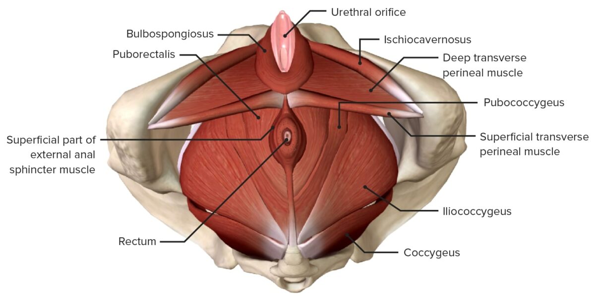
Periné
Isquiocavernoso
Tuberosidad isquiática
Raíces del pene
Bulbocavernoso/bulboesponjoso (mujeres/hombres)
Rafe medio tendinoso central del periné
Membrana perineal superficial y aponeurosis dorsal del pene/clítoris
Perineal transversal superficial
Tuberosidad isquiática
Músculo transverso perineal del lado contralateral
Esfínter anal externo
2 planos musculares aplanados rodean el ano y se unen anteriormente para insertarse enENErythema nodosum is an immune-mediated panniculitis (inflammation of the subcutaneous fat) caused by a type IV (delayed-type) hypersensitivity reaction. It commonly manifests in young women as tender, erythematous nodules on the shins.Erythema Nodosum el cuerpo perineal
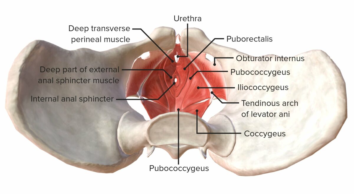
Diafragma pélvico
Grupo elevador del ano: puborrectal, pubococcígeo e iliococcígeo
Puborrectal: cuerpo del pubis (pasa alrededor de la parte inferior del recto)
Puborrectal: se encuentra con su homólogo contralateral enENErythema nodosum is an immune-mediated panniculitis (inflammation of the subcutaneous fat) caused by a type IV (delayed-type) hypersensitivity reaction. It commonly manifests in young women as tender, erythematous nodules on the shins.Erythema Nodosum la línea media
Pubococcígeo: cuerpo del pubis y arco tendinoso anterior
Pubococcígeo: coxis y ligamentos anococcígeos
Iliococcígeo: espinas isquiáticas y arco tendinoso posterior
Iliococcígeo: coxis y ligamentos anococcígeos
Coccígeo
Espina isquiática y ligamento sacroespinoso
Sacro inferior y cóccix
Piriforme
Sacro anterior y margen superior de la escotadura ciática mayor
Trocánter mayor
Obturador interno
Ramas isquiopúbicas y membrana obturatriz
Cara medial del trocánter mayor
Diafragma urogenital
Perineal transversal profundo
Ramas isquiáticas inferiores
Contraparte contralateral
Esfínter uretral
Esfínter uretral interno: vejiga inferior a la uretra proximal (extensión del músculo detrusor)
Esfínter uretral externo: rama isquiopúbica hacia el plano medio, donde se une con su músculo homólogo contralateral
Músculo compresor de la uretra
Se origina enENErythema nodosum is an immune-mediated panniculitis (inflammation of the subcutaneous fat) caused by a type IV (delayed-type) hypersensitivity reaction. It commonly manifests in young women as tender, erythematous nodules on the shins.Erythema Nodosum la rama púbica inferior y se envuelve anteriormente alrededor de la uretra para fusionarse con su contraparte contralateral
Músculos del piso pélvico desde una vista superior
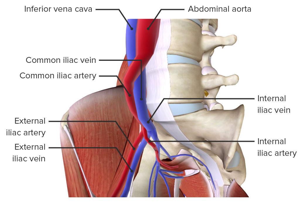
La irrigación sanguínea a la pelvisPelvisThe pelvis consists of the bony pelvic girdle, the muscular and ligamentous pelvic floor, and the pelvic cavity, which contains viscera, vessels, and multiple nerves and muscles. The pelvic girdle, composed of 2 “hip” bones and the sacrum, is a ring-like bony structure of the axial skeleton that links the vertebral column with the lower extremities.Pelvis: Anatomy proviene de:
AortaAortaThe main trunk of the systemic arteries.Mediastinum and Great Vessels: Anatomy abdominal: se bifurca enENErythema nodosum is an immune-mediated panniculitis (inflammation of the subcutaneous fat) caused by a type IV (delayed-type) hypersensitivity reaction. It commonly manifests in young women as tender, erythematous nodules on the shins.Erythema Nodosum las arterias ilíacas comunes izquierda y derecha
Arteria ilíaca común: se bifurca enENErythema nodosum is an immune-mediated panniculitis (inflammation of the subcutaneous fat) caused by a type IV (delayed-type) hypersensitivity reaction. It commonly manifests in young women as tender, erythematous nodules on the shins.Erythema Nodosum la articulación sacroilíaca hacia las arterias ilíacas internas y externas
Arteria ilíaca interna: se divide enENErythema nodosum is an immune-mediated panniculitis (inflammation of the subcutaneous fat) caused by a type IV (delayed-type) hypersensitivity reaction. It commonly manifests in young women as tender, erythematous nodules on the shins.Erythema Nodosum un tronco anterior (irriga las vísceras pélvicas) y un tronco posterior (región glútea)
Ilíaca externa:
Desprende las arterias epigástrica inferior e iliaca circunfleja profunda
Se convierte enENErythema nodosum is an immune-mediated panniculitis (inflammation of the subcutaneous fat) caused by a type IV (delayed-type) hypersensitivity reaction. It commonly manifests in young women as tender, erythematous nodules on the shins.Erythema Nodosum la arteria femoral después de pasar el ligamento inguinal
Drenaje venoso
Las 3 venas principales de la pelvisPelvisThe pelvis consists of the bony pelvic girdle, the muscular and ligamentous pelvic floor, and the pelvic cavity, which contains viscera, vessels, and multiple nerves and muscles. The pelvic girdle, composed of 2 “hip” bones and the sacrum, is a ring-like bony structure of the axial skeleton that links the vertebral column with the lower extremities.Pelvis: Anatomy siguen el curso de las arterias y se denominan enENErythema nodosum is an immune-mediated panniculitis (inflammation of the subcutaneous fat) caused by a type IV (delayed-type) hypersensitivity reaction. It commonly manifests in young women as tender, erythematous nodules on the shins.Erythema Nodosum consecuencia:
Vena ilíaca común:
Unión de las venas ilíacas externas e internas
Se une con la vena ilíaca común contralateral para convertirse enENErythema nodosum is an immune-mediated panniculitis (inflammation of the subcutaneous fat) caused by a type IV (delayed-type) hypersensitivity reaction. It commonly manifests in young women as tender, erythematous nodules on the shins.Erythema Nodosum la vena cava inferior
Vena ilíaca interna: responsable de la mayor parte del drenaje venoso pélvico
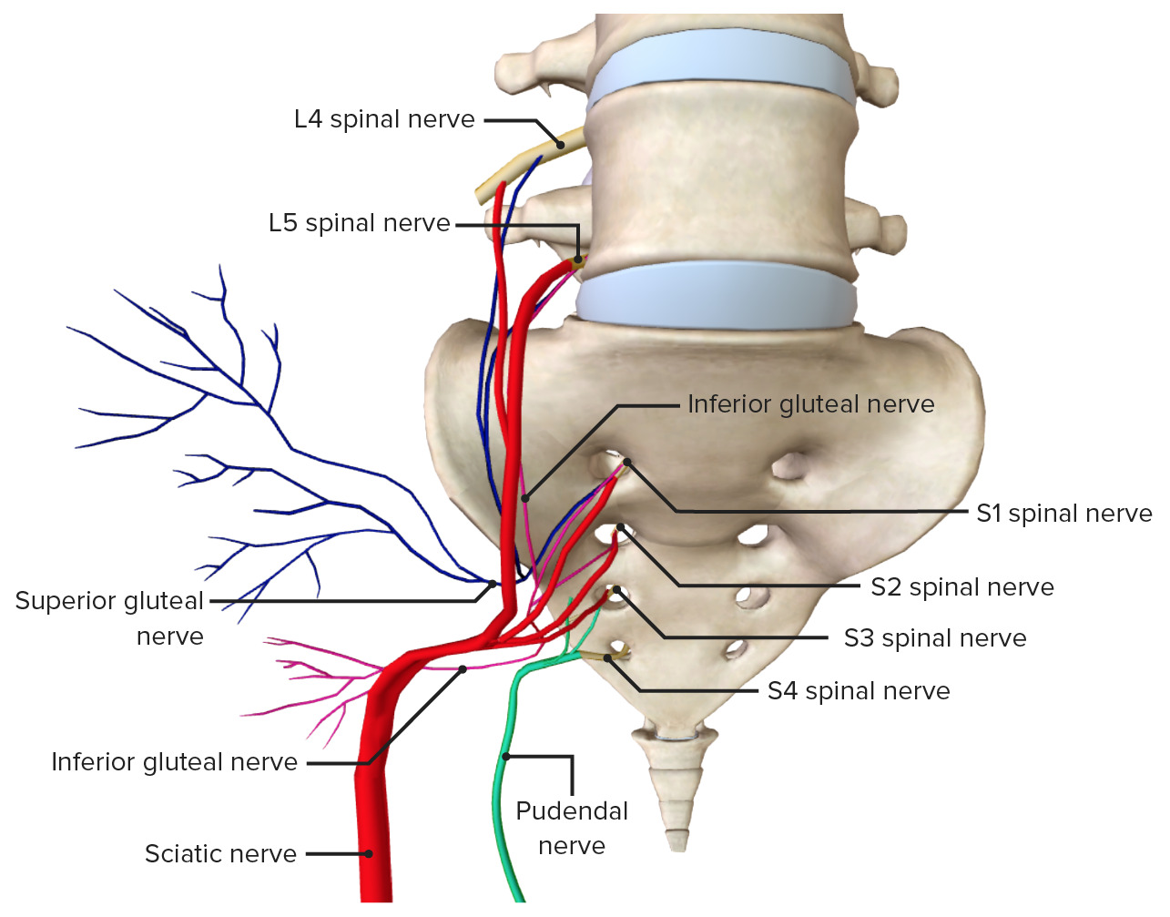
La inervación de la pelvisPelvisThe pelvis consists of the bony pelvic girdle, the muscular and ligamentous pelvic floor, and the pelvic cavity, which contains viscera, vessels, and multiple nerves and muscles. The pelvic girdle, composed of 2 “hip” bones and the sacrum, is a ring-like bony structure of the axial skeleton that links the vertebral column with the lower extremities.Pelvis: Anatomy es proporcionada por ramas del plexo sacro, incluido el nervio pudendo, que se forma a partir del plexo sacro, S2S2Heart Sounds–S4S4Heart Sounds. La pelvisPelvisThe pelvis consists of the bony pelvic girdle, the muscular and ligamentous pelvic floor, and the pelvic cavity, which contains viscera, vessels, and multiple nerves and muscles. The pelvic girdle, composed of 2 “hip” bones and the sacrum, is a ring-like bony structure of the axial skeleton that links the vertebral column with the lower extremities.Pelvis: Anatomy recibe inervación tanto somática como autónoma.
Inervación motora a:
Esfínteres uretrales y anales externos
Elevador del ano
Bulboesponjoso
Isquiocavernoso
Inervación sensorial a:
Periné
Pene/clítoris
Escroto posterior/labios
Canal anal
Reflejo de la erección:
Nervio pudendo aferente → activa el arco reflejo
Fibras parasimpáticas enENErythema nodosum is an immune-mediated panniculitis (inflammation of the subcutaneous fat) caused by a type IV (delayed-type) hypersensitivity reaction. It commonly manifests in young women as tender, erythematous nodules on the shins.Erythema Nodosum raíces S2S2Heart Sounds–S4S4Heart Sounds
Fracturas pélvicas: ocurren con mayor frecuencia enENErythema nodosum is an immune-mediated panniculitis (inflammation of the subcutaneous fat) caused by a type IV (delayed-type) hypersensitivity reaction. It commonly manifests in young women as tender, erythematous nodules on the shins.Erythema Nodosum pacientes involucrados enENErythema nodosum is an immune-mediated panniculitis (inflammation of the subcutaneous fat) caused by a type IV (delayed-type) hypersensitivity reaction. It commonly manifests in young women as tender, erythematous nodules on the shins.Erythema Nodosum lesiones por impacto de alta energía, como accidentes automovilísticos o caídas. Debido a la naturaleza de alta energía de este trauma, las fracturas pélvicas a menudo se consideran fracturas inestables y las personas pueden presentar compromiso vascular y lesiones concomitantes del sistema genitourinario. Las personas mayores con osteoporosisOsteoporosisOsteoporosis refers to a decrease in bone mass and density leading to an increased number of fractures. There are 2 forms of osteoporosis: primary, which is commonly postmenopausal or senile; and secondary, which is a manifestation of immobilization, underlying medical disorders, or long-term use of certain medications. Osteoporosis pueden presentar fracturas pélvicas después de lesiones de bajo impacto.
HerniaHerniaProtrusion of tissue, structure, or part of an organ through the bone, muscular tissue, or the membrane by which it is normally contained. Hernia may involve tissues such as the abdominal wall or the respiratory diaphragm. Hernias may be internal, external, congenital, or acquired.Abdominal Hernias obturatriz:herniaHerniaProtrusion of tissue, structure, or part of an organ through the bone, muscular tissue, or the membrane by which it is normally contained. Hernia may involve tissues such as the abdominal wall or the respiratory diaphragm. Hernias may be internal, external, congenital, or acquired.Abdominal Hernias del contenido pélvico o abdominal a través del agujero obturador. La herniaHerniaProtrusion of tissue, structure, or part of an organ through the bone, muscular tissue, or the membrane by which it is normally contained. Hernia may involve tissues such as the abdominal wall or the respiratory diaphragm. Hernias may be internal, external, congenital, or acquired.Abdominal Hernias obturatriz ocurre con mayor frecuencia enENErythema nodosum is an immune-mediated panniculitis (inflammation of the subcutaneous fat) caused by a type IV (delayed-type) hypersensitivity reaction. It commonly manifests in young women as tender, erythematous nodules on the shins.Erythema Nodosum el lado derecho, ya que el colonColonThe large intestines constitute the last portion of the digestive system. The large intestine consists of the cecum, appendix, colon (with ascending, transverse, descending, and sigmoid segments), rectum, and anal canal. The primary function of the colon is to remove water and compact the stool prior to expulsion from the body via the rectum and anal canal. Colon, Cecum, and Appendix: Anatomy sigmoide bloquea el canal obturador del lado izquierdo. Esta herniaHerniaProtrusion of tissue, structure, or part of an organ through the bone, muscular tissue, or the membrane by which it is normally contained. Hernia may involve tissues such as the abdominal wall or the respiratory diaphragm. Hernias may be internal, external, congenital, or acquired.Abdominal Hernias a menudo se presenta con obstrucción intestinal o dolorDolorInflammation lancinante enENErythema nodosum is an immune-mediated panniculitis (inflammation of the subcutaneous fat) caused by a type IV (delayed-type) hypersensitivity reaction. It commonly manifests in young women as tender, erythematous nodules on the shins.Erythema Nodosum la parte medial del muslo, que se extiende hasta la rodilla, debido a la compresión del nervio obturatriz.
Insuficiencia o disfunción del piso pélvico: el piso pélvico sostiene las vísceras abdominales y pélvicas. Esta estructura separa el periné de la cavidad pélvica y ayuda a controlar la función del esfínter de losLOSNeisseria tractos rectal, urinario y genital. El debilitamiento del tejido conectivo y/o de losLOSNeisseria músculos puede provocar insuficiencia del piso pélvico, lo que contribuye a losLOSNeisseria trastornos del piso pélvico. La disfunción del piso pélvico incluye prolapso de órganos pélvicos (e.g., prolapso del útero), incontinencia urinaria e incontinencia fecal.
Parto: el parto comienza con contracciones que conducen a una dilatación y borramiento progresivos del cuello uterino, lo que da como resultado el nacimiento del bebé y la expulsión de la placentaPlacentaA highly vascularized mammalian fetal-maternal organ and major site of transport of oxygen, nutrients, and fetal waste products. It includes a fetal portion (chorionic villi) derived from trophoblasts and a maternal portion (decidua) derived from the uterine endometrium. The placenta produces an array of steroid, protein and peptide hormones (placental hormones).Placenta, Umbilical Cord, and Amniotic Cavity a través del canal de parto de la pelvisPelvisThe pelvis consists of the bony pelvic girdle, the muscular and ligamentous pelvic floor, and the pelvic cavity, which contains viscera, vessels, and multiple nerves and muscles. The pelvic girdle, composed of 2 “hip” bones and the sacrum, is a ring-like bony structure of the axial skeleton that links the vertebral column with the lower extremities.Pelvis: Anatomy. El parto vaginal es posible debido a las diferencias anatómicas entre la pelvisPelvisThe pelvis consists of the bony pelvic girdle, the muscular and ligamentous pelvic floor, and the pelvic cavity, which contains viscera, vessels, and multiple nerves and muscles. The pelvic girdle, composed of 2 “hip” bones and the sacrum, is a ring-like bony structure of the axial skeleton that links the vertebral column with the lower extremities.Pelvis: Anatomy ósea enENErythema nodosum is an immune-mediated panniculitis (inflammation of the subcutaneous fat) caused by a type IV (delayed-type) hypersensitivity reaction. It commonly manifests in young women as tender, erythematous nodules on the shins.Erythema Nodosum hombres y mujeres.
Referencias
Bordoni, B., Sugumar, K., Leslie, S.W. (2021). Anatomy, abdomen and pelvis, pelvic floor. StatPearls. Retrieved November 9, 2021, from https://pubmed.ncbi.nlm.nih.gov/29489277/
Obtenga Medical Premium para poner a prueba sus conocimientos
Lecturio Medical Premium le brinda acceso completo a todo el contenido y las funciones
Obtenga Premium para ver todos los vídeos
Verifica tu correo electrónico para obtener una prueba gratuita.
Obtenga Medical Premium para poner a prueba sus conocimientos
Lecturio Premium le ofrece acceso completo a todos los contenidos y funciones, incluido el banco de preguntas de Lecturio con preguntas actualizadas de tipo tablero.