El examen abdominal es la parte del examen físico que evalúa el abdomen en EN Erythema nodosum is an immune-mediated panniculitis (inflammation of the subcutaneous fat) caused by a type IV (delayed-type) hypersensitivity reaction. It commonly manifests in young women as tender, erythematous nodules on the shins. Erythema Nodosum busca de signos de enfermedad. El examen abdominal consiste en EN Erythema nodosum is an immune-mediated panniculitis (inflammation of the subcutaneous fat) caused by a type IV (delayed-type) hypersensitivity reaction. It commonly manifests in young women as tender, erythematous nodules on the shins. Erythema Nodosum la inspección, auscultación, percusión y palpación. Junto con la información de los LOS Neisseria antecedentes, el médico utiliza la información obtenida en EN Erythema nodosum is an immune-mediated panniculitis (inflammation of the subcutaneous fat) caused by a type IV (delayed-type) hypersensitivity reaction. It commonly manifests in young women as tender, erythematous nodules on the shins. Erythema Nodosum la exploración física del abdomen para elaborar un diagnóstico diferencial y un plan de tratamiento para el paciente.
Last updated: Dec 15, 2025
1eros pasos:
Los LOS Neisseria componentes del examen abdominal:
En EN Erythema nodosum is an immune-mediated panniculitis (inflammation of the subcutaneous fat) caused by a type IV (delayed-type) hypersensitivity reaction. It commonly manifests in young women as tender, erythematous nodules on the shins. Erythema Nodosum un orden diferente (auscultación antes de percusión), los LOS Neisseria mismos elementos componen las otras secciones del examen físico, pero tienen diferentes grados de importancia.
El examen pélvico, genital y rectal deben complementar al AL Amyloidosis examen abdominal para un diagnóstico completo de la patología abdominal.
Anatomía:
El abdomen se divide en EN Erythema nodosum is an immune-mediated panniculitis (inflammation of the subcutaneous fat) caused by a type IV (delayed-type) hypersensitivity reaction. It commonly manifests in young women as tender, erythematous nodules on the shins. Erythema Nodosum 4 cuadrantes: superior derecho, inferior derecho, superior izquierdo e inferior izquierdo. Los LOS Neisseria cuadrantes se relacionan a órganos y estructuras específicas.
Alternativamente, el abdomen también puede dividirse en EN Erythema nodosum is an immune-mediated panniculitis (inflammation of the subcutaneous fat) caused by a type IV (delayed-type) hypersensitivity reaction. It commonly manifests in young women as tender, erythematous nodules on the shins. Erythema Nodosum 9 regiones: hipocondríaco derecho, epigástrico, hipocondríaco izquierdo, lumbar derecho, umbilical, lumbar izquierdo, ilíaco derecho, hipogástrico e ilíaco izquierdo. Estas 9 regiones se utilizan a menudo para una localización más precisa.
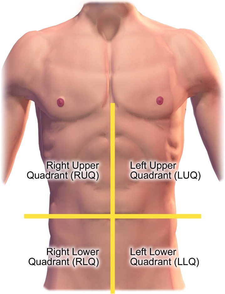
Cuadrantes del abdomen
Imagen: “Quadrants of the abdomen” por BruceBlaus. Licencia: CC BY 3.0, recortada por Lecturio.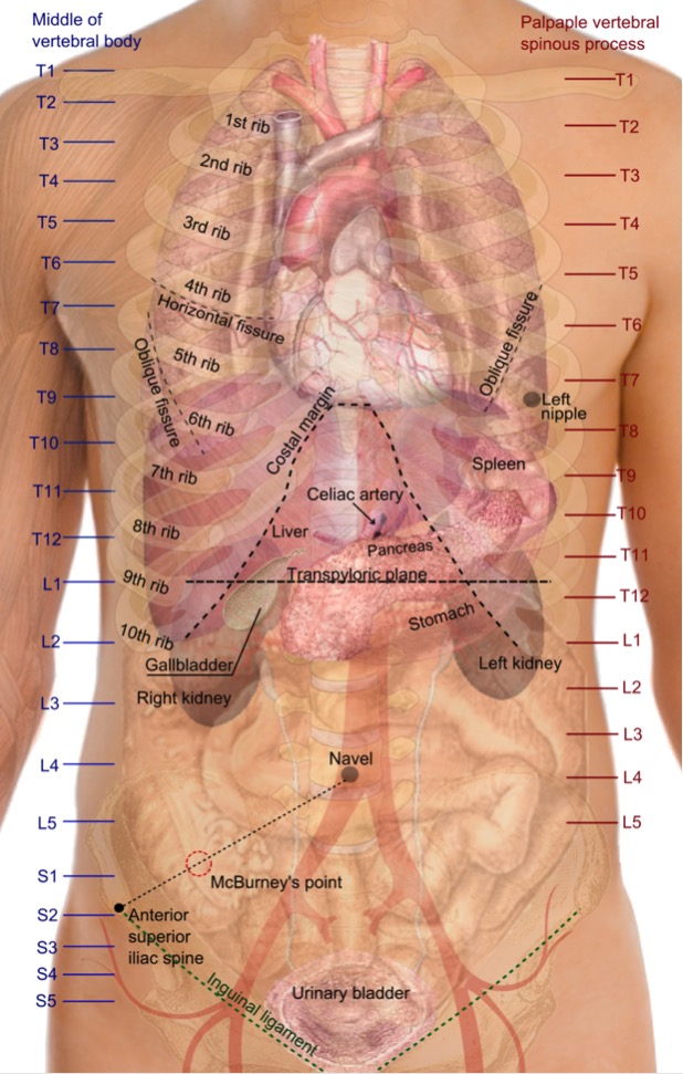
Proyecciones superficiales de los órganos del tronco: la localización de los órganos se deducen principalmente utilizando los niveles de las vértebras, las costillas y el ilium.
Imagen: “Surface projections of the organs of the trunk” por Mikael Häggström. Licencia: Dominio Público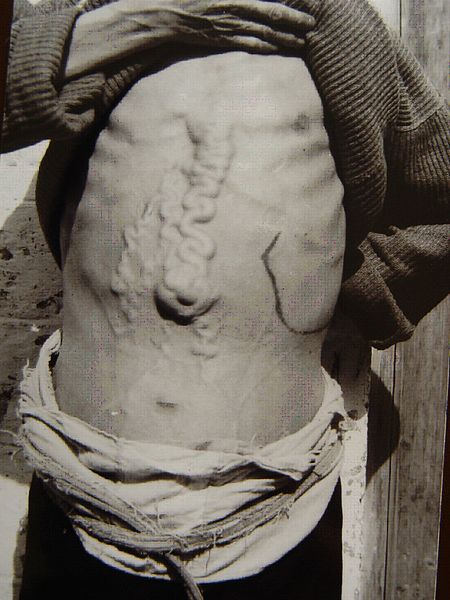
Cabeza de medusa en la hipertensión portal
Imagen: “Dilatation of abdominal collateral veins” por Chen M. Licencia: CC BY 2.0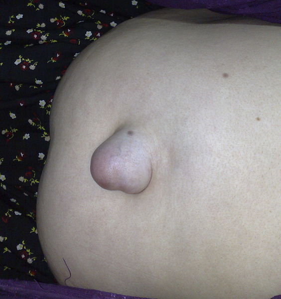
Hernia umbilical
Imagen: “Umbilical hernia” por Saltanat. Licencia: CC0 1.0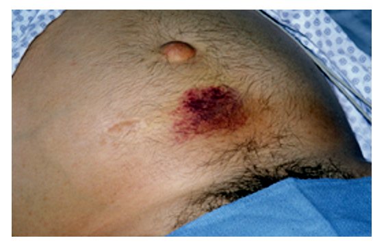
Equimosis periumbilical, compatible con el signo de Cullen (pancreatitis hemorrágica)
Imagen: “Acute pancreatitis with Cullen’s sign” por Herbert L. Fred, MD. Licencia: CC BY 2.0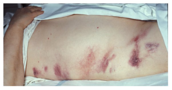
Equimosis alrededor de los flancos, compatible con el signo de Grey Turner (pancreatitis hemorrágica)
Imagen por Herbert L. Fred, MD and Hendrik A. van Dijk. Licencia: CC BY 2.0Dado que las manipulaciones mecánicas del abdomen pueden alterar la regularidad de los LOS Neisseria sonidos intestinales, la auscultación se realiza antes de la percusión o la palpación.
Pasos:
Hallazgos:
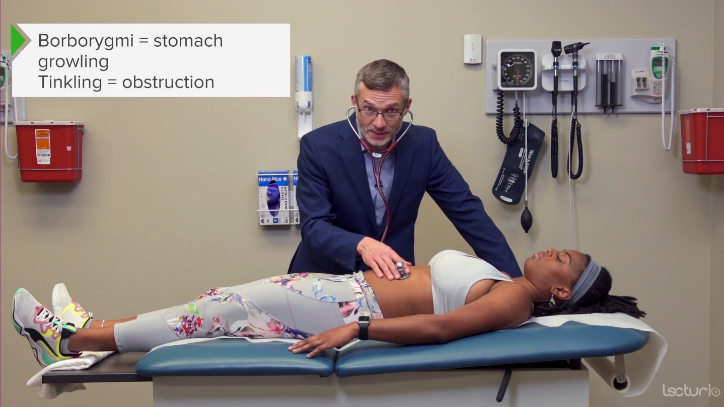
Auscultación del abdomen: se escucha en busca de ruidos intestinales
Imagen por Lecturio. Licencia: CC BY-NC-SA 4.0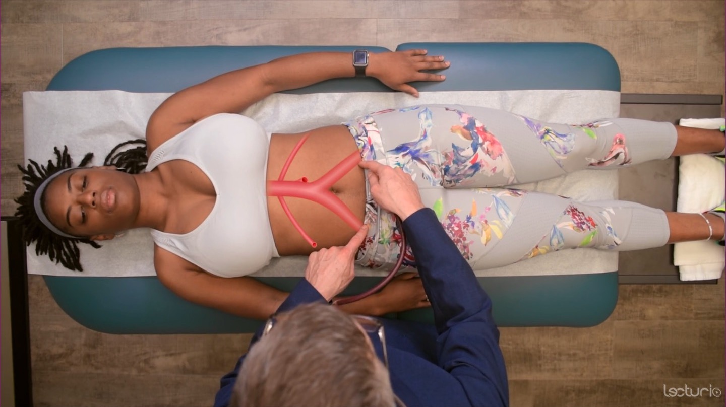
Auscultación del abdomen: se escucha en busca de soplos sobre las arterias aorta, ilíacas y renales
Imagen por Lecturio. Licencia: CC BY-NC-SA 4.0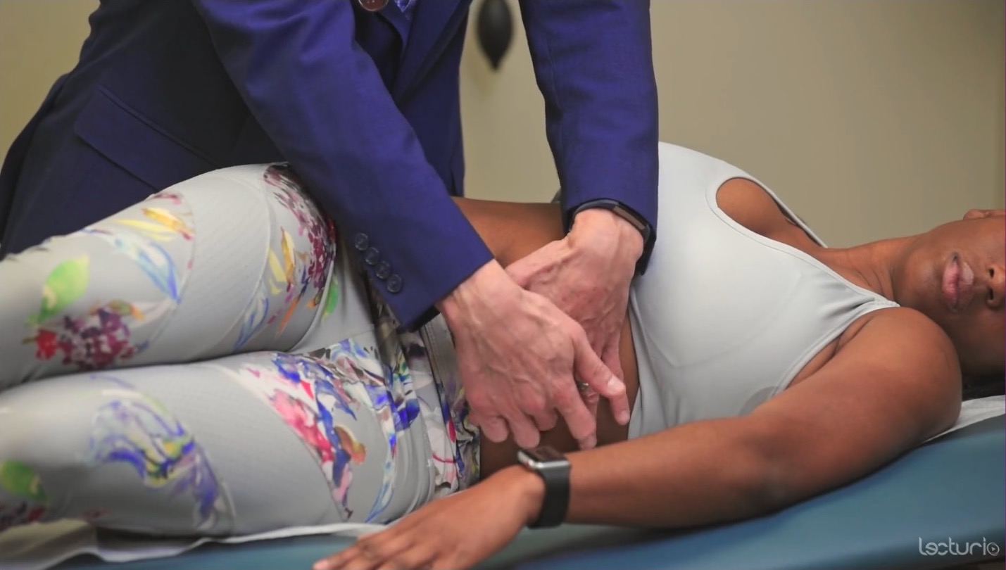
Evaluación de la matidez cambiante
Imagen por Lecturio. Licencia: CC BY-NC-SA 4.0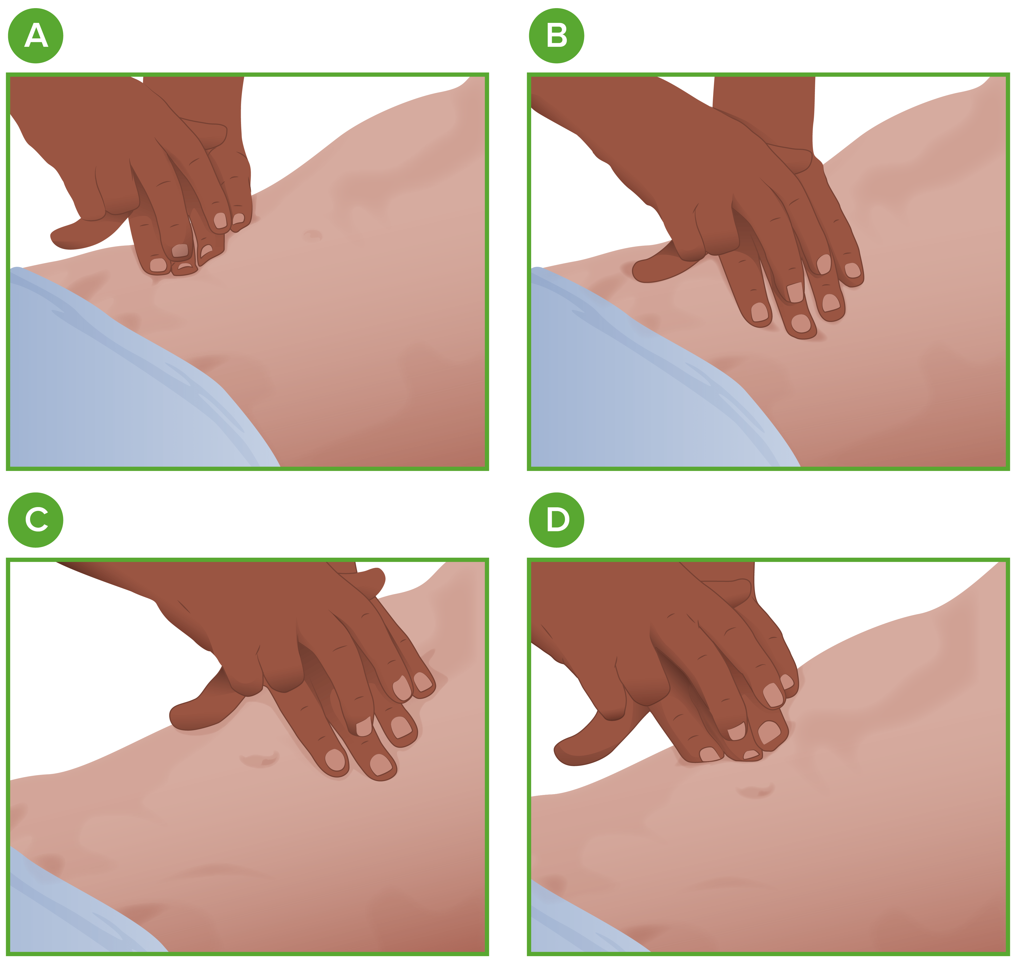
Percusión en el examen abdominal:
El examinador debe percutir los 4 cuadrantes (A, B, C y D) con 1 dedo medio firmemente sobre la pared abdominal del individuo y el 2do dedo medio golpeando la articulación interfalángica distal.
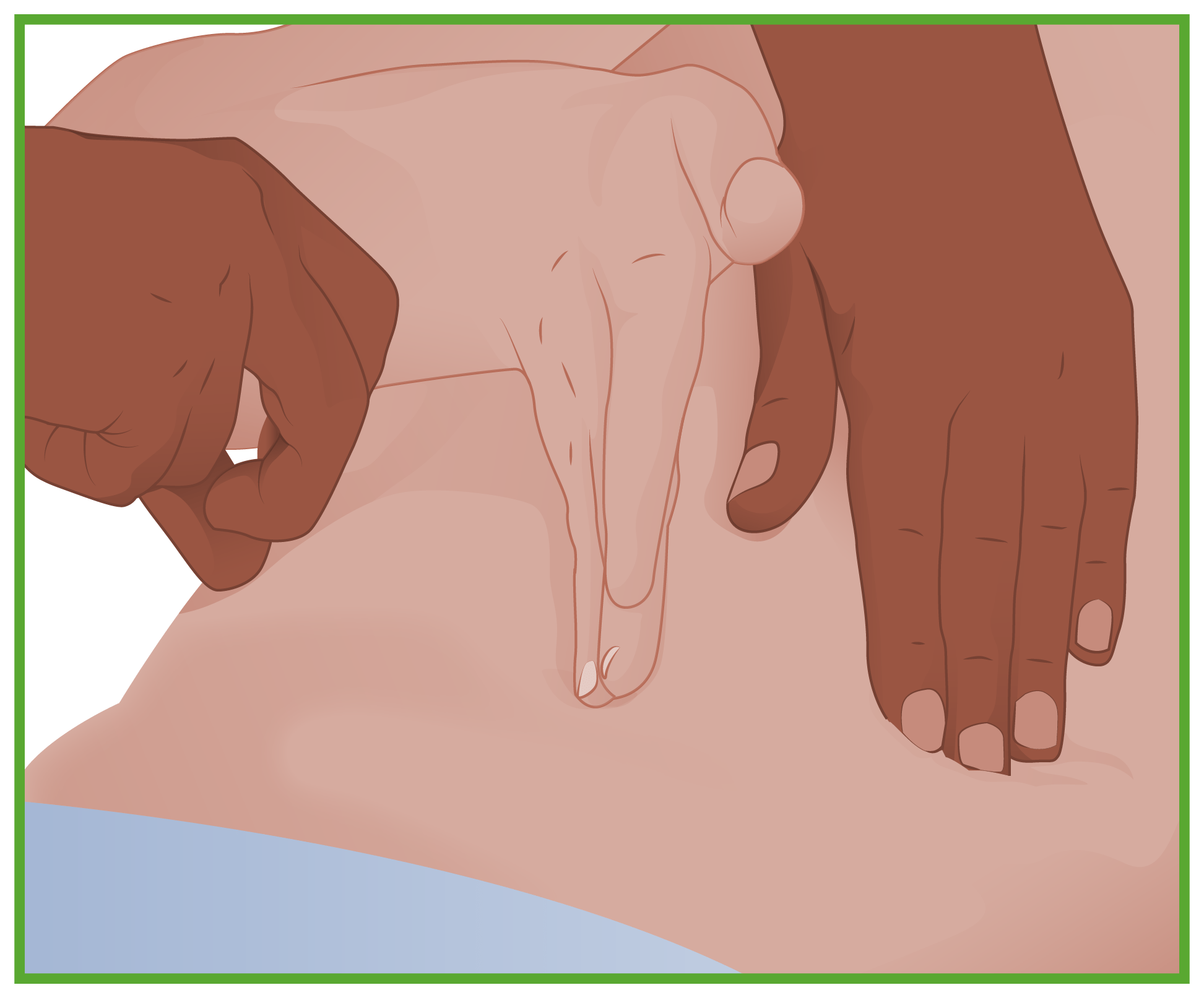
Prueba de la onda de líquido:
El individuo (u otro colega) coloca su mano en la línea media del abdomen. A continuación, el examinador da un golpecito en un lado. La prueba es positiva si el examinador siente el golpe en el otro lado.
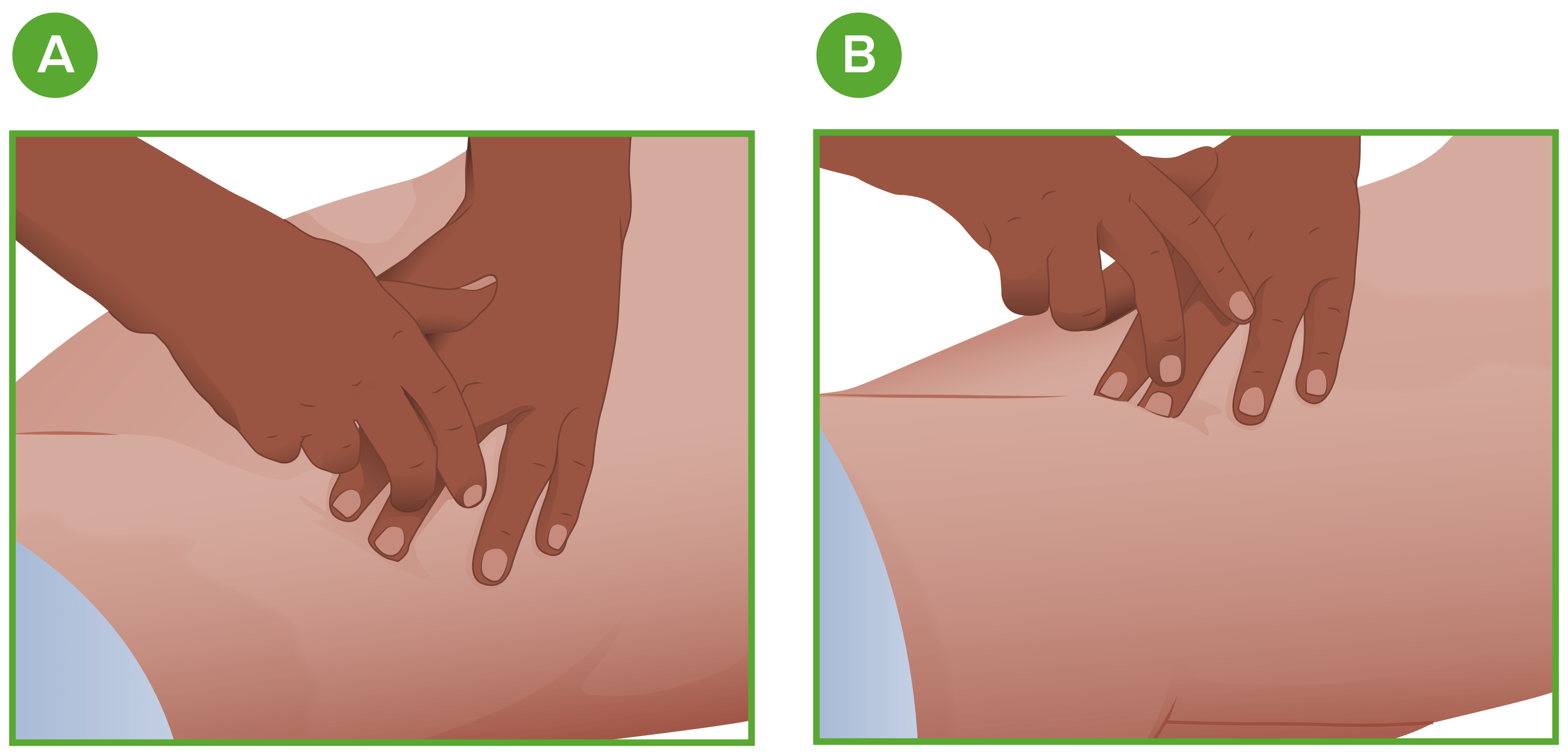
Examen de matidez cambiante:
A: El examinador percute lateralmente desde la línea media.
B: El individuo se inclina de lado; el examinador vuelve a percutir.
Evaluando el tamaño hepático por medio de la percusión
Imagen por Lecturio. Licencia: CC BY-NC-SA 4.0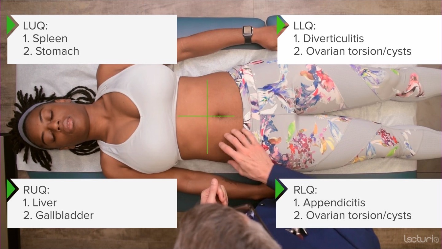
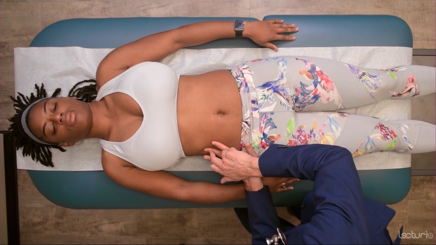
Palpación de la sensibilidad abdominal en los 4 cuadrantes
Imagen por Lecturio. Licencia: CC BY-NC-SA 4.0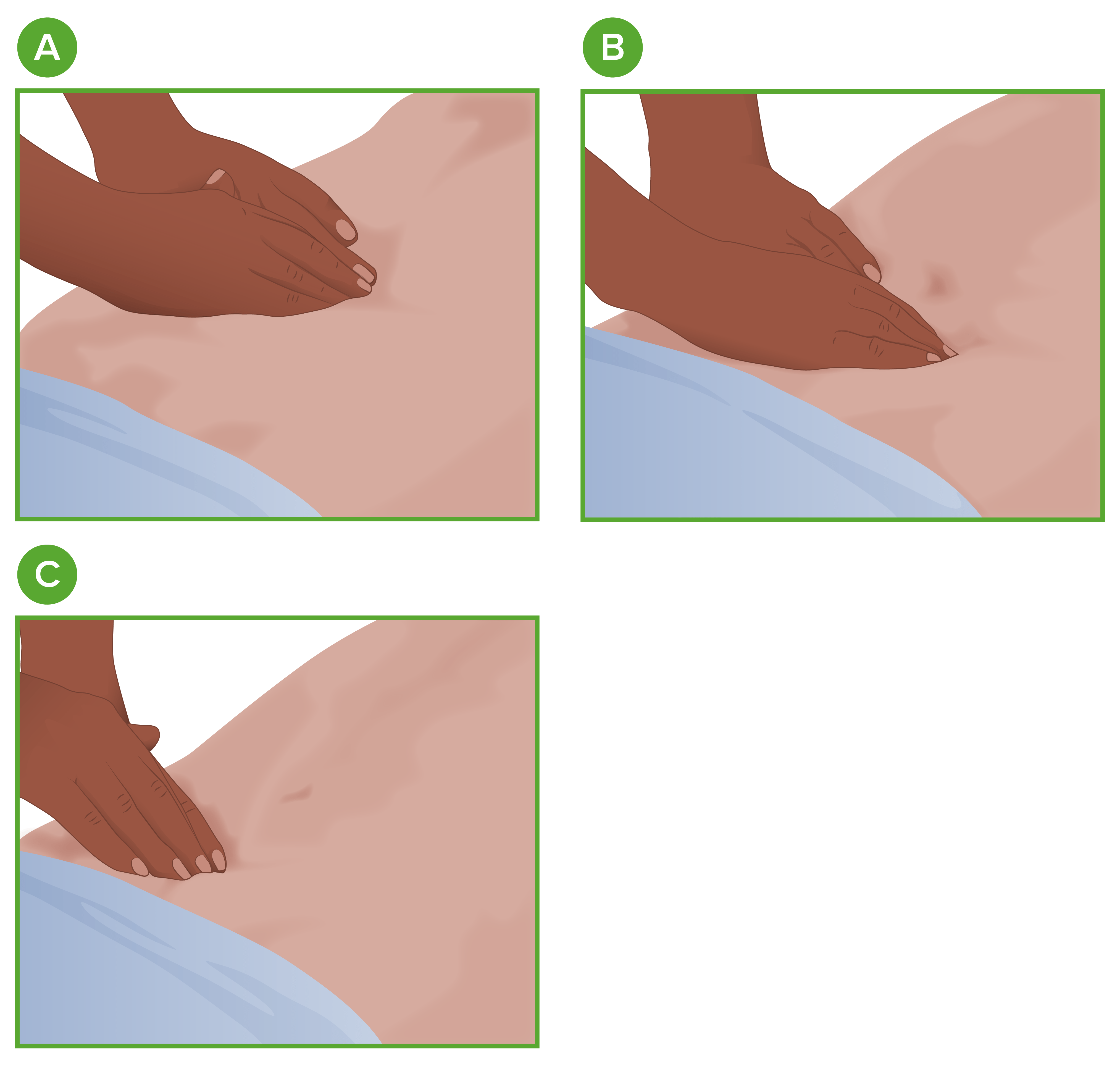
Palpación abdominal
A: Región epigástrica
B: Región umbilical
C: Región suprapúbica