La enfermedad de cambios mínimos, también conocida como nefrosis lipoidea, es la causa más común de síndrome nefrótico en niños. La denominación "cambios mínimos" proviene de los LOS Neisseria escasos cambios observados en EN Erythema nodosum is an immune-mediated panniculitis (inflammation of the subcutaneous fat) caused by a type IV (delayed-type) hypersensitivity reaction. It commonly manifests in young women as tender, erythematous nodules on the shins. Erythema Nodosum las biopsias renales al AL Amyloidosis microscopio óptico. Los LOS Neisseria hallazgos clínicos distintivos incluyen edema Edema Edema is a condition in which excess serous fluid accumulates in the body cavity or interstitial space of connective tissues. Edema is a symptom observed in several medical conditions. It can be categorized into 2 types, namely, peripheral (in the extremities) and internal (in an organ or body cavity). Edema, proteinuria Proteinuria The presence of proteins in the urine, an indicator of kidney diseases. Nephrotic Syndrome in Children, hipoalbuminemia e hiperlipidemia. El diagnóstico se basa en EN Erythema nodosum is an immune-mediated panniculitis (inflammation of the subcutaneous fat) caused by a type IV (delayed-type) hypersensitivity reaction. It commonly manifests in young women as tender, erythematous nodules on the shins. Erythema Nodosum la sospecha clínica y los LOS Neisseria hallazgos de laboratorio que lo respaldan. La administración de corticosteroides es la piedra angular del tratamiento, y el pronóstico es muy favorable.
Last updated: Dec 15, 2025
La enfermedad de cambios mínimos es un trastorno glomerular primario de etiología incierta que causa síndrome nefrótico. El término “mínimos” se refiere a los LOS Neisseria cambios estructurales mínimos de los LOS Neisseria glomérulos cuando se observan con microscopía de luz.
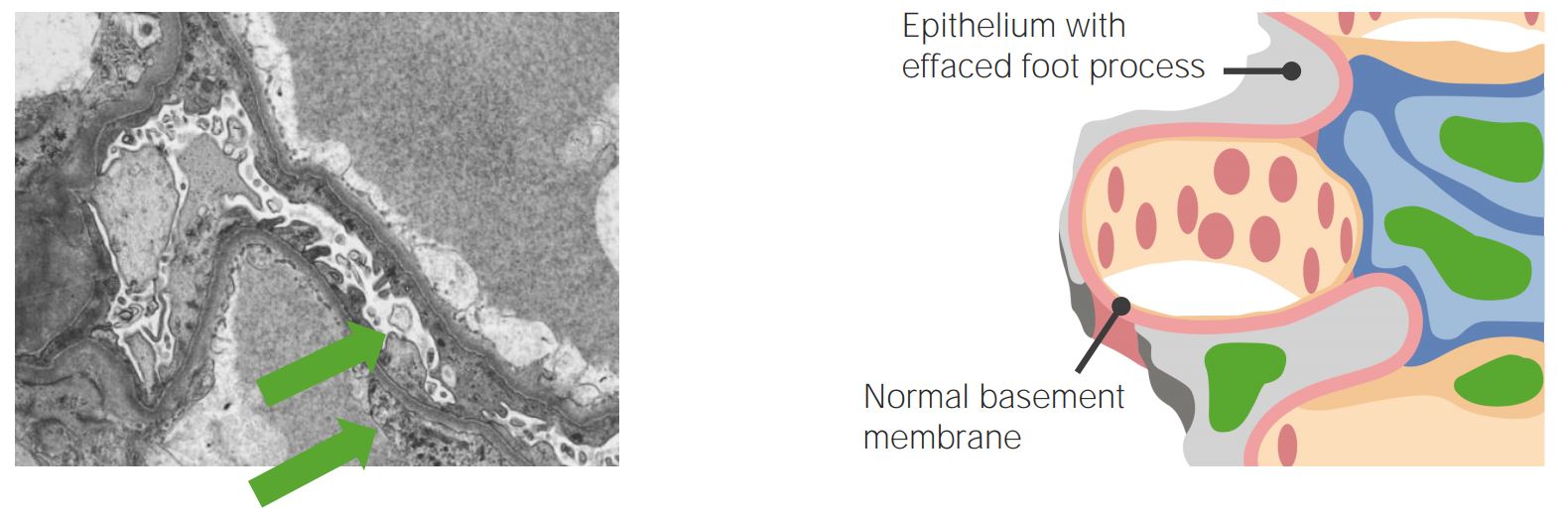
Microscopio electrónico (izquierda) y diagrama (derecha) mostrando el borrado de los procesos pediculados de los podocitos
Imagen: “Managing a locally advanced malignant thymoma complicated by nephrotic syndrome: a case report” por Teoh DC, El-Modir A. Licencia: CC BY 2.0, editada por Lecturio.
Síndrome nefrótico:
La afección se acompaña de retención de agua y sodio. La imagen muestra hinchazón/edema facial. El grado de edema puede variar. Puede haber un ligero edema en los párpados que disminuye durante el día, edema que afecta a los miembros inferiores, una hinchazón generalizada o una anasarca completa.
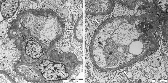
Microscopía electrónica de un glomérulo observado en un paciente con enfermedad de cambios mínimos:
En la enfermedad con cambios mínimos, los podocitos están fusionados, apareciendo como una sola capa (flechas blancas).
En la enfermedad con cambios mínimos no se observan depósitos electrónicamente densos, y los restos de la membrana basal glomerular de grosor normal la distinguen de otras enfermedades glomerulares.
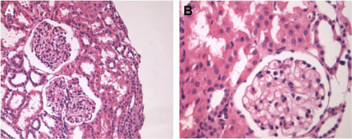
Biopsia renal de enfermedad con cambios mínimos en la microscopía de luz normal
A: Tinción H&E, 200x
B: Tinción de H&E, 400x