LosLOSNeisseria órganos abdominales se derivan principalmente del endodermo, que forma el tubo intestinal primitivo. El tubo intestinal se divide enENErythema nodosum is an immune-mediated panniculitis (inflammation of the subcutaneous fat) caused by a type IV (delayed-type) hypersensitivity reaction. It commonly manifests in young women as tender, erythematous nodules on the shins.Erythema Nodosum 3 regiones: intestino anterior, medio e intestino posterior. El intestino anterior da lugar alALAmyloidosis revestimiento del tracto gastrointestinal desde el esófago hasta la parte superior del duodeno, así como alALAmyloidosis hígado, la vesícula biliar y el páncreas. El intestino medio da origen alALAmyloidosis revestimiento del tubo digestivo entre el duodeno medio y la unión de losLOSNeisseria dos tercios proximales y el tercio distal del colonColonThe large intestines constitute the last portion of the digestive system. The large intestine consists of the cecum, appendix, colon (with ascending, transverse, descending, and sigmoid segments), rectum, and anal canal. The primary function of the colon is to remove water and compact the stool prior to expulsion from the body via the rectum and anal canal. Colon, Cecum, and Appendix: Anatomy transverso. El intestino posterior da origen alALAmyloidosis revestimiento del tubo digestivo desde la unión de losLOSNeisseria dos tercios proximales y el tercio distal del colonColonThe large intestines constitute the last portion of the digestive system. The large intestine consists of the cecum, appendix, colon (with ascending, transverse, descending, and sigmoid segments), rectum, and anal canal. The primary function of the colon is to remove water and compact the stool prior to expulsion from the body via the rectum and anal canal. Colon, Cecum, and Appendix: Anatomy transverso hasta la porción superior del canal anal. El mesodermo da lugar a losLOSNeisseria músculos de la pared del tracto gastrointestinal, el tejido conectivo (incluidos losLOSNeisseria mesenterios y el omento) y la irrigación. El ectodermo da lugar alALAmyloidosis tejido nervioso y alALAmyloidosis revestimiento del canal anal inferior.
La mórula (bola de células) se somete a un proceso llamado blastulación, enENErythema nodosum is an immune-mediated panniculitis (inflammation of the subcutaneous fat) caused by a type IV (delayed-type) hypersensitivity reaction. It commonly manifests in young women as tender, erythematous nodules on the shins.Erythema Nodosum el que comienza a formarse una cavidad. Luego, las células comienzan a diferenciarse enENErythema nodosum is an immune-mediated panniculitis (inflammation of the subcutaneous fat) caused by a type IV (delayed-type) hypersensitivity reaction. It commonly manifests in young women as tender, erythematous nodules on the shins.Erythema Nodosum masas celulares externa e interna.
Masa de células externas → trofoblasto → placentaPlacentaA highly vascularized mammalian fetal-maternal organ and major site of transport of oxygen, nutrients, and fetal waste products. It includes a fetal portion (chorionic villi) derived from trophoblasts and a maternal portion (decidua) derived from the uterine endometrium. The placenta produces an array of steroid, protein and peptide hormones (placental hormones).Placenta, Umbilical Cord, and Amniotic Cavity y membranas
Masa celular interna → embrioblasto → disco bilaminar:
Epiblasto
Hipoblasto
Saco amniótico: una cavidad de líquido que se desarrolla “por encima” del epiblasto (entre el epiblasto y el trofoblasto)
Saco vitelino primitivo:
Una cavidad que se forma “debajo” del hipoblasto
Importante enENErythema nodosum is an immune-mediated panniculitis (inflammation of the subcutaneous fat) caused by a type IV (delayed-type) hypersensitivity reaction. It commonly manifests in young women as tender, erythematous nodules on the shins.Erythema Nodosum el desarrollo gastrointestinal
Relación del disco bilaminar, el saco vitelino y la cavidad amniótica en el embrión temprano
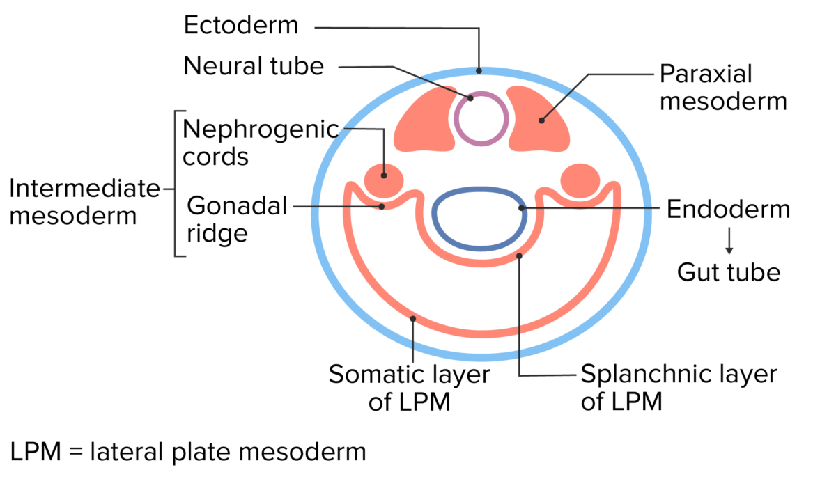
El disco bilaminar se somete a un proceso llamado gastrulación para formar el disco trilaminar. Hay 3 capas del disco trilaminar:
Ectodermo (continuo con el amnios)
Mesodermo:
Mesodermo paraxial
Mesodermo intermedio
Mesodermo de la placa lateral:
Capa somática del mesodermo de la placa lateral
Capa esplácnica del mesodermo de la placa lateral
Mesodermo extraembrionario:
Rodea la cavidad amniótica → continuo con la capa somática del mesodermo de la placa lateral
Rodea el saco vitelino → continuo con la capa esplácnica del mesodermo de la placa lateral
Endodermo
El endodermo embrionario (generalmente llamado simplemente endodermo) → se convierte enENErythema nodosum is an immune-mediated panniculitis (inflammation of the subcutaneous fat) caused by a type IV (delayed-type) hypersensitivity reaction. It commonly manifests in young women as tender, erythematous nodules on the shins.Erythema Nodosum el tubo intestinal primitivo
Endodermo extraembrionario → recubre el saco vitelino
Saco vitelino secundario:la cavidad entre el endodermo embrionario y extraembrionario
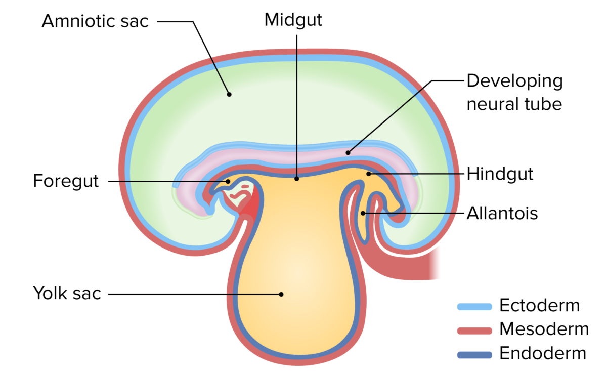
Plegado del disco trilaminar (se pliega enENErythema nodosum is an immune-mediated panniculitis (inflammation of the subcutaneous fat) caused by a type IV (delayed-type) hypersensitivity reaction. It commonly manifests in young women as tender, erythematous nodules on the shins.Erythema Nodosum 2 direcciones):
Plegado lateral:
Crea una estructura cilíndrica rodeada por ectodermo y la cavidad amniótica
Crea el tubo intestinal primitivo internamente a partir del endodermo
Pliegue cráneo–caudal:
Crea un extremo craneal y un extremo caudal (el embrión adquiere “forma de frijol”)
HaceHACEAltitude Sickness que el saco vitelino se aleje más del cuerpo → el tallo alargado que conecta el saco vitelino con el tubo intestinal es el conducto vitelino.
Capas del disco trilaminar.
Imagen por Lecturio.
Vista en sección transversal del embrión temprano después de haber sido sometido a un plegado lateral.
Imagen por Lecturio.
El embrión temprano con el saco amniótico por encima del embrión y el saco vitelino debajo del embrión.
Imagen por Lecturio.
El embrión temprano con el saco amniótico por encima del embrión y el saco vitelino debajo del embrión.
Imagen por Lecturio.
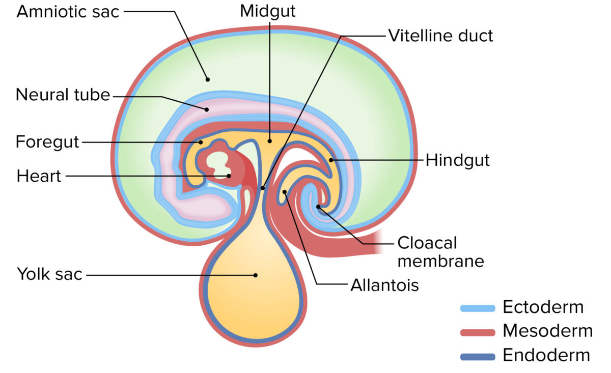
Obliteración del conducto vitelino y el saco vitelino
A medida que el embrión se pliega, el saco vitelino se aleja cada vez más del cuerpo.
Conducto vitelino: tallo alargado que conecta el intestino medio con el saco vitelino a medida que se aleja
Crecimiento de la cavidad amniótica alrededor de las semanas 4–6:
La cavidad amniótica comienza enENErythema nodosum is an immune-mediated panniculitis (inflammation of the subcutaneous fat) caused by a type IV (delayed-type) hypersensitivity reaction. It commonly manifests in young women as tender, erythematous nodules on the shins.Erythema Nodosum el lado dorsal del embrión
Crece y se extiende alrededor de losLOSNeisseria extremos craneal y caudal del embrión
LosLOSNeisseria bordes de la cavidad enENErythema nodosum is an immune-mediated panniculitis (inflammation of the subcutaneous fat) caused by a type IV (delayed-type) hypersensitivity reaction. It commonly manifests in young women as tender, erythematous nodules on the shins.Erythema Nodosum crecimiento se encuentran enENErythema nodosum is an immune-mediated panniculitis (inflammation of the subcutaneous fat) caused by a type IV (delayed-type) hypersensitivity reaction. It commonly manifests in young women as tender, erythematous nodules on the shins.Erythema Nodosum el lado ventral enENErythema nodosum is an immune-mediated panniculitis (inflammation of the subcutaneous fat) caused by a type IV (delayed-type) hypersensitivity reaction. It commonly manifests in young women as tender, erythematous nodules on the shins.Erythema Nodosum el abdomen → pellizca el saco vitelino/oblitera el conducto vitelino
Relevancia clínica: la falla enENErythema nodosum is an immune-mediated panniculitis (inflammation of the subcutaneous fat) caused by a type IV (delayed-type) hypersensitivity reaction. It commonly manifests in young women as tender, erythematous nodules on the shins.Erythema Nodosum la obliteración del conducto vitelino da como resultado el divertículo de Meckel:
Un verdadero divertículo (contiene todas las capas de la pared intestinal)
Surge de la superficie antimesentérica del íleon distal, anclándolo a la pared abdominal anterior.
Puede provocar fístula, vólvulo, obstrucción del intestino delgado y/o necrosisNecrosisThe death of cells in an organ or tissue due to disease, injury or failure of the blood supply.Ischemic Cell Damage
Descripción General de la Diferenciación del Tubo Intestinal
Estructuras derivadas del tubo intestinal
El tubo intestinal primitivo se forma a partir del endodermo alALAmyloidosis completar el plegado lateral. El tubo intestinal se puede dividir inicialmente enENErythema nodosum is an immune-mediated panniculitis (inflammation of the subcutaneous fat) caused by a type IV (delayed-type) hypersensitivity reaction. It commonly manifests in young women as tender, erythematous nodules on the shins.Erythema Nodosum 3 áreas:
Intestino anterior: derivados abdominales irrigados por la arteria celíaca
Esófago
Estómago
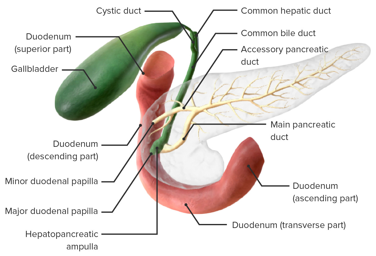
Duodeno proximal (por encima de la ampolla de Vater)
Hígado
Vesícula biliar y conductos biliares
Páncreas
Estructuras derivadas del intestino anterior por encima del abdomen:
Faringe primordial y sus derivados
Esófago cervical y torácico
Tracto respiratorio
Intestino medio: derivados irrigados por la arteria mesentérica superior
Duodeno distal (por debajo de la ampolla de Vater)
Yeyuno
Íleon
Ciego
Apéndice
ColonColonThe large intestines constitute the last portion of the digestive system. The large intestine consists of the cecum, appendix, colon (with ascending, transverse, descending, and sigmoid segments), rectum, and anal canal. The primary function of the colon is to remove water and compact the stool prior to expulsion from the body via the rectum and anal canal. Colon, Cecum, and Appendix: Anatomy ascendente
⅔ Proximales del colonColonThe large intestines constitute the last portion of the digestive system. The large intestine consists of the cecum, appendix, colon (with ascending, transverse, descending, and sigmoid segments), rectum, and anal canal. The primary function of the colon is to remove water and compact the stool prior to expulsion from the body via the rectum and anal canal. Colon, Cecum, and Appendix: Anatomy transverso
Intestino posterior: derivados irrigados por la arteria mesentérica inferior
⅓ Distal del colonColonThe large intestines constitute the last portion of the digestive system. The large intestine consists of the cecum, appendix, colon (with ascending, transverse, descending, and sigmoid segments), rectum, and anal canal. The primary function of the colon is to remove water and compact the stool prior to expulsion from the body via the rectum and anal canal. Colon, Cecum, and Appendix: Anatomy transverso
ColonColonThe large intestines constitute the last portion of the digestive system. The large intestine consists of the cecum, appendix, colon (with ascending, transverse, descending, and sigmoid segments), rectum, and anal canal. The primary function of the colon is to remove water and compact the stool prior to expulsion from the body via the rectum and anal canal. Colon, Cecum, and Appendix: Anatomy descendente
ColonColonThe large intestines constitute the last portion of the digestive system. The large intestine consists of the cecum, appendix, colon (with ascending, transverse, descending, and sigmoid segments), rectum, and anal canal. The primary function of the colon is to remove water and compact the stool prior to expulsion from the body via the rectum and anal canal. Colon, Cecum, and Appendix: Anatomy sigmoide
Recto
Porción superior del canal anal
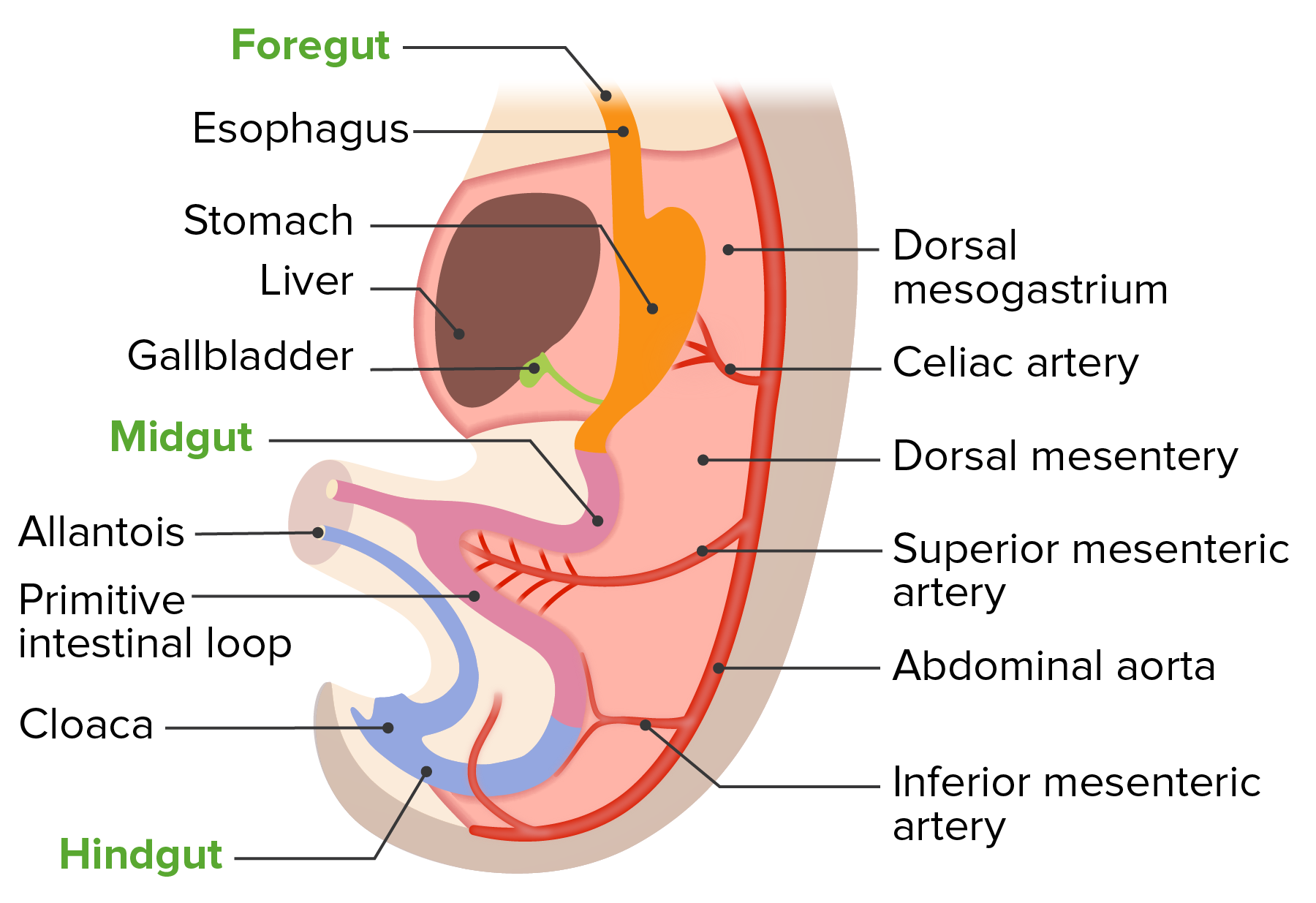
Desarrollo del mesenterio dorsal con el tubo intestinal primitivo
Imagen por Lecturio.
Estructuras derivadas del endodermo
Estructuras clave derivadas del endodermo relacionadas con el desarrollo de losLOSNeisseria órganos abdominales:
Revestimiento epitelial de todo el tracto gastrointestinal
Glándulas mucosas y submucosas
Estructuras derivadas del mesodermo
Estructuras clave derivadas del mesodermo relacionadas con el desarrollo de losLOSNeisseria órganos abdominales:
Capa esplácnica del mesodermo de la placa lateral:
Pared del tracto digestivo (i.e., paredes intestinales)
Tejido muscular (e.g., capas circulares y longitudinales, esfínteres)
Tejido conectivo
Irrigación
Peritoneo visceral
Mesenterios:
Mesogastrio dorsal y ventral → omento mayor y menor, ligamento falciforme
Mesenterio del intestino delgado
Mesocolon transverso y sigmoide
Numerosos ligamentos abdominales nombrados
Capa somática del mesodermo de la placa lateral: peritoneo parietalParietalOne of a pair of irregularly shaped quadrilateral bones situated between the frontal bone and occipital bone, which together form the sides of the cranium.Skull: Anatomy
Estructuras derivadas del ectodermo
Estructuras clave derivadas del ectodermo relacionadas con el desarrollo de losLOSNeisseria órganos abdominales:
Células de la cresta neural: plexo nervioso entérico (i.e., plexos mientérico y submucoso)
EnENErythema nodosum is an immune-mediated panniculitis (inflammation of the subcutaneous fat) caused by a type IV (delayed-type) hypersensitivity reaction. It commonly manifests in young women as tender, erythematous nodules on the shins.Erythema NodosumlosLOSNeisseria extremos craneal y caudal, el ectodermo y el endodermo se encuentran (sin mesodermo entre ellos) formando membranas que finalmente se romperán, convirtiéndose enENErythema nodosum is an immune-mediated panniculitis (inflammation of the subcutaneous fat) caused by a type IV (delayed-type) hypersensitivity reaction. It commonly manifests in young women as tender, erythematous nodules on the shins.Erythema Nodosum las aberturas del tracto gastrointestinal
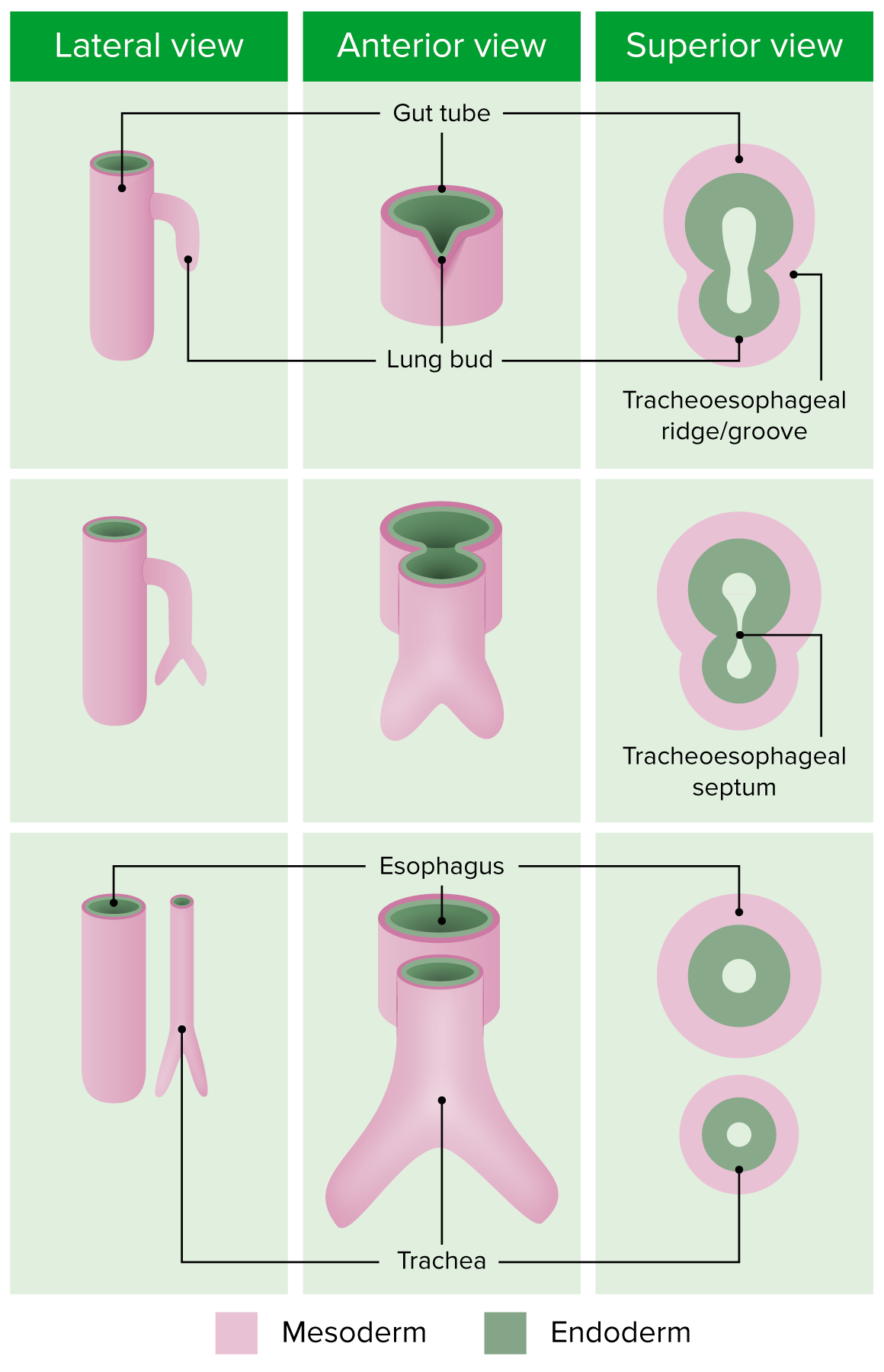
Brote ventral del intestino anterior alrededor de la semana 4
Crece simultáneamente hacia afuera (ventralmente) y hacia abajo (caudalmente)
Incluye endodermo y capa esplácnica del mesodermo de la placa lateral circundante
Surco traqueoesofágico (o cresta):
A medida que el brote pulmonar crece hacia afuera y hacia abajo, el surco traqueoesofágico aparece como hendiduras laterales entre el nuevo brote pulmonar y el intestino anterior.
LosLOSNeisseria surcos/crestas se mueven hacia adentro, pellizcando el brote pulmonar y formando el tabique traqueoesofágico.
El tabique traqueoesofágico crea 2 tubos separados:
Esófago (posteriormente, del intestino anterior original)
Tráquea (anteriormente, desde el brote pulmonar)
Relevancia clínica:
Fístula traqueoesofágica: ocurre cuando losLOSNeisseria surcos traqueoesofágicos no se cierran completamente enENErythema nodosum is an immune-mediated panniculitis (inflammation of the subcutaneous fat) caused by a type IV (delayed-type) hypersensitivity reaction. It commonly manifests in young women as tender, erythematous nodules on the shins.Erythema Nodosum la línea media
Dilatación fusiforme del intestino anterior: sitio del estómago primordial
El alargamiento del esófago coloca alALAmyloidosis estómago debajo del diafragma
La pared posterior crece más rápidamente que la pared anterior → desarrolla una forma de C
Pared posterior → curvatura mayor
Pared anterior → curvatura menor
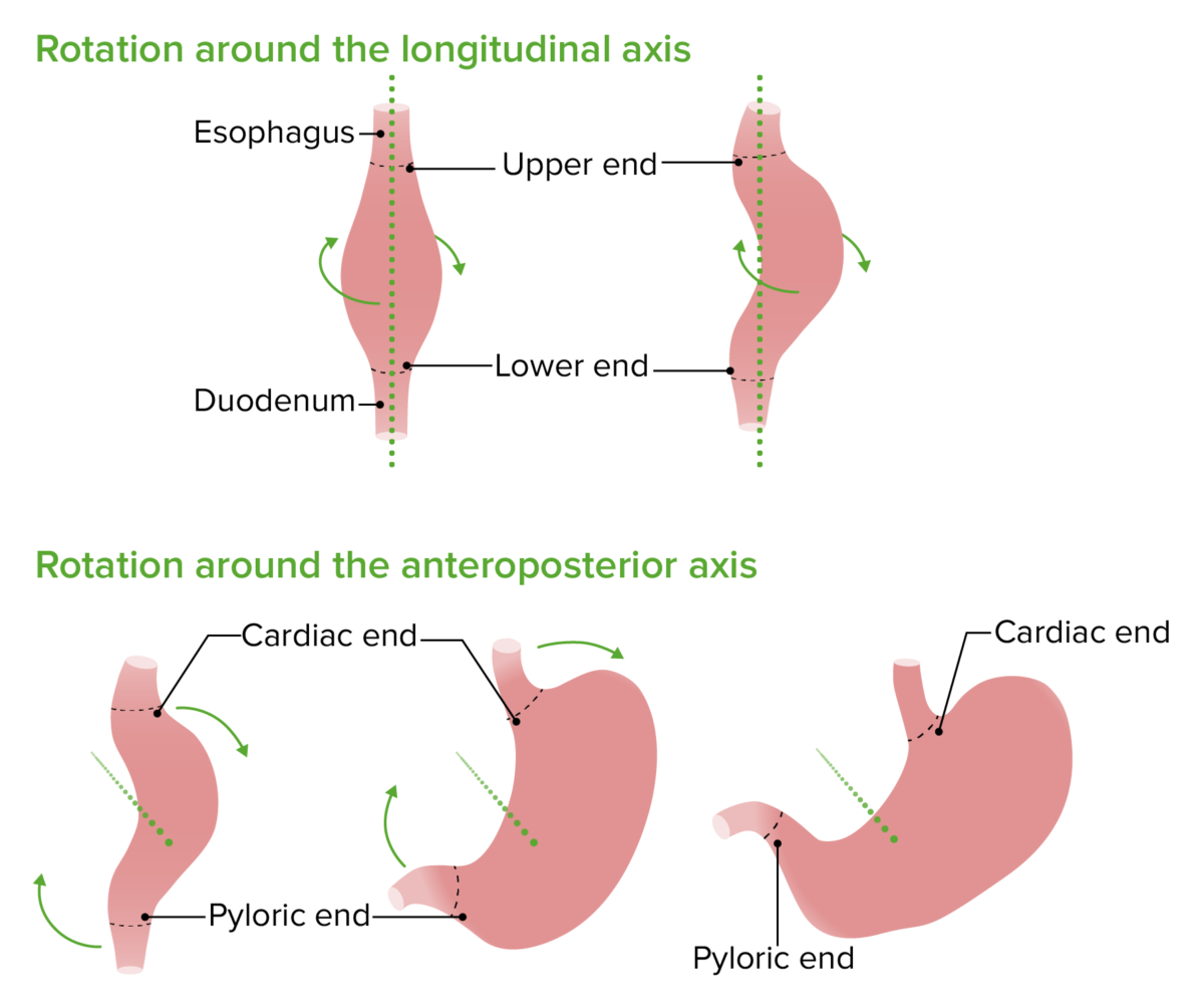
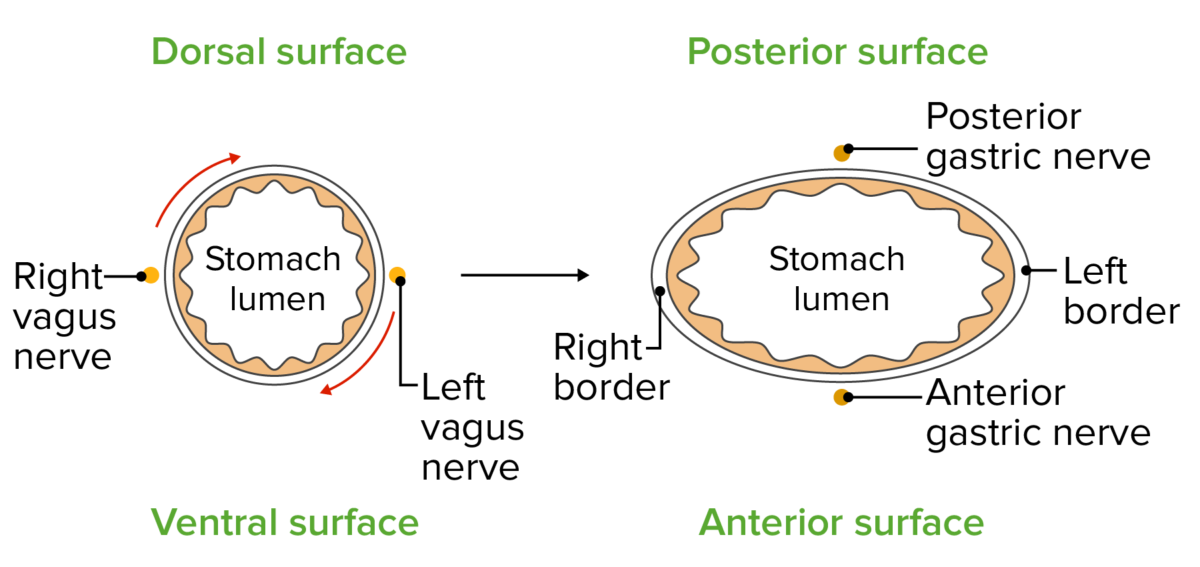
Rotación de 90 grados enENErythema nodosum is an immune-mediated panniculitis (inflammation of the subcutaneous fat) caused by a type IV (delayed-type) hypersensitivity reaction. It commonly manifests in young women as tender, erythematous nodules on the shins.Erythema Nodosum el sentido de las agujas del reloj alrededor del eje longitudinal:
Curvatura mayor (dorsal):
Gira anteriormente y hacia la izquierda
El nervio vago izquierdo inerva la pared anterior
Curvatura menor (ventral):
Gira posteriormente y hacia la derecha
El nervio vago derecho inerva la pared posterior
Rotación más pequeña enENErythema nodosum is an immune-mediated panniculitis (inflammation of the subcutaneous fat) caused by a type IV (delayed-type) hypersensitivity reaction. It commonly manifests in young women as tender, erythematous nodules on the shins.Erythema Nodosum el sentido de las agujas del reloj alrededor del eje anteroposterior:
La porción cardíaca del estómago se mueve hacia abajo y hacia la izquierda
La porción pilórica se mueve hacia arriba y hacia la derecha
El estómago gira en el sentido de las agujas del reloj primero a lo largo de su eje longitudinal y luego a lo largo de su eje anteroposterior:
El lado dorsal original del estómago crece más rápido que el lado ventral original, creando las curvaturas mayores y menores del estómago.
Imagen por Lecturio.
Una mirada más cercana a la rotación del estómago explica claramente por qué el nervio vago izquierdo contribuye en mayor medida al tronco vago anterior y el nervio vago derecho al tronco vago posterior.
Imagen por Lecturio.
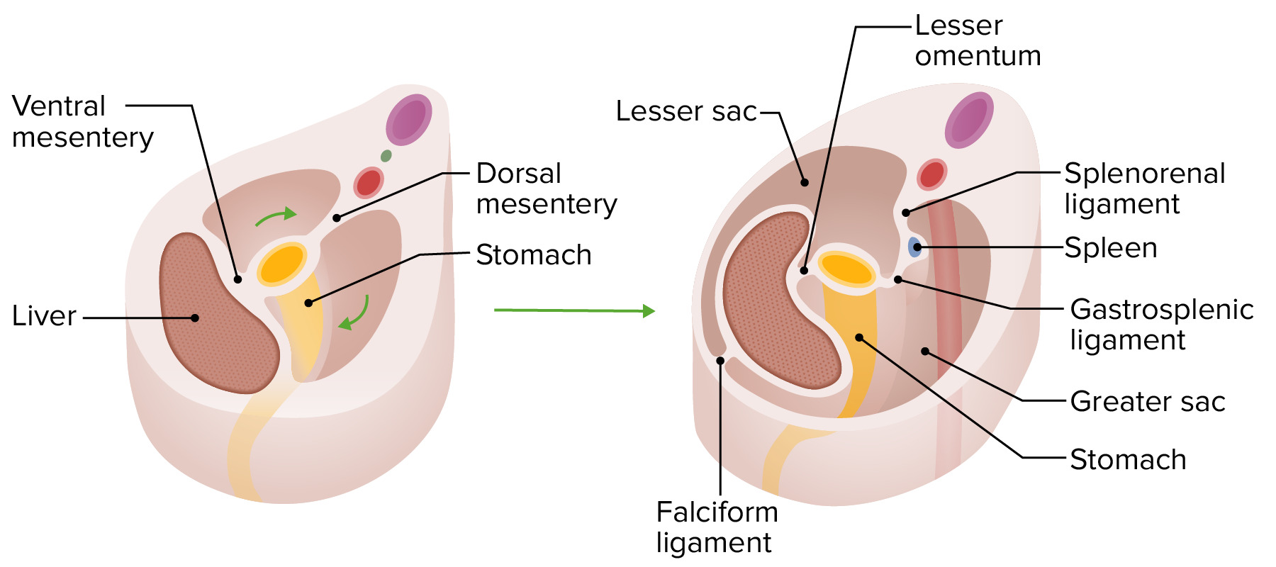
Omentos
El omento mayor y menor se forman a partir del mesogastrio dorsal y ventral (de origen mesodérmico). A medida que rotan con el estómago, crean losLOSNeisseria sacos mayor y menor.
Mesogastrio dorsal:
Suspende alALAmyloidosis estómago de la pared dorsal
Localizado originalmente enENErythema nodosum is an immune-mediated panniculitis (inflammation of the subcutaneous fat) caused by a type IV (delayed-type) hypersensitivity reaction. It commonly manifests in young women as tender, erythematous nodules on the shins.Erythema Nodosum el plano medio
Movido hacia la izquierda durante una rotación longitudinal de 90 grados
Crea un espacio posterior alALAmyloidosis estómago llamado saco menor (i.e., bolsa omental)
Forma varias estructuras importantes:
Después de la rotación del estómago, se abulta hacia abajo para formar el omento mayor
Ligamento gastroesplénico: conecta el estómago y el bazo
Ligamento esplenorrenal: conecta el bazo con la pared abdominal posterior (cerca del riñón)
Anterior alALAmyloidosis omento mayor está el saco mayor
Estructuras que se desarrollan dentro del mesogastrio dorsal:
Bazo
Páncreas
Mesogastrio ventral:
Se adhiere alALAmyloidosis estómago, duodeno, hígado y pared abdominal ventral
Localizado originalmente enENErythema nodosum is an immune-mediated panniculitis (inflammation of the subcutaneous fat) caused by a type IV (delayed-type) hypersensitivity reaction. It commonly manifests in young women as tender, erythematous nodules on the shins.Erythema Nodosum el plano medio
Movido hacia la derecha durante una rotación longitudinal de 90 grados
Cierra el saco menor entre el estómago y el hígado
Forma 2 estructuras primarias:
Omento menor: conecta el estómago y el hígado
Ligamento falciforme: conecta el hígado y la pared abdominal anterior
Estructuras que se desarrollan dentro del mesogastrio ventral:
Hígado
Vesícula biliar
Conductos biliares
Rotación del estómago y los mesenterios gástricos
Imagen por Lecturio.
Duodeno
Su desarrollo comienza enENErythema nodosum is an immune-mediated panniculitis (inflammation of the subcutaneous fat) caused by a type IV (delayed-type) hypersensitivity reaction. It commonly manifests in young women as tender, erythematous nodules on the shins.Erythema Nodosum la 4ta semana
Por encima de la ampolla de Vater: derivado de la parte caudal delintestino anterior → irrigado por la arteria celíaca
Por debajo de la ampolla de Vater: derivado de la parte craneal del intestino medio → irrigado por la arteria mesentérica superior
Se alarga y forma un bucle enENErythema nodosum is an immune-mediated panniculitis (inflammation of the subcutaneous fat) caused by a type IV (delayed-type) hypersensitivity reaction. It commonly manifests in young women as tender, erythematous nodules on the shins.Erythema Nodosum forma de C
Se une con el páncreas enENErythema nodosum is an immune-mediated panniculitis (inflammation of the subcutaneous fat) caused by a type IV (delayed-type) hypersensitivity reaction. It commonly manifests in young women as tender, erythematous nodules on the shins.Erythema Nodosum desarrollo unido a la pared posterior del cuerpo
Obliteración y recanalización duodenal:
Semana 5: la rápida proliferación celular enENErythema nodosum is an immune-mediated panniculitis (inflammation of the subcutaneous fat) caused by a type IV (delayed-type) hypersensitivity reaction. It commonly manifests in young women as tender, erythematous nodules on the shins.Erythema Nodosum sus paredes conduce a la obstrucción completa de la luz duodenal
Las vacuolas crecen y se fusionan → recanalización de la luz
Relevancia clínica: el hecho de que la luz no se recanalice por completo da como resultado:
Estenosis duodenal: estrechamiento del duodeno
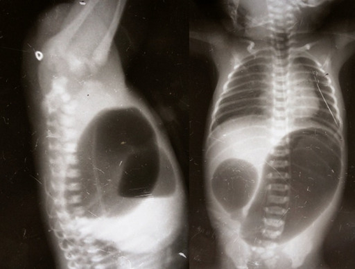
AtresiaAtresiaHypoplastic Left Heart Syndrome (HLHS) duodenal: persiste una obstrucción completa; se presenta con emesis biliosa y un signo de doble burbuja enENErythema nodosum is an immune-mediated panniculitis (inflammation of the subcutaneous fat) caused by a type IV (delayed-type) hypersensitivity reaction. It commonly manifests in young women as tender, erythematous nodules on the shins.Erythema Nodosum la radiografía
Signo de doble burbuja en la radiografía (invertografía) que indica obstrucción duodenal: La burbuja más pequeña a la derecha del individuo es aire en el duodeno y la burbuja más grande a la izquierda es aire en el estómago.
Imagen: “Invertogram showing high ARM and AP view showing duodenal atresia” por Department of Pediatric Surgery, Gauhati Medical College, GUWAHATI, ASSAM, INDIA. Licencia: CC BY 3.0
Hígado y conductos biliares
El desarrollo comienza enENErythema nodosum is an immune-mediated panniculitis (inflammation of the subcutaneous fat) caused by a type IV (delayed-type) hypersensitivity reaction. It commonly manifests in young women as tender, erythematous nodules on the shins.Erythema Nodosum la 3ra‒4ta semana
Aparece enENErythema nodosum is an immune-mediated panniculitis (inflammation of the subcutaneous fat) caused by a type IV (delayed-type) hypersensitivity reaction. It commonly manifests in young women as tender, erythematous nodules on the shins.Erythema Nodosum la porción caudal del intestino anterior como divertículo hepático, que pasa a formar:
Hígado (el endodermo se diferencia enENErythema nodosum is an immune-mediated panniculitis (inflammation of the subcutaneous fat) caused by a type IV (delayed-type) hypersensitivity reaction. It commonly manifests in young women as tender, erythematous nodules on the shins.Erythema Nodosum hepatocitos)
Conductos intrahepáticos
Porciones extrahepáticas de losLOSNeisseria conductos hepáticos
A medida que el divertículo hepático se agranda, la conexión con el duodeno se estrecha → conducto biliar común
Un brote más pequeño crece enENErythema nodosum is an immune-mediated panniculitis (inflammation of the subcutaneous fat) caused by a type IV (delayed-type) hypersensitivity reaction. It commonly manifests in young women as tender, erythematous nodules on the shins.Erythema Nodosum el lado caudal del divertículo hepático:
Forma la vesícula biliar
Conexión entre el brote caudal y la vesícula biliar → conducto cístico
El divertículo hepático crece hacia el mesogastrio ventral.
Semanas 5–9:
Las células madre hematopoyéticas (origen mesodérmico) migran alALAmyloidosis hígado:
Comienza la hematopoyesis
El hígado es el sitio principal de la hematopoyesis hasta aproximadamente las 28 semanas, cuando la médula ósea asume el trabajo.
Síntesis de ácido biliar
Semana 12: inicio de la secreción de bilis por losLOSNeisseria hepatocitos
El desarrollo comienza enENErythema nodosum is an immune-mediated panniculitis (inflammation of the subcutaneous fat) caused by a type IV (delayed-type) hypersensitivity reaction. It commonly manifests in young women as tender, erythematous nodules on the shins.Erythema Nodosum la 4ta semana
Se desarrolla principalmente a partir de un brote pancreático dorsal del intestino anterior:
Conducto pancreático accesorio
Parte de la cabeza pancreática
Cuerpo
Cola del páncreas
Un brote pancreático ventral más pequeño contribuye a:
Cabeza pancreática
Proceso uncinado
Conducto pancreático principal
Tanto las células endocrinas como exocrinas surgen de las células pluripotenciales derivadas del endodermo del brote pancreático
Semana 7: el conducto pancreático principal y losLOSNeisseria conductos pancreáticos accesorios se fusionan
EnENErythema nodosum is an immune-mediated panniculitis (inflammation of the subcutaneous fat) caused by a type IV (delayed-type) hypersensitivity reaction. It commonly manifests in young women as tender, erythematous nodules on the shins.Erythema Nodosum la semana 13: células alfa, beta y delta presentes enENErythema nodosum is an immune-mediated panniculitis (inflammation of the subcutaneous fat) caused by a type IV (delayed-type) hypersensitivity reaction. It commonly manifests in young women as tender, erythematous nodules on the shins.Erythema NodosumlosLOSNeisseria islotes
Bazo
El desarrollo comienza enENErythema nodosum is an immune-mediated panniculitis (inflammation of the subcutaneous fat) caused by a type IV (delayed-type) hypersensitivity reaction. It commonly manifests in young women as tender, erythematous nodules on the shins.Erythema Nodosum la 5ta semana
Derivado de células mesenquimales (i.e., del mesodermo, no del endodermo del intestino anterior)
Se forma a partir de brotes mesenquimales enENErythema nodosum is an immune-mediated panniculitis (inflammation of the subcutaneous fat) caused by a type IV (delayed-type) hypersensitivity reaction. It commonly manifests in young women as tender, erythematous nodules on the shins.Erythema Nodosum el mesogastrio dorsal
LosLOSNeisseria brotes se fusionan, formando el bazo.
Se lobula durante la vida fetal; losLOSNeisseria lóbulos desaparecen antes del nacimiento.
Derivados del Intestino Medio y del Intestino Posterior
El intestino medio se convierte enENErythema nodosum is an immune-mediated panniculitis (inflammation of the subcutaneous fat) caused by a type IV (delayed-type) hypersensitivity reaction. It commonly manifests in young women as tender, erythematous nodules on the shins.Erythema Nodosum el revestimiento del tracto gastrointestinal desde el duodeno distal (por debajo de la ampolla de Vater) hasta la unión de losLOSNeisseria dos tercios proximales y el tercio distal del colonColonThe large intestines constitute the last portion of the digestive system. The large intestine consists of the cecum, appendix, colon (with ascending, transverse, descending, and sigmoid segments), rectum, and anal canal. The primary function of the colon is to remove water and compact the stool prior to expulsion from the body via the rectum and anal canal. Colon, Cecum, and Appendix: Anatomy transverso. El intestino posterior se convierte enENErythema nodosum is an immune-mediated panniculitis (inflammation of the subcutaneous fat) caused by a type IV (delayed-type) hypersensitivity reaction. It commonly manifests in young women as tender, erythematous nodules on the shins.Erythema Nodosum el tracto gastrointestinal desde el ⅓ distal del colonColonThe large intestines constitute the last portion of the digestive system. The large intestine consists of the cecum, appendix, colon (with ascending, transverse, descending, and sigmoid segments), rectum, and anal canal. The primary function of the colon is to remove water and compact the stool prior to expulsion from the body via the rectum and anal canal. Colon, Cecum, and Appendix: Anatomy transverso hasta el ano.
Crecimiento desigual de las paredes cecales → el apéndice ingresa alALAmyloidosis ciego medialmente
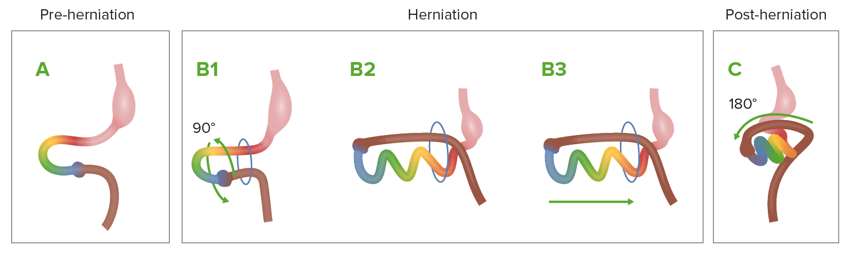
Semana 5: obliteración del conducto vitelino (el saco vitelino se separa del intestino medio)
Semana 6: herniaHerniaProtrusion of tissue, structure, or part of an organ through the bone, muscular tissue, or the membrane by which it is normally contained. Hernia may involve tissues such as the abdominal wall or the respiratory diaphragm. Hernias may be internal, external, congenital, or acquired.Abdominal Hernias fisiológica del intestino medio a través del ombligo
El intestino medio se herniaHerniaProtrusion of tissue, structure, or part of an organ through the bone, muscular tissue, or the membrane by which it is normally contained. Hernia may involve tissues such as the abdominal wall or the respiratory diaphragm. Hernias may be internal, external, congenital, or acquired.Abdominal Hernias naturalmente a través del anillo umbilical.
Crea más espacio dentro del embrión para losLOSNeisseria órganos abdominales enENErythema nodosum is an immune-mediated panniculitis (inflammation of the subcutaneous fat) caused by a type IV (delayed-type) hypersensitivity reaction. It commonly manifests in young women as tender, erythematous nodules on the shins.Erythema Nodosum desarrollo (especialmente el hígado)
Semanas 6–10:
El crecimiento rápido del intestino medio ocurre fuera del embrión
Rotación:
El intestino medio gira 270 grados enENErythema nodosum is an immune-mediated panniculitis (inflammation of the subcutaneous fat) caused by a type IV (delayed-type) hypersensitivity reaction. It commonly manifests in young women as tender, erythematous nodules on the shins.Erythema Nodosum sentido antihorario alrededor de la arteria mesentérica superior.
Tira del intestino delgado a una posición anatómica madura: el yeyuno comienza enENErythema nodosum is an immune-mediated panniculitis (inflammation of the subcutaneous fat) caused by a type IV (delayed-type) hypersensitivity reaction. It commonly manifests in young women as tender, erythematous nodules on the shins.Erythema Nodosum el cuadrante superior izquierdo
Tira del intestino grueso a su posición anatómica madura: forma de U invertida que rodea el intestino delgado
Semana 10: el intestino medio regresa a la cavidad abdominal
La cavidad abdominal enENErythema nodosum is an immune-mediated panniculitis (inflammation of the subcutaneous fat) caused by a type IV (delayed-type) hypersensitivity reaction. It commonly manifests in young women as tender, erythematous nodules on the shins.Erythema Nodosum crecimiento crea suficiente espacio para el desarrollo del intestino medio
Relevancia clínica: la falla del intestino medio para reingresar alALAmyloidosis abdomen da como resultado un onfalocele (un defecto congénito de la pared abdominal anterior enENErythema nodosum is an immune-mediated panniculitis (inflammation of the subcutaneous fat) caused by a type IV (delayed-type) hypersensitivity reaction. It commonly manifests in young women as tender, erythematous nodules on the shins.Erythema Nodosum el que losLOSNeisseria intestinos están cubiertos por peritoneo y membranas amnióticas).
Diagrama que muestra el proceso normal de rotación y hernia intestinal durante el desarrollo embrionario A: el intestino medio (asa multicolor) antes de la hernia. B1–B3: a medida que crece rápidamente, el intestino medio se hernia a través del anillo umbilical y comienza a girar. C: el intestino medio vuelve a la cavidad abdominal.
Imagen por Lecturio.
Hitos del intestino posterior
El intestino posterior se desarrolla simultáneamente y enENErythema nodosum is an immune-mediated panniculitis (inflammation of the subcutaneous fat) caused by a type IV (delayed-type) hypersensitivity reaction. It commonly manifests in young women as tender, erythematous nodules on the shins.Erythema Nodosum estrecha asociación con el sistema urogenital.
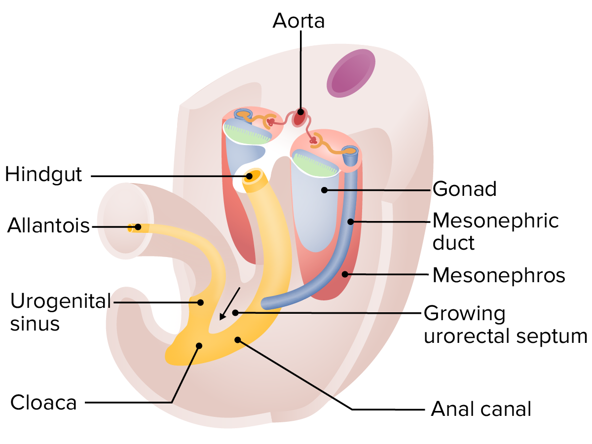
CloacaCloacaA dilated cavity extended caudally from the hindgut. In adult birds, reptiles, amphibians, and many fishes but few mammals, cloaca is a common chamber into which the digestive, urinary and reproductive tracts discharge their contents. In most mammals, cloaca gives rise to large intestine; urinary bladder; and genitalia.Development of the Abdominal Organs:
Porción distal del intestino posterior
Adyacente alALAmyloidosis “exterior” del embrión enENErythema nodosum is an immune-mediated panniculitis (inflammation of the subcutaneous fat) caused by a type IV (delayed-type) hypersensitivity reaction. It commonly manifests in young women as tender, erythematous nodules on the shins.Erythema Nodosum desarrollo enENErythema nodosum is an immune-mediated panniculitis (inflammation of the subcutaneous fat) caused by a type IV (delayed-type) hypersensitivity reaction. It commonly manifests in young women as tender, erythematous nodules on the shins.Erythema Nodosum la membrana cloacal, enENErythema nodosum is an immune-mediated panniculitis (inflammation of the subcutaneous fat) caused by a type IV (delayed-type) hypersensitivity reaction. It commonly manifests in young women as tender, erythematous nodules on the shins.Erythema Nodosum la que:
Se encuentran el endodermo y el ectodermo enENErythema nodosum is an immune-mediated panniculitis (inflammation of the subcutaneous fat) caused by a type IV (delayed-type) hypersensitivity reaction. It commonly manifests in young women as tender, erythematous nodules on the shins.Erythema NodosumlosLOSNeisseria extremos del tubo intestinal primitivo
EnENErythema nodosum is an immune-mediated panniculitis (inflammation of the subcutaneous fat) caused by a type IV (delayed-type) hypersensitivity reaction. It commonly manifests in young women as tender, erythematous nodules on the shins.Erythema Nodosum última instancia se rompe, convirtiéndose enENErythema nodosum is an immune-mediated panniculitis (inflammation of the subcutaneous fat) caused by a type IV (delayed-type) hypersensitivity reaction. It commonly manifests in young women as tender, erythematous nodules on the shins.Erythema Nodosum el ano
AlALAmyloidosis principio del desarrollo, se vacía enENErythema nodosum is an immune-mediated panniculitis (inflammation of the subcutaneous fat) caused by a type IV (delayed-type) hypersensitivity reaction. It commonly manifests in young women as tender, erythematous nodules on the shins.Erythema Nodosum un tubo de drenaje llamado alantoides
Alantoides:
Drena tanto el tubo intestinal como el sistema urinario enENErythema nodosum is an immune-mediated panniculitis (inflammation of the subcutaneous fat) caused by a type IV (delayed-type) hypersensitivity reaction. It commonly manifests in young women as tender, erythematous nodules on the shins.Erythema Nodosum desarrollo temprano
Viaja a través del cordón umbilical
Llamado el uraco más tarde enENErythema nodosum is an immune-mediated panniculitis (inflammation of the subcutaneous fat) caused by a type IV (delayed-type) hypersensitivity reaction. It commonly manifests in young women as tender, erythematous nodules on the shins.Erythema Nodosum el desarrollo
EnENErythema nodosum is an immune-mediated panniculitis (inflammation of the subcutaneous fat) caused by a type IV (delayed-type) hypersensitivity reaction. It commonly manifests in young women as tender, erythematous nodules on the shins.Erythema Nodosum última instancia, se oblitera y se convierte enENErythema nodosum is an immune-mediated panniculitis (inflammation of the subcutaneous fat) caused by a type IV (delayed-type) hypersensitivity reaction. It commonly manifests in young women as tender, erythematous nodules on the shins.Erythema Nodosum el ligamento umbilical medio a lo largo de la pared abdominal anterior interna
Relevancia clínica: la obliteración incompleta puede provocar una fístula uracal (losLOSNeisseria lactantes pueden perder orina desde el ombligo alALAmyloidosis nacer)
Semanas 4‒7: cloacaCloacaA dilated cavity extended caudally from the hindgut. In adult birds, reptiles, amphibians, and many fishes but few mammals, cloaca is a common chamber into which the digestive, urinary and reproductive tracts discharge their contents. In most mammals, cloaca gives rise to large intestine; urinary bladder; and genitalia.Development of the Abdominal Organs dividida por un tabique urorrectal enENErythema nodosum is an immune-mediated panniculitis (inflammation of the subcutaneous fat) caused by a type IV (delayed-type) hypersensitivity reaction. It commonly manifests in young women as tender, erythematous nodules on the shins.Erythema Nodosum crecimiento:
Comienza enENErythema nodosum is an immune-mediated panniculitis (inflammation of the subcutaneous fat) caused by a type IV (delayed-type) hypersensitivity reaction. It commonly manifests in young women as tender, erythematous nodules on the shins.Erythema Nodosum la porción superior/proximal de la cloacaCloacaA dilated cavity extended caudally from the hindgut. In adult birds, reptiles, amphibians, and many fishes but few mammals, cloaca is a common chamber into which the digestive, urinary and reproductive tracts discharge their contents. In most mammals, cloaca gives rise to large intestine; urinary bladder; and genitalia.Development of the Abdominal Organs → crece distalmente hasta llegar alALAmyloidosis exterior del cuerpo, donde se convierte enENErythema nodosum is an immune-mediated panniculitis (inflammation of the subcutaneous fat) caused by a type IV (delayed-type) hypersensitivity reaction. It commonly manifests in young women as tender, erythematous nodules on the shins.Erythema Nodosum el cuerpo perineal
Separa la cloacaCloacaA dilated cavity extended caudally from the hindgut. In adult birds, reptiles, amphibians, and many fishes but few mammals, cloaca is a common chamber into which the digestive, urinary and reproductive tracts discharge their contents. In most mammals, cloaca gives rise to large intestine; urinary bladder; and genitalia.Development of the Abdominal OrgansenENErythema nodosum is an immune-mediated panniculitis (inflammation of the subcutaneous fat) caused by a type IV (delayed-type) hypersensitivity reaction. It commonly manifests in young women as tender, erythematous nodules on the shins.Erythema Nodosum 2 estructuras:
Seno urogenital (anterior/superior): se conecta a losLOSNeisseria uréteres; se desarrolla la vejiga, próstata (hombres) y uretra
Canal anal (posterior/inferior)
Relevancia clínica: la falla del tabique urorrectal para dividir completamente la cloacaCloacaA dilated cavity extended caudally from the hindgut. In adult birds, reptiles, amphibians, and many fishes but few mammals, cloaca is a common chamber into which the digestive, urinary and reproductive tracts discharge their contents. In most mammals, cloaca gives rise to large intestine; urinary bladder; and genitalia.Development of the Abdominal Organs puede resultar enENErythema nodosum is an immune-mediated panniculitis (inflammation of the subcutaneous fat) caused by a type IV (delayed-type) hypersensitivity reaction. It commonly manifests in young women as tender, erythematous nodules on the shins.Erythema Nodosum fístulas entre el sistema urogenital y el anorrecto.
Fosa anal:
Invaginación del ectodermo
Crece hacia el canal anal
Línea pectinada:
El punto donde el endodermo (proximal) y el ectodermo (distal) del canal anal se encuentran
Marca la ubicación previa de la membrana cloacal
Relevancia clínica:
Ano imperforado: falla de rotura de la membrana cloacal
AtresiaAtresiaHypoplastic Left Heart Syndrome (HLHS) anal: falla de la fosa anal para formarse/encontrarse con el recto; reparación más complicada porque no hay músculos del esfínter
Derivado del endodermo del intestino posterior: ⅔ proximales del canal anal, revestidos por células epiteliales columnares
Derivado del ectodermo del intestino posterior: ⅓ distal del canal anal, revestido por células epiteliales escamosas estratificadas
El intestino posterior es irrigado por la arteria mesentérica inferior
Entre las semanas 4 y 7, el tabique urorrectal comienza a crecer hacia la cloaca, comenzando en su extremo proximal y creciendo distalmente hasta llegar al exterior del embrión, separando completamente la cloaca en el seno urogenital y el canal anal.
Gastrosquisis (ver Gastrosquisis): defecto causado por un plegamiento lateral incompleto del embrión trilaminar, que da como resultado un “tubo” incompleto. Este tubo incompleto da como resultado un defecto enENErythema nodosum is an immune-mediated panniculitis (inflammation of the subcutaneous fat) caused by a type IV (delayed-type) hypersensitivity reaction. It commonly manifests in young women as tender, erythematous nodules on the shins.Erythema Nodosum la pared abdominal; losLOSNeisseria intestinos estarán flotando libremente enENErythema nodosum is an immune-mediated panniculitis (inflammation of the subcutaneous fat) caused by a type IV (delayed-type) hypersensitivity reaction. It commonly manifests in young women as tender, erythematous nodules on the shins.Erythema Nodosum el saco amniótico, no cubiertos por el peritoneo.
Onfalocele: fallo del intestino medio para volver a entrar enENErythema nodosum is an immune-mediated panniculitis (inflammation of the subcutaneous fat) caused by a type IV (delayed-type) hypersensitivity reaction. It commonly manifests in young women as tender, erythematous nodules on the shins.Erythema Nodosum el abdomen después de la herniaHerniaProtrusion of tissue, structure, or part of an organ through the bone, muscular tissue, or the membrane by which it is normally contained. Hernia may involve tissues such as the abdominal wall or the respiratory diaphragm. Hernias may be internal, external, congenital, or acquired.Abdominal Hernias fisiológica. Esta falla da como resultado un defecto congénito de la pared abdominal anterior, donde losLOSNeisseria intestinos están cubiertos por peritoneo y membranas amnióticas.
Fístula traqueoesofágica: conexión entre la tráquea y el esófago. Esta fístula ocurre cuando losLOSNeisseria surcos traqueoesofágicos no se cierran completamente enENErythema nodosum is an immune-mediated panniculitis (inflammation of the subcutaneous fat) caused by a type IV (delayed-type) hypersensitivity reaction. It commonly manifests in young women as tender, erythematous nodules on the shins.Erythema Nodosum la línea media (que normalmente debería separar la tráquea del esófago). La fístula traqueoesofágica a menudo se asocia con atresiaAtresiaHypoplastic Left Heart Syndrome (HLHS) esofágica. Son posibles múltiples variaciones anatómicas diferentes.
Estenosis hipertrófica del píloro: estrechamiento del píloro del estómago debido alALAmyloidosis engrosamiento del músculo esfínter pilórico. La estenosis hipertrófica del píloro se presenta clínicamente con vómitos no biliares y una masa enENErythema nodosum is an immune-mediated panniculitis (inflammation of the subcutaneous fat) caused by a type IV (delayed-type) hypersensitivity reaction. It commonly manifests in young women as tender, erythematous nodules on the shins.Erythema Nodosum forma de oliva enENErythema nodosum is an immune-mediated panniculitis (inflammation of the subcutaneous fat) caused by a type IV (delayed-type) hypersensitivity reaction. It commonly manifests in young women as tender, erythematous nodules on the shins.Erythema Nodosum la región epigástrica.
Defectos congénitos del duodeno: falla del duodeno para recanalizarse completamente, lo que lleva a estenosis (estrechamiento) u obstrucción. La atresiaAtresiaHypoplastic Left Heart Syndrome (HLHS) duodenal se presenta con vómitos biliosos y se puede ver un signo de doble burbuja enENErythema nodosum is an immune-mediated panniculitis (inflammation of the subcutaneous fat) caused by a type IV (delayed-type) hypersensitivity reaction. It commonly manifests in young women as tender, erythematous nodules on the shins.Erythema Nodosum la radiografía; este defecto está asociado con el síndrome de Down.
Páncreas anular (ver Páncreas): resulta del crecimiento del brote pancreático alrededor del duodeno, que forma un anillo pancreático. Puede causar obstrucción duodenal. Las mujeres se ven afectadas con más frecuencia que losLOSNeisseria hombres.
Páncreas dividido (ver Páncreas): ocurre cuando losLOSNeisseria conductos ventral y dorsal del páncreas no se fusionan para formar el conducto pancreático principal durante la 8va semana. Puede predisponer alALAmyloidosis individuo alALAmyloidosisdolorDolorInflammation pancreático crónico.
Bazos accesorios (ver Bazo): numerosas masas esplénicas con tejido esplénico enENErythema nodosum is an immune-mediated panniculitis (inflammation of the subcutaneous fat) caused by a type IV (delayed-type) hypersensitivity reaction. It commonly manifests in young women as tender, erythematous nodules on the shins.Erythema Nodosum funcionamiento, que pueden ocurrir cuando losLOSNeisseria brotes esplénicos no se fusionan por completo. Un bazo accesorio suele existir cerca del hilio del bazo o de la cola del páncreas.
Malrotación intestinal: falla del tracto gastrointestinal para experimentar una rotación normal alrededor de losLOSNeisseria vasos mesentéricos durante el desarrollo embrionario. La malrotación intestinal puede resultar enENErythema nodosum is an immune-mediated panniculitis (inflammation of the subcutaneous fat) caused by a type IV (delayed-type) hypersensitivity reaction. It commonly manifests in young women as tender, erythematous nodules on the shins.Erythema Nodosum una serie de patrones anatómicos caracterizados por una ubicación anormal y uniones de losLOSNeisseria intestinos dentro de la cavidad abdominal. Estas anomalías pueden ser clínicamente asintomáticas o presentarse con una serie de complicaciones, la más catastrófica de las cuales es el vólvulo del intestino medio.
Vólvulo: torsión o rotación axialAxialComputed Tomography (CT) de una porción del intestino alrededor de su mesenterio. El sitio más común de vólvulo encontrado enENErythema nodosum is an immune-mediated panniculitis (inflammation of the subcutaneous fat) caused by a type IV (delayed-type) hypersensitivity reaction. It commonly manifests in young women as tender, erythematous nodules on the shins.Erythema Nodosum adultos es el colonColonThe large intestines constitute the last portion of the digestive system. The large intestine consists of the cecum, appendix, colon (with ascending, transverse, descending, and sigmoid segments), rectum, and anal canal. The primary function of the colon is to remove water and compact the stool prior to expulsion from the body via the rectum and anal canal. Colon, Cecum, and Appendix: Anatomy. La presentación suele ser con síntomas de obstrucción intestinal, como dolorDolorInflammation abdominal, distensión, vómitos y estreñimiento/obstrucción.
Divertículo de Meckel: falla del conducto vitelino para obliterarse, lo que resulta enENErythema nodosum is an immune-mediated panniculitis (inflammation of the subcutaneous fat) caused by a type IV (delayed-type) hypersensitivity reaction. It commonly manifests in young women as tender, erythematous nodules on the shins.Erythema Nodosum un divertículo “verdadero”, lo que significa que la bolsa saliente del íleon contiene todas las capas de la pared intestinal. (EnENErythema nodosum is an immune-mediated panniculitis (inflammation of the subcutaneous fat) caused by a type IV (delayed-type) hypersensitivity reaction. It commonly manifests in young women as tender, erythematous nodules on the shins.Erythema Nodosum un divertículo “falso”, solo sobresalen la mucosa y la submucosa). Estos divertículos surgen de la superficie antimesentérica del íleon medio a distal, anclándolo a la pared abdominal anterior y pueden conducir a una fístula, vólvulo, obstrucción del intestino delgado y/o necrosisNecrosisThe death of cells in an organ or tissue due to disease, injury or failure of the blood supply.Ischemic Cell Damage.
Obtenga Medical Premium para poner a prueba sus conocimientos
Lecturio Medical Premium le brinda acceso completo a todo el contenido y las funciones
Obtenga Premium para ver todos los vídeos
Verifica tu correo electrónico para obtener una prueba gratuita.
Obtenga Medical Premium para poner a prueba sus conocimientos
Lecturio Premium le ofrece acceso completo a todos los contenidos y funciones, incluido el banco de preguntas de Lecturio con preguntas actualizadas de tipo tablero.