Domina Conceptos Médicos

Estudia para la escuela de medicina y tus examenes con Lecturio.

Linfocitos B: Tipos y Funciones

Los LOS Neisseria linfocitos B, también conocidos como células B, son componentes importantes del sistema inmunitario adaptativo. En EN Erythema nodosum is an immune-mediated panniculitis (inflammation of the subcutaneous fat) caused by a type IV (delayed-type) hypersensitivity reaction. It commonly manifests in young women as tender, erythematous nodules on the shins. Erythema Nodosum la médula ósea, las células madre hematopoyéticas pasan por una serie de pasos para convertirse en EN Erythema nodosum is an immune-mediated panniculitis (inflammation of the subcutaneous fat) caused by a type IV (delayed-type) hypersensitivity reaction. It commonly manifests in young women as tender, erythematous nodules on the shins. Erythema Nodosum linfocitos B maduros vírgenes. Las células migran a los LOS Neisseria órganos linfoides secundarios para su activación y posterior maduración. El proceso implica la estimulación con antígenos, con o sin la ayuda de los LOS Neisseria linfocitos T. La activación independiente de los LOS Neisseria linfocitos T genera una respuesta inmunitaria de corta duración (a través de las células plasmáticas), y esto se observa con antígenos como los LOS Neisseria lipopolisacáridos bacterianos. La activación dependiente de linfocitos T, por otro lado, produce tanto células plasmáticas como linfocitos B de memoria. Los LOS Neisseria linfocitos B activados luego proliferan en EN Erythema nodosum is an immune-mediated panniculitis (inflammation of the subcutaneous fat) caused by a type IV (delayed-type) hypersensitivity reaction. It commonly manifests in young women as tender, erythematous nodules on the shins. Erythema Nodosum los LOS Neisseria centros germinales, pero no todas se convierten en EN Erythema nodosum is an immune-mediated panniculitis (inflammation of the subcutaneous fat) caused by a type IV (delayed-type) hypersensitivity reaction. It commonly manifests in young women as tender, erythematous nodules on the shins. Erythema Nodosum linfocitos B efectores. A través de la hipermutación somática, los LOS Neisseria linfocitos B se someten a mecanismos adicionales para aumentar la afinidad del anticuerpo por el antígeno. Solo aquellos con receptores de linfocito B de alta afinidad avanzan posteriormente hacia la diferenciación terminal. Luego, los LOS Neisseria linfocitos B pasan por un cambio de clase (de IgM IgM A class of immunoglobulin bearing mu chains (immunoglobulin mu-chains). Igm can fix complement. The name comes from its high molecular weight and originally being called a macroglobulin. Immunoglobulins: Types and Functions a otra clase de Ig Ig X-linked Agammaglobulinemia) bajo la influencia de las citoquinas. Después del cambio de clase, los LOS Neisseria linfocitos B se convierten en EN Erythema nodosum is an immune-mediated panniculitis (inflammation of the subcutaneous fat) caused by a type IV (delayed-type) hypersensitivity reaction. It commonly manifests in young women as tender, erythematous nodules on the shins. Erythema Nodosum células plasmáticas (que producen anticuerpos) o linfocitos B de memoria (que establecen una respuesta inmunitaria secundaria robusta).

Last updated: Dec 15, 2025

Editorial responsibility: Stanley Oiseth, Lindsay Jones, Evelin Maza

Desarrollo de Linfocitos B

Definición

Los LOS Neisseria linfocitos B (derivados de la bursa), o células B, son un tipo de linfocito que surge del progenitor linfoide común.

  • Participa en EN Erythema nodosum is an immune-mediated panniculitis (inflammation of the subcutaneous fat) caused by a type IV (delayed-type) hypersensitivity reaction. It commonly manifests in young women as tender, erythematous nodules on the shins. Erythema Nodosum la inmunidad adaptativa humoral
  • Funciones:
    • Los LOS Neisseria linfocitos B se diferencian en EN Erythema nodosum is an immune-mediated panniculitis (inflammation of the subcutaneous fat) caused by a type IV (delayed-type) hypersensitivity reaction. It commonly manifests in young women as tender, erythematous nodules on the shins. Erythema Nodosum células plasmáticas → producen anticuerpos (que previenen la infección al AL Amyloidosis inhibir que los LOS Neisseria microbios se adhieran a las células diana)
    • Los LOS Neisseria linfocitos B se diferencian en EN Erythema nodosum is an immune-mediated panniculitis (inflammation of the subcutaneous fat) caused by a type IV (delayed-type) hypersensitivity reaction. It commonly manifests in young women as tender, erythematous nodules on the shins. Erythema Nodosum linfocitos B de memoria → se activan contra la reinfección

Desarrollo

  • Comienza transitoriamente en EN Erythema nodosum is an immune-mediated panniculitis (inflammation of the subcutaneous fat) caused by a type IV (delayed-type) hypersensitivity reaction. It commonly manifests in young women as tender, erythematous nodules on the shins. Erythema Nodosum el hígado fetal antes del nacimiento y continúa en EN Erythema nodosum is an immune-mediated panniculitis (inflammation of the subcutaneous fat) caused by a type IV (delayed-type) hypersensitivity reaction. It commonly manifests in young women as tender, erythematous nodules on the shins. Erythema Nodosum la médula ósea durante toda la vida.
  • En EN Erythema nodosum is an immune-mediated panniculitis (inflammation of the subcutaneous fat) caused by a type IV (delayed-type) hypersensitivity reaction. It commonly manifests in young women as tender, erythematous nodules on the shins. Erythema Nodosum la médula ósea: células madre hematopoyéticas → progenitor linfoide común
  • Para producir un linfocito B maduro funcional a partir del progenitor linfoide común:
    • La molécula de Ig Ig X-linked Agammaglobulinemia de superficie celular (una parte del receptor Receptor Receptors are proteins located either on the surface of or within a cell that can bind to signaling molecules known as ligands (e.g., hormones) and cause some type of response within the cell. Receptors de linfocito B) debe expresarse.
    • El ADN de la línea germinal no tiene los LOS Neisseria genes Genes A category of nucleic acid sequences that function as units of heredity and which code for the basic instructions for the development, reproduction, and maintenance of organisms. DNA Types and Structure completos que codifican una Ig Ig X-linked Agammaglobulinemia completa.
    • Se necesitan reordenamientos de genes Genes A category of nucleic acid sequences that function as units of heredity and which code for the basic instructions for the development, reproduction, and maintenance of organisms. DNA Types and Structure (que unen diferentes segmentos de genes Genes A category of nucleic acid sequences that function as units of heredity and which code for the basic instructions for the development, reproduction, and maintenance of organisms. DNA Types and Structure) dentro de los LOS Neisseria linfocitos B para ensamblar la molécula de Ig Ig X-linked Agammaglobulinemia.
    • Este proceso también produce un repertorio de diversos linfocitos B; estos, en EN Erythema nodosum is an immune-mediated panniculitis (inflammation of the subcutaneous fat) caused by a type IV (delayed-type) hypersensitivity reaction. It commonly manifests in young women as tender, erythematous nodules on the shins. Erythema Nodosum efecto, crean protección contra diferentes tipos de infecciones.
  • Molécula de superficie celular ( Ig Ig X-linked Agammaglobulinemia):
    • Tiene cadenas pesadas (μ, δ, γ, α o ε) unidas por puentes disulfuro a cadenas ligeras (κ o λ)
    • Los LOS Neisseria genes Genes A category of nucleic acid sequences that function as units of heredity and which code for the basic instructions for the development, reproduction, and maintenance of organisms. DNA Types and Structure de cadena pesada (que se encuentran dentro de un mismo locus Locus Specific regions that are mapped within a genome. Genetic loci are usually identified with a shorthand notation that indicates the chromosome number and the position of a specific band along the P or Q arm of the chromosome where they are found. For example the locus 6p21 is found within band 21 of the P-arm of chromosome 6. Many well known genetic loci are also known by common names that are associated with a genetic function or hereditary disease. Basic Terms of Genetics, IgH), se ensamblan a partir de 4 segmentos de genes Genes A category of nucleic acid sequences that function as units of heredity and which code for the basic instructions for the development, reproduction, and maintenance of organisms. DNA Types and Structure:
      • Región variable Variable Variables represent information about something that can change. The design of the measurement scales, or of the methods for obtaining information, will determine the data gathered and the characteristics of that data. As a result, a variable can be qualitative or quantitative, and may be further classified into subgroups. Types of Variables (V)
      • Segmento de diversidad (D)
      • Región de acoplamiento (J, por su inicial en EN Erythema nodosum is an immune-mediated panniculitis (inflammation of the subcutaneous fat) caused by a type IV (delayed-type) hypersensitivity reaction. It commonly manifests in young women as tender, erythematous nodules on the shins. Erythema Nodosum inglés)
      • Región constante (C)
    • Los LOS Neisseria genes Genes A category of nucleic acid sequences that function as units of heredity and which code for the basic instructions for the development, reproduction, and maintenance of organisms. DNA Types and Structure de cadena ligera (que se encuentran como 2 loci de genes Genes A category of nucleic acid sequences that function as units of heredity and which code for the basic instructions for the development, reproduction, and maintenance of organisms. DNA Types and Structure separados: el locus Locus Specific regions that are mapped within a genome. Genetic loci are usually identified with a shorthand notation that indicates the chromosome number and the position of a specific band along the P or Q arm of the chromosome where they are found. For example the locus 6p21 is found within band 21 of the P-arm of chromosome 6. Many well known genetic loci are also known by common names that are associated with a genetic function or hereditary disease. Basic Terms of Genetics κ y el locus Locus Specific regions that are mapped within a genome. Genetic loci are usually identified with a shorthand notation that indicates the chromosome number and the position of a specific band along the P or Q arm of the chromosome where they are found. For example the locus 6p21 is found within band 21 of the P-arm of chromosome 6. Many well known genetic loci are also known by common names that are associated with a genetic function or hereditary disease. Basic Terms of Genetics λ) provienen de 3 segmentos de genes Genes A category of nucleic acid sequences that function as units of heredity and which code for the basic instructions for the development, reproduction, and maintenance of organisms. DNA Types and Structure:
      • Región variable Variable Variables represent information about something that can change. The design of the measurement scales, or of the methods for obtaining information, will determine the data gathered and the characteristics of that data. As a result, a variable can be qualitative or quantitative, and may be further classified into subgroups. Types of Variables (V)
      • Región de acoplamiento (J)
      • Región constante (C)
Receptor de células B (BCR)

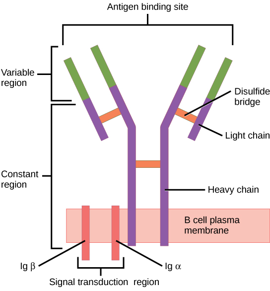
El receptor de linfocitos B consiste en la molécula de Ig y la molécula de señalización:
La Ig contiene 2 cadenas pesadas idénticas y 2 cadenas ligeras idénticas unidas por un puente disulfuro. La Ig unida a la membrana está anclada a la superficie celular.

Imagen: “Figure 42 02 06” por OpenStax. Licencia: CC BY 4.0

Estadios

Para alcanzar la funcionalidad, el linfocito B pasa por estadios en EN Erythema nodosum is an immune-mediated panniculitis (inflammation of the subcutaneous fat) caused by a type IV (delayed-type) hypersensitivity reaction. It commonly manifests in young women as tender, erythematous nodules on the shins. Erythema Nodosum la médula ósea y los LOS Neisseria órganos linfoides secundarios:

  • En EN Erythema nodosum is an immune-mediated panniculitis (inflammation of the subcutaneous fat) caused by a type IV (delayed-type) hypersensitivity reaction. It commonly manifests in young women as tender, erythematous nodules on the shins. Erythema Nodosum los LOS Neisseria estadios iniciales que ocurren en EN Erythema nodosum is an immune-mediated panniculitis (inflammation of the subcutaneous fat) caused by a type IV (delayed-type) hypersensitivity reaction. It commonly manifests in young women as tender, erythematous nodules on the shins. Erythema Nodosum la médula ósea, el objetivo es construir el receptor Receptor Receptors are proteins located either on the surface of or within a cell that can bind to signaling molecules known as ligands (e.g., hormones) and cause some type of response within the cell. Receptors (que no requiere antígeno).
  • Cuando se libera a los LOS Neisseria órganos linfoides secundarios, un antígeno (con o sin ayuda de linfocitos T) activará los LOS Neisseria linfocitos B para continuar el proceso de maduración.
Tabla: Estadios del desarrollo de los LOS Neisseria linfocitos B
Estadio de maduración Genes Genes A category of nucleic acid sequences that function as units of heredity and which code for the basic instructions for the development, reproduction, and maintenance of organisms. DNA Types and Structure Ig Ig X-linked Agammaglobulinemia Receptor Receptor Receptors are proteins located either on the surface of or within a cell that can bind to signaling molecules known as ligands (e.g., hormones) and cause some type of response within the cell. Receptors de linfocitos B Eventos asociados
Independiente de antígeno
Célula pre-pro-B ADN de línea germinal Ninguno Sin expresión de cadenas pesadas o ligeras
Célula pro-B IgH con recombinación D-J Ninguno Comienza a expresar CD19, CD34 y sistema del antígeno leucocitario humano (HLA, por sus siglas en EN Erythema nodosum is an immune-mediated panniculitis (inflammation of the subcutaneous fat) caused by a type IV (delayed-type) hypersensitivity reaction. It commonly manifests in young women as tender, erythematous nodules on the shins. Erythema Nodosum inglés)-DR (antígeno de histocompatibilidad de clase II)
Célula pre-B IgH con recombinación V-D-J Se forma el pre-receptor de linfocito B:
  • Cadena pesada presente
  • Cadena ligera sustituta presente
Aparecen otros marcadores (CD79, CD10, CD20, CD40 CD40 Members of the tumor necrosis factor receptor superfamily with specificity for CD40 ligand. They are found on mature B-lymphocytes, some epithelial cells; and lymphoid dendritic cells. Evidence suggests that CD40-dependent activation of B-cells is important for generation of memory B-cells within the germinal centers. Mutations in the CD40 antigen gene result in hyper-igm immunodeficiency syndrome, type 3. Signaling of the receptor occurs through its association with tnf receptor-associated factors. Hyper-IgM Syndrome y la desoxinucleotidil transferasa terminal entre ellos).
Linfocito B inmaduro
  • IgH con recombinación V-D-J
  • Cadena ligera con recombinación V-J
Receptor Receptor Receptors are proteins located either on the surface of or within a cell that can bind to signaling molecules known as ligands (e.g., hormones) and cause some type of response within the cell. Receptors de linfocito B maduro (molécula de IgM IgM A class of immunoglobulin bearing mu chains (immunoglobulin mu-chains). Igm can fix complement. The name comes from its high molecular weight and originally being called a macroglobulin. Immunoglobulins: Types and Functions) La expresión de HLA-DR, CD19, CD20 y CD40 CD40 Members of the tumor necrosis factor receptor superfamily with specificity for CD40 ligand. They are found on mature B-lymphocytes, some epithelial cells; and lymphoid dendritic cells. Evidence suggests that CD40-dependent activation of B-cells is important for generation of memory B-cells within the germinal centers. Mutations in the CD40 antigen gene result in hyper-igm immunodeficiency syndrome, type 3. Signaling of the receptor occurs through its association with tnf receptor-associated factors. Hyper-IgM Syndrome continúa, pero no la de los LOS Neisseria otros marcadores (e.g., CD10, CD34 y desoxinucleotidil transferasa terminal).
Linfocito B maduro (virgen)
  • IgH con recombinación V-D-J
  • Cadena ligera con recombinación V-J
Con receptor Receptor Receptors are proteins located either on the surface of or within a cell that can bind to signaling molecules known as ligands (e.g., hormones) and cause some type of response within the cell. Receptors de linfocitos B maduro ( IgM IgM A class of immunoglobulin bearing mu chains (immunoglobulin mu-chains). Igm can fix complement. The name comes from its high molecular weight and originally being called a macroglobulin. Immunoglobulins: Types and Functions) → salida de médula ósea Todos expresan CD19 y CD20.
Dependiente de antígeno
Linfocito B maduro ( en EN Erythema nodosum is an immune-mediated panniculitis (inflammation of the subcutaneous fat) caused by a type IV (delayed-type) hypersensitivity reaction. It commonly manifests in young women as tender, erythematous nodules on the shins. Erythema Nodosum tejidos linfoides secundarios) Receptor Receptor Receptors are proteins located either on the surface of or within a cell that can bind to signaling molecules known as ligands (e.g., hormones) and cause some type of response within the cell. Receptors de linfocito B maduro (expresa IgM IgM A class of immunoglobulin bearing mu chains (immunoglobulin mu-chains). Igm can fix complement. The name comes from its high molecular weight and originally being called a macroglobulin. Immunoglobulins: Types and Functions e IgD IgD An immunoglobulin which accounts for less than 1% of plasma immunoglobulin. It is found on the membrane of many circulating B lymphocytes. Immunoglobulins: Types and Functions al AL Amyloidosis estar dentro de los LOS Neisseria tejidos linfoides secundarios) Las células pueden descansar o puede ocurrir la activación de los LOS Neisseria linfocitos B: los LOS Neisseria linfocitos B interactúan con el antígeno exógeno y/o los LOS Neisseria linfocitos T colaboradores.
Linfocito B activado Cambio de clase Una vez activado, puede cambiar a IgE IgE An immunoglobulin associated with mast cells. Overexpression has been associated with allergic hypersensitivity. Immunoglobulins: Types and Functions, IgG IgG The major immunoglobulin isotype class in normal human serum. There are several isotype subclasses of igg, for example, igg1, igg2a, and igg2b. Hypersensitivity Pneumonitis, IgA IgA Represents 15-20% of the human serum immunoglobulins, mostly as the 4-chain polymer in humans or dimer in other mammals. Secretory iga is the main immunoglobulin in secretions. Immunoglobulins: Types and Functions o permanecer como IgM IgM A class of immunoglobulin bearing mu chains (immunoglobulin mu-chains). Igm can fix complement. The name comes from its high molecular weight and originally being called a macroglobulin. Immunoglobulins: Types and Functions
Linfocito B de memoria
  • Linfocito B activado → algunos se convierten en EN Erythema nodosum is an immune-mediated panniculitis (inflammation of the subcutaneous fat) caused by a type IV (delayed-type) hypersensitivity reaction. It commonly manifests in young women as tender, erythematous nodules on the shins. Erythema Nodosum linfocitos B de memoria
  • Circula, listo para reaccionar a la estimulación del antígeno y generar células plasmáticas
Célula plasmática
  • Linfocito B activado → algunos se convierten en EN Erythema nodosum is an immune-mediated panniculitis (inflammation of the subcutaneous fat) caused by a type IV (delayed-type) hypersensitivity reaction. It commonly manifests in young women as tender, erythematous nodules on the shins. Erythema Nodosum células plasmáticas
  • Células grandes que secretan anticuerpos que combaten la infección.
  • Migra a la médula ósea
D: segmento de diversidad
J: región de acoplamiento
V: región variable
Differentiation stages of the B cell

Estadios de diferenciación del linfocito B:
En estadios independientes del antígeno, la producción de linfocitos B comienza con la célula madre hematopoyética, que se convierte en un progenitor linfoide común y luego en una célula pre-pro-B o célula progenitora B. Los siguientes pasos incluyen la recombinación de genes para ensamblar la molécula de Ig. Las cadenas pesadas de Ig comienzan con la recombinación de los segmentos de diversidad y acoplamiento para formar la célula pro-B. En el siguiente paso (células pre-B), se completa la recombinación de la cadena pesada de Ig (región variable, segmento de diversidad, región de acoplamiento) y se forma el receptor de célula pre-B. Se produce la recombinación de la cadena ligera (kappa (κ) o lambda (λ)), lo que da como resultado la expresión de una molécula de anticuerpo IgM completa por parte de un linfocito B inmaduro. Sigue la formación del linfocito B maduro (virgen) con IgM e IgD.
Los estadios dependientes de antígeno ocurren en tejidos linfoides secundarios. Una vez que el linfocito B maduro produce IgM e IgD, puede tener lugar un cambio de clase para producir IgE, IgG e IgA. Los linfocitos B se activan y se convierten en células plasmáticas o linfocitos B de memoria.

Imagen por Lecturio.

Activación de Linfocitos B

El linfocito B migra desde la médula ósea a los LOS Neisseria órganos linfoides secundarios. Este proceso toma una serie de medidas para producir un linfocito B diferenciado funcional: activación por un antígeno, proliferación, maduración por afinidad, cambio de clase y diferenciación ( en EN Erythema nodosum is an immune-mediated panniculitis (inflammation of the subcutaneous fat) caused by a type IV (delayed-type) hypersensitivity reaction. It commonly manifests in young women as tender, erythematous nodules on the shins. Erythema Nodosum células plasmáticas o linfocitos B de memoria).

Proceso inicial de activación

  • Los LOS Neisseria linfocitos B vírgenes migran a los LOS Neisseria órganos linfoides secundarios, principalmente los LOS Neisseria ganglios linfáticos y el bazo.
    • En EN Erythema nodosum is an immune-mediated panniculitis (inflammation of the subcutaneous fat) caused by a type IV (delayed-type) hypersensitivity reaction. It commonly manifests in young women as tender, erythematous nodules on the shins. Erythema Nodosum los LOS Neisseria ganglios linfáticos:
      • Los LOS Neisseria linfocitos B están en EN Erythema nodosum is an immune-mediated panniculitis (inflammation of the subcutaneous fat) caused by a type IV (delayed-type) hypersensitivity reaction. It commonly manifests in young women as tender, erythematous nodules on the shins. Erythema Nodosum la corteza.
      • Los LOS Neisseria linfocitos T están en EN Erythema nodosum is an immune-mediated panniculitis (inflammation of the subcutaneous fat) caused by a type IV (delayed-type) hypersensitivity reaction. It commonly manifests in young women as tender, erythematous nodules on the shins. Erythema Nodosum la paracorteza.
      • La entrada de linfocitos B en EN Erythema nodosum is an immune-mediated panniculitis (inflammation of the subcutaneous fat) caused by a type IV (delayed-type) hypersensitivity reaction. It commonly manifests in young women as tender, erythematous nodules on the shins. Erythema Nodosum el tejido se realiza mediante la unión a un endotelio especializado (vénulas de endotelio alto).
    • Una vez en EN Erythema nodosum is an immune-mediated panniculitis (inflammation of the subcutaneous fat) caused by a type IV (delayed-type) hypersensitivity reaction. It commonly manifests in young women as tender, erythematous nodules on the shins. Erythema Nodosum los LOS Neisseria órganos linfoides secundarios, se expresan IgM IgM A class of immunoglobulin bearing mu chains (immunoglobulin mu-chains). Igm can fix complement. The name comes from its high molecular weight and originally being called a macroglobulin. Immunoglobulins: Types and Functions e IgD IgD An immunoglobulin which accounts for less than 1% of plasma immunoglobulin. It is found on the membrane of many circulating B lymphocytes. Immunoglobulins: Types and Functions de superficie.
  • Los LOS Neisseria linfocitos B son células en EN Erythema nodosum is an immune-mediated panniculitis (inflammation of the subcutaneous fat) caused by a type IV (delayed-type) hypersensitivity reaction. It commonly manifests in young women as tender, erythematous nodules on the shins. Erythema Nodosum reposo que sufren apoptosis Apoptosis A regulated cell death mechanism characterized by distinctive morphologic changes in the nucleus and cytoplasm, including the endonucleolytic cleavage of genomic DNA, at regularly spaced, internucleosomal sites, I.e., DNA fragmentation. It is genetically-programmed and serves as a balance to mitosis in regulating the size of animal tissues and in mediating pathologic processes associated with tumor growth. Ischemic Cell Damage si no son activadas (por el antígeno).
  • Se necesitan dos señales para la activación de los LOS Neisseria linfocitos B:
    • Señal 1: unión del antígeno al AL Amyloidosis receptor Receptor Receptors are proteins located either on the surface of or within a cell that can bind to signaling molecules known as ligands (e.g., hormones) and cause some type of response within the cell. Receptors de linfocito B (cuantos más receptores de linfocitos B entrecrucen con el antígeno, más fuerte es la señal)
    • Señal 2:
      • Las fuentes inflamatorias o antígenos presentan una amenaza para el huésped.
      • Sin la señal 2, los LOS Neisseria linfocitos B no se activan (esto evita la activación involuntaria por parte de antígenos inofensivos).
Sección histológica del ganglio linfático que muestra la corteza, la paracorteza y la médula

Sección histológica del ganglio linfático que muestra la corteza, la paracorteza y la médula

Imagen por Geoffrey Meyer, editada por Lecturio.
Estructura y regiones funcionales de un ganglio linfático

Estructura y regiones funcionales de un ganglio linfático: comprenden una cápsula fibrosa rica en colágeno y un seno subcapsular subyacente.
Las células se segregan en (1) la corteza (que consta de linfocitos B, linfocitos T colaboradores foliculares y células dendríticas foliculares dispuestos en folículos primarios, en los que los linfocitos B examinan los antígenos presentados en la red estromal de células dendríticas foliculares); y (2) la paracorteza (aloja los linfocitos T, las células dendríticas y células reticulares fibroblásticas que forman redes de células estromales y fibras reticulares).
La médula interna está compuesta de tejidos linfáticos (cordones medulares) separados por senos medulares que consisten en linfa.

Imagen: “The structure of the lymph node” por Colbeck, Ager, Gallimore y Jones. Licencia: CC BY 4.0

Selección clonal

  • Desafío antigénico:
    • La interacción ocurre solo entre el antígeno y el linfocito B con el “ajuste” apropiado o la mejor “pareja” (basado en EN Erythema nodosum is an immune-mediated panniculitis (inflammation of the subcutaneous fat) caused by a type IV (delayed-type) hypersensitivity reaction. It commonly manifests in young women as tender, erythematous nodules on the shins. Erythema Nodosum el receptor Receptor Receptors are proteins located either on the surface of or within a cell that can bind to signaling molecules known as ligands (e.g., hormones) and cause some type of response within the cell. Receptors de linfocitos B específico).
    • Esta es una forma de selección positiva, con el linfocito B recién unido activado para responder.
    • Una vez que se produce la unión, ese linfocito B se divide y forma un clon.
  • El clon seleccionado experimentará expansión clonal (o proliferación) con la ayuda de linfocitos T.

Tipos de activación

La activación de los LOS Neisseria linfocitos B por presentación de antígeno puede tener diferentes caminos:

  • Dependiente de linfocitos T:
    • El antígeno circulante interactúa con el receptor Receptor Receptors are proteins located either on the surface of or within a cell that can bind to signaling molecules known as ligands (e.g., hormones) and cause some type of response within the cell. Receptors de linfocitos B.
    • El antígeno sufre endocitosis y degradación.
    • Luego, los LOS Neisseria componentes peptídicos forman complejos con moléculas del CMH II de la superficie celular.
    • Rol de los LOS Neisseria linfocitos T:
      • Los LOS Neisseria linfocitos T colaboradores foliculares (Tfh, por sus siglas en EN Erythema nodosum is an immune-mediated panniculitis (inflammation of the subcutaneous fat) caused by a type IV (delayed-type) hypersensitivity reaction. It commonly manifests in young women as tender, erythematous nodules on the shins. Erythema Nodosum inglés) CD4+ especializados previamente activados por células dendríticas (presentando el mismo antígeno).
      • Los LOS Neisseria Tfh reconocen y se unen al AL Amyloidosis complejo antígeno-CMH II.
      • Luego, los LOS Neisseria Tfh expresan el ligando CD40 CD40 Members of the tumor necrosis factor receptor superfamily with specificity for CD40 ligand. They are found on mature B-lymphocytes, some epithelial cells; and lymphoid dendritic cells. Evidence suggests that CD40-dependent activation of B-cells is important for generation of memory B-cells within the germinal centers. Mutations in the CD40 antigen gene result in hyper-igm immunodeficiency syndrome, type 3. Signaling of the receptor occurs through its association with tnf receptor-associated factors. Hyper-IgM Syndrome, que se une al AL Amyloidosis CD40 CD40 Members of the tumor necrosis factor receptor superfamily with specificity for CD40 ligand. They are found on mature B-lymphocytes, some epithelial cells; and lymphoid dendritic cells. Evidence suggests that CD40-dependent activation of B-cells is important for generation of memory B-cells within the germinal centers. Mutations in the CD40 antigen gene result in hyper-igm immunodeficiency syndrome, type 3. Signaling of the receptor occurs through its association with tnf receptor-associated factors. Hyper-IgM Syndrome de los LOS Neisseria linfocitos B, lo que lleva a la activación y proliferación de los LOS Neisseria linfocitos B.
    • Los LOS Neisseria linfocitos B activados entran y proliferan en EN Erythema nodosum is an immune-mediated panniculitis (inflammation of the subcutaneous fat) caused by a type IV (delayed-type) hypersensitivity reaction. It commonly manifests in young women as tender, erythematous nodules on the shins. Erythema Nodosum los LOS Neisseria centros germinales, donde continúan el proceso que lleva a la diferenciación.
    • Un ejemplo de ello es la vacuna neumocócica conjugada 13-valente:
      • El conjugado de polisacárido-proteína induce una respuesta inmunitaria dependiente de linfocitos T.
      • Forma anticuerpos específicos del serotipo neumocócico y linfocitos B de memoria, lo que crea una memoria inmunológica.
  • Independiente de linfocitos T:
    • La activación de los LOS Neisseria linfocitos B no siempre necesita la ayuda de los LOS Neisseria linfocitos T.
    • Algunos antígenos, como los LOS Neisseria polisacáridos de una célula bacteriana, pueden estimular directamente a los LOS Neisseria linfocitos B y unirse a muchos receptores de IgM IgM A class of immunoglobulin bearing mu chains (immunoglobulin mu-chains). Igm can fix complement. The name comes from its high molecular weight and originally being called a macroglobulin. Immunoglobulins: Types and Functions para lograr una fuerte señal 1.
    • La señal 2 pueden ser derivados del complemento C3b unidos a la célula bacteriana o patrones moleculares asociados a patógenos.
    • Pasando por alto la ayuda de los LOS Neisseria linfocitos T, estas respuestas son de corta duración, generadas principalmente por la producción de IgM IgM A class of immunoglobulin bearing mu chains (immunoglobulin mu-chains). Igm can fix complement. The name comes from its high molecular weight and originally being called a macroglobulin. Immunoglobulins: Types and Functions (cambio de clase limitado y sin linfocitos B de memoria).
    • Un ejemplo es la vacuna neumocócica polisacárida 23:
      • Transporta polisacáridos de superficie de 23 serotipos de Streptococcus Streptococcus Streptococcus is one of the two medically important genera of gram-positive cocci, the other being Staphylococcus. Streptococci are identified as different species on blood agar on the basis of their hemolytic pattern and sensitivity to optochin and bacitracin. There are many pathogenic species of streptococci, including S. pyogenes, S. agalactiae, S. pneumoniae, and the viridans streptococci. Streptococcus pneumoniae
      • La señal 1 es el polisacárido y la señal 2 es el adyuvante (no hay péptidos/proteínas para ser reconocidos por los LOS Neisseria linfocitos T colaboradores).
      • Con las 2 señales, la activación y proliferación de los LOS Neisseria linfocitos B ocurre independientemente de los LOS Neisseria linfocitos T.
Unión de células T y B

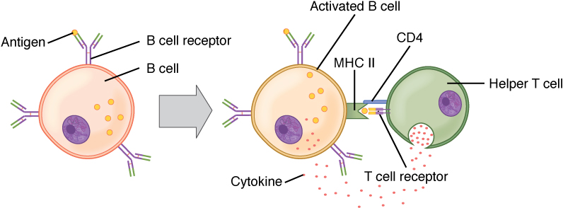
Activación de linfocitos B (dependiente de linfocitos T):
El antígeno circulante interactúa con el receptor de linfocito B. El antígeno se somete a endocitosis y se degrada y los componentes peptídicos forman un complejo con moléculas del CMH II de la superficie celular. Los linfocitos T colaboradores foliculares (Tfh) (linfocitos T colaboradores CD4+ especializados) reconocen y se unen al complejo antígeno-CMH II. Los Tfh liberan citocinas, lo que lleva a la activación y proliferación de linfocitos B. Los linfocitos B activados ingresan a los centros germinales, donde continúan el proceso, lo que lleva a la diferenciación.

Imagen:T and B cell binding” por OpenStax College. Licencia: CC BY 3.0

Maduración y Diferenciación de Linfocitos B

Maduración de la afinidad

  • Mientras que el linfocito B se ha activado, se llevan a cabo procesos en la zona oscura del centro germinal para “ajustar” aún más la afinidad del anticuerpo por el antígeno.
  • La maduración por afinidad es el mecanismo por el cual los LOS Neisseria linfocitos B, después de una estimulación repetida, aumentan su afinidad por un antígeno específico que se les presenta.
  • La hipermutación somática facilita el aumento de la afinidad:
    • Una mutación programada que involucra las regiones variables de los LOS Neisseria genes Genes A category of nucleic acid sequences that function as units of heredity and which code for the basic instructions for the development, reproduction, and maintenance of organisms. DNA Types and Structure de cadena ligera y pesada de Ig Ig X-linked Agammaglobulinemia, que ocurre después de la activación dependiente de antígeno
    • Impulsado por enzimas modificadoras del ADN:
      • Desaminasa de citidina inducida por activación
      • Uracilo nucleósido glicosilasa
    • Produce un receptor Receptor Receptors are proteins located either on the surface of or within a cell that can bind to signaling molecules known as ligands (e.g., hormones) and cause some type of response within the cell. Receptors de linfocitos B con capacidad mejorada para reconocer y unirse al AL Amyloidosis antígeno
  • Selección:
    • Después de la mutación, los LOS Neisseria linfocitos B con receptor Receptor Receptors are proteins located either on the surface of or within a cell that can bind to signaling molecules known as ligands (e.g., hormones) and cause some type of response within the cell. Receptors de linfocitos B de alta afinidad ahora se mueven a la zona clara y acceden al AL Amyloidosis antígeno presentado por las células dendríticas foliculares.
      • Alta afinidad con el antígeno → es más probable que se seleccionen para presentar y recibir señales de supervivencia de los LOS Neisseria Tfh
      • Los LOS Neisseria linfocitos B con menor afinidad no recibirán señales de supervivencia y morirán por apoptosis Apoptosis A regulated cell death mechanism characterized by distinctive morphologic changes in the nucleus and cytoplasm, including the endonucleolytic cleavage of genomic DNA, at regularly spaced, internucleosomal sites, I.e., DNA fragmentation. It is genetically-programmed and serves as a balance to mitosis in regulating the size of animal tissues and in mediating pathologic processes associated with tumor growth. Ischemic Cell Damage.
  • Este proceso no solo permite la diversidad, sino que también permite que solo los LOS Neisseria linfocitos B más optimizados sobrevivan y se diferencien.

Recombinación de cambio de clase

  • Los LOS Neisseria linfocitos B supervivientes (con alta afinidad por el antígeno) luego se someten a un cambio de clase, un paso que también requiere desaminasa de citidina inducida por activación.
    • La región constante de la cadena pesada puede cambiar el segmento μ a uno de los LOS Neisseria otros segmentos de la cadena pesada (γ, ε o α).
    • La composición de la cadena pesada determina la clase de Ig Ig X-linked Agammaglobulinemia:
      • µ: IgM IgM A class of immunoglobulin bearing mu chains (immunoglobulin mu-chains). Igm can fix complement. The name comes from its high molecular weight and originally being called a macroglobulin. Immunoglobulins: Types and Functions
      • δ: IgD IgD An immunoglobulin which accounts for less than 1% of plasma immunoglobulin. It is found on the membrane of many circulating B lymphocytes. Immunoglobulins: Types and Functions
      • γ: IgG IgG The major immunoglobulin isotype class in normal human serum. There are several isotype subclasses of igg, for example, igg1, igg2a, and igg2b. Hypersensitivity Pneumonitis
      • α: IgA IgA Represents 15-20% of the human serum immunoglobulins, mostly as the 4-chain polymer in humans or dimer in other mammals. Secretory iga is the main immunoglobulin in secretions. Immunoglobulins: Types and Functions
      • ε: IgE IgE An immunoglobulin associated with mast cells. Overexpression has been associated with allergic hypersensitivity. Immunoglobulins: Types and Functions
    • El cambio está influenciado por las citocinas.
      • TGF-β: cambia preferentemente a IgA IgA Represents 15-20% of the human serum immunoglobulins, mostly as the 4-chain polymer in humans or dimer in other mammals. Secretory iga is the main immunoglobulin in secretions. Immunoglobulins: Types and Functions
      • IL-4: IgE IgE An immunoglobulin associated with mast cells. Overexpression has been associated with allergic hypersensitivity. Immunoglobulins: Types and Functions
      • IFN-γ, IL-4: IgG IgG The major immunoglobulin isotype class in normal human serum. There are several isotype subclasses of igg, for example, igg1, igg2a, and igg2b. Hypersensitivity Pneumonitis
    • La región constante de la cadena pesada de Ig Ig X-linked Agammaglobulinemia cambia, pero la región variable Variable Variables represent information about something that can change. The design of the measurement scales, or of the methods for obtaining information, will determine the data gathered and the characteristics of that data. As a result, a variable can be qualitative or quantitative, and may be further classified into subgroups. Types of Variables permanece sin cambios.
    • Dado que la región variable Variable Variables represent information about something that can change. The design of the measurement scales, or of the methods for obtaining information, will determine the data gathered and the characteristics of that data. As a result, a variable can be qualitative or quantitative, and may be further classified into subgroups. Types of Variables está intacta, la especificidad del anticuerpo no cambia.
  • Después del cambio de clase, los LOS Neisseria linfocitos B salen de los LOS Neisseria centros germinales y se diferencian terminalmente en EN Erythema nodosum is an immune-mediated panniculitis (inflammation of the subcutaneous fat) caused by a type IV (delayed-type) hypersensitivity reaction. It commonly manifests in young women as tender, erythematous nodules on the shins. Erythema Nodosum células plasmáticas o linfocitos B de memoria.
Procesos de activación y maduración de células B en el centro germinal

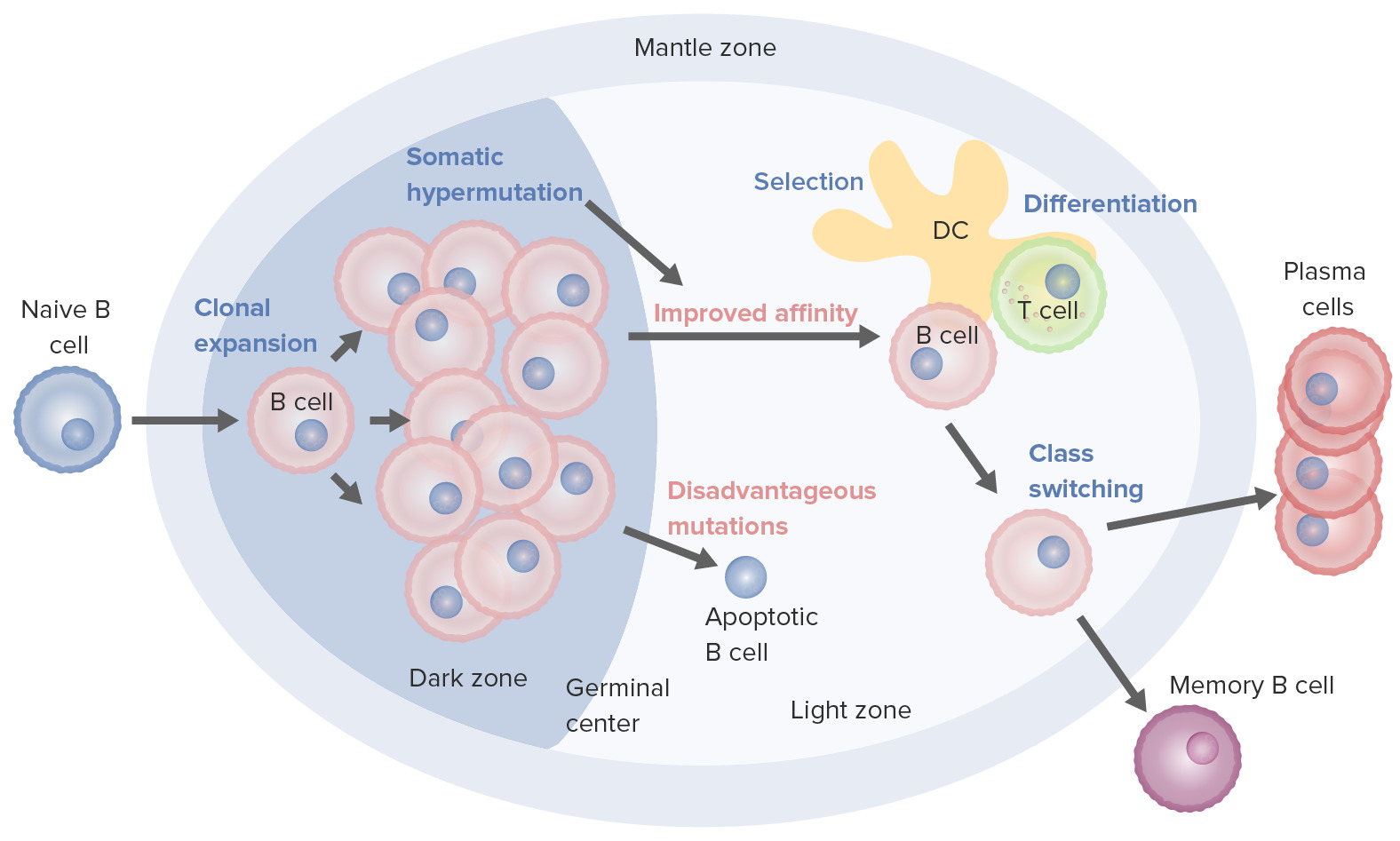
Procesos de activación y maduración de los linfocitos B que tienen lugar en el centro germinal:
Al activarse, el linfocito B se mueve desde la zona del manto y entra al centro germinal. Ocurre la proliferación de linfocitos B (expansión clonal) y la afinidad del anticuerpo por el antígeno aumenta a través del proceso de hipermutación somática. Los ciclos repetidos de proliferación e hipermutación afinan el receptor de linfocito B. Sin embargo, no todos los linfocitos B continúan diferenciándose, especialmente si la afinidad es débil. La apoptosis ocurre si la unión antígeno-anticuerpo no se optimiza. Aquellos con fuerte afinidad sobreviven (selección), con la ayuda de señales de supervivencia de células dendríticas foliculares y linfocitos T. Estos linfocitos B seleccionados pasan al cambio de clase y la diferenciación en células plasmáticas o linfocitos B de memoria.

Imagen por Lecturio. Licencia: CC BY-NC-SA 4.0
Tinción de hematoxilina y eosina del centro germinal del tejido linfoide secundario

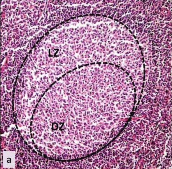
Centro germinal: histología del centro germinal de un tejido linfoide secundario
LZ: zona clara (en inglés)
DZ: zona oscura (en inglés)

Imagen:Haematoxylin and eosin stain” por Petra Korać et al. Licencia: CC BY 4.0, recortada por Lecturio.

Células plasmáticas y linfocitos B de memoria

  • Células plasmáticas:
    • Células grandes (hasta 20 micras de diámetro)
    • Producen anticuerpos
    • Migran a la médula ósea
  • Linfocitos B de memoria:
    • Reaccionan a la estimulación antigénica ( en EN Erythema nodosum is an immune-mediated panniculitis (inflammation of the subcutaneous fat) caused by a type IV (delayed-type) hypersensitivity reaction. It commonly manifests in young women as tender, erythematous nodules on the shins. Erythema Nodosum respuesta a la reinfección)
    • Generan células plasmáticas, que tienen anticuerpos de alta afinidad en EN Erythema nodosum is an immune-mediated panniculitis (inflammation of the subcutaneous fat) caused by a type IV (delayed-type) hypersensitivity reaction. It commonly manifests in young women as tender, erythematous nodules on the shins. Erythema Nodosum respuestas inmunitarias secundarias
Summary of B cell development to differentiation

Resumen del desarrollo de los linfocitos B hasta la diferenciación (desde la médula ósea hasta el órgano linfoide secundario):

Desarrollo de linfocitos B:
En la médula ósea, los linfocitos B se convierten en linfocitos B inmaduros, un proceso en el que se ensambla el receptor de linfocitos B. Luego, el linfocito B migra a los órganos linfoides secundarios, donde se produce la activación.

Activación de linfocitos B:
El antígeno se une al linfocito B con el receptor de “mejor compatibilidad”. Una vía de activación es independiente de los linfocitos T, por lo que el linfocito B activado se activa para diferenciarse en una célula plasmática de vida corta (que produce anticuerpos) sin la ayuda del linfocito T. En la activación dependiente de linfocitos T, el linfocito T reconoce el antígeno CMH II y desencadena la proliferación del linfocito B en el centro germinal del tejido linfoide.

Proliferación y maduración:
El proceso va seguido de una hipermutación somática (una mutación programada para ajustar aún más la afinidad del anticuerpo por el antígeno). Los ciclos repetidos de proliferación e hipermutación refinan el receptor de linfocito B. Solo aquellos con la mejor afinidad serán seleccionados y sobrevivirán; aquellos con baja afinidad sufrirán apoptosis. Los linfocitos B supervivientes luego pasan por una recombinación de cambio de clase, en la que se cambia la composición de la cadena pesada (IgM a otros isotipos) con la ayuda de citoquinas.

Diferenciación:
Estos linfocitos B luego se diferencian en células plasmáticas y linfocitos B de memoria, dejando el centro germinal.

Imagen por Lecturio.

Diversidad de Anticuerpos

Desde la producción inicial de linfocitos B, muchos procesos permiten a los LOS Neisseria humanos producir diferentes moléculas de anticuerpos que son significativamente más que la cantidad de genes Genes A category of nucleic acid sequences that function as units of heredity and which code for the basic instructions for the development, reproduction, and maintenance of organisms. DNA Types and Structure en EN Erythema nodosum is an immune-mediated panniculitis (inflammation of the subcutaneous fat) caused by a type IV (delayed-type) hypersensitivity reaction. It commonly manifests in young women as tender, erythematous nodules on the shins. Erythema Nodosum el genoma.

Se estima que se generan miles de millones de anticuerpos, frente a unos 30 000 genes Genes A category of nucleic acid sequences that function as units of heredity and which code for the basic instructions for the development, reproduction, and maintenance of organisms. DNA Types and Structure.

El sistema inmunitario tiene mecanismos únicos para crear diversidad de anticuerpos, que incluyen:

  • Tener múltiples segmentos V, D, J:
    • Como se mencionó en EN Erythema nodosum is an immune-mediated panniculitis (inflammation of the subcutaneous fat) caused by a type IV (delayed-type) hypersensitivity reaction. It commonly manifests in young women as tender, erythematous nodules on the shins. Erythema Nodosum la discusión sobre el desarrollo temprano de linfocitos B, las cadenas pesadas y las cadenas ligeras tienen múltiples segmentos.
    • V, D, J, C para cadena pesada
    • V, J, C para cadena ligera
  • Reordenamientos de los LOS Neisseria segmentos V, D, J:
    • Las secuencias de ADN (llamadas secuencias de señal de recombinación) flanquean cada segmento de gen.
    • Estas secuencias son sitios de reconocimiento para el proceso de acoplamiento.
    • El complejo enzimático recombinasa RAG1 RAG1 Immunoglobulins: Types and Functions y RAG2 RAG2 Immunoglobulins: Types and Functions ( genes Genes A category of nucleic acid sequences that function as units of heredity and which code for the basic instructions for the development, reproduction, and maintenance of organisms. DNA Types and Structure activadores de recombinación 1 y 2) reconoce las secuencias de señal de recombinación y cataliza el proceso de acoplamiento.
    • La deficiencia en EN Erythema nodosum is an immune-mediated panniculitis (inflammation of the subcutaneous fat) caused by a type IV (delayed-type) hypersensitivity reaction. It commonly manifests in young women as tender, erythematous nodules on the shins. Erythema Nodosum RAG1 RAG1 Immunoglobulins: Types and Functions o RAG2 RAG2 Immunoglobulins: Types and Functions puede producir linfocitos B no funcionales.
    • ***Como se mencionó anteriormente en EN Erythema nodosum is an immune-mediated panniculitis (inflammation of the subcutaneous fat) caused by a type IV (delayed-type) hypersensitivity reaction. It commonly manifests in young women as tender, erythematous nodules on the shins. Erythema Nodosum la sección anterior, después de los LOS Neisseria segmentos de la cadena pesada, los LOS Neisseria segmentos de la cadena ligera también se recombinan.
  • Diversidad de acoplamiento:
    • El acoplamiento de segmentos de genes Genes A category of nucleic acid sequences that function as units of heredity and which code for the basic instructions for the development, reproduction, and maintenance of organisms. DNA Types and Structure de anticuerpos puede ser imprecisa.
    • Se pueden eliminar y/o insertar varios nucleótidos desde los LOS Neisseria extremos de los LOS Neisseria segmentos del gen recombinante.
  • Diversidad combinatoria: la diversidad se crea mediante el emparejamiento aleatorio de las cadenas pesada y ligera.
  • Hipermutación somática:
    • Las mutaciones puntuales ocurren con la estimulación repetida de antígenos (de respuestas primarias a secundarias).
    • Aumenta la afinidad por el antígeno
    • Crea diversidad adicional al AL Amyloidosis anticuerpo

Relevancia Clínica

  • Agammaglobulinemia ligada al AL Amyloidosis cromosoma X: resulta de mutaciones en EN Erythema nodosum is an immune-mediated panniculitis (inflammation of the subcutaneous fat) caused by a type IV (delayed-type) hypersensitivity reaction. It commonly manifests in young women as tender, erythematous nodules on the shins. Erythema Nodosum el gen del cromosoma X que codifica la tirosina quinasa de Bruton, que es esencial para el desarrollo y la maduración de los LOS Neisseria linfocitos B. La enfermedad se caracteriza por la ausencia de linfocitos B, lo que da lugar a infecciones recurrentes, principalmente por bacterias y virus Virus Viruses are infectious, obligate intracellular parasites composed of a nucleic acid core surrounded by a protein capsid. Viruses can be either naked (non-enveloped) or enveloped. The classification of viruses is complex and based on many factors, including type and structure of the nucleoid and capsid, the presence of an envelope, the replication cycle, and the host range. Virology encapsulados, que afectan a los LOS Neisseria pulmones, los LOS Neisseria senos paranasales y la piel, así como al AL Amyloidosis SNC. El tratamiento consiste en EN Erythema nodosum is an immune-mediated panniculitis (inflammation of the subcutaneous fat) caused by a type IV (delayed-type) hypersensitivity reaction. It commonly manifests in young women as tender, erythematous nodules on the shins. Erythema Nodosum la administración de inmunoglobulina.
  • Inmunodeficiencia común variable Variable Variables represent information about something that can change. The design of the measurement scales, or of the methods for obtaining information, will determine the data gathered and the characteristics of that data. As a result, a variable can be qualitative or quantitative, and may be further classified into subgroups. Types of Variables: también conocida como inmunodeficiencia humoral. La inmunodeficiencia común variable Variable Variables represent information about something that can change. The design of the measurement scales, or of the methods for obtaining information, will determine the data gathered and the characteristics of that data. As a result, a variable can be qualitative or quantitative, and may be further classified into subgroups. Types of Variables es un trastorno del sistema inmunológico caracterizado por niveles séricos reducidos de IgG IgG The major immunoglobulin isotype class in normal human serum. There are several isotype subclasses of igg, for example, igg1, igg2a, and igg2b. Hypersensitivity Pneumonitis, IgA IgA Represents 15-20% of the human serum immunoglobulins, mostly as the 4-chain polymer in humans or dimer in other mammals. Secretory iga is the main immunoglobulin in secretions. Immunoglobulins: Types and Functions e IgM IgM A class of immunoglobulin bearing mu chains (immunoglobulin mu-chains). Igm can fix complement. The name comes from its high molecular weight and originally being called a macroglobulin. Immunoglobulins: Types and Functions. Las causas subyacentes de la inmunodeficiencia común variable Variable Variables represent information about something that can change. The design of the measurement scales, or of the methods for obtaining information, will determine the data gathered and the characteristics of that data. As a result, a variable can be qualitative or quantitative, and may be further classified into subgroups. Types of Variables se desconocen en EN Erythema nodosum is an immune-mediated panniculitis (inflammation of the subcutaneous fat) caused by a type IV (delayed-type) hypersensitivity reaction. It commonly manifests in young women as tender, erythematous nodules on the shins. Erythema Nodosum gran medida. Los LOS Neisseria pacientes con esta afección son propensos a infecciones en EN Erythema nodosum is an immune-mediated panniculitis (inflammation of the subcutaneous fat) caused by a type IV (delayed-type) hypersensitivity reaction. It commonly manifests in young women as tender, erythematous nodules on the shins. Erythema Nodosum el tracto gastrointestinal y en EN Erythema nodosum is an immune-mediated panniculitis (inflammation of the subcutaneous fat) caused by a type IV (delayed-type) hypersensitivity reaction. It commonly manifests in young women as tender, erythematous nodules on the shins. Erythema Nodosum las vías respiratorias superiores e inferiores. La inmunodeficiencia común variable Variable Variables represent information about something that can change. The design of the measurement scales, or of the methods for obtaining information, will determine the data gathered and the characteristics of that data. As a result, a variable can be qualitative or quantitative, and may be further classified into subgroups. Types of Variables también se asocia con un mayor riesgo de desarrollar trastornos autoinmunes, enfermedades granulomatosas y malignidad. El tratamiento es la terapia de reemplazo con inmunoglobulina.
  • Síndrome de hiper IgM IgM A class of immunoglobulin bearing mu chains (immunoglobulin mu-chains). Igm can fix complement. The name comes from its high molecular weight and originally being called a macroglobulin. Immunoglobulins: Types and Functions: caracterizado por niveles normales o elevados de IgM IgM A class of immunoglobulin bearing mu chains (immunoglobulin mu-chains). Igm can fix complement. The name comes from its high molecular weight and originally being called a macroglobulin. Immunoglobulins: Types and Functions con niveles disminuidos o ausentes de otras Ig Ig X-linked Agammaglobulinemia. Hay tipos de síndrome de hiper IgM IgM A class of immunoglobulin bearing mu chains (immunoglobulin mu-chains). Igm can fix complement. The name comes from its high molecular weight and originally being called a macroglobulin. Immunoglobulins: Types and Functions ligado al AL Amyloidosis cromosoma X y autosómico recesivo. Los LOS Neisseria pacientes presentan infecciones sinopulmonares recurrentes, diarrea crónica e hiperplasia linfoide. El diagnóstico se verifica mediante pruebas genéticas. El tratamiento incluye terapia de reemplazo con inmunoglobulina y antibióticos profilácticos. El trasplante de células madre hematopoyéticas es otra opción.
  • Deficiencia de IgA IgA Represents 15-20% of the human serum immunoglobulins, mostly as the 4-chain polymer in humans or dimer in other mammals. Secretory iga is the main immunoglobulin in secretions. Immunoglobulins: Types and Functions: caracterizada por niveles bajos de IgA IgA Represents 15-20% of the human serum immunoglobulins, mostly as the 4-chain polymer in humans or dimer in other mammals. Secretory iga is the main immunoglobulin in secretions. Immunoglobulins: Types and Functions, con niveles normales de IgG IgG The major immunoglobulin isotype class in normal human serum. There are several isotype subclasses of igg, for example, igg1, igg2a, and igg2b. Hypersensitivity Pneumonitis e IgM IgM A class of immunoglobulin bearing mu chains (immunoglobulin mu-chains). Igm can fix complement. The name comes from its high molecular weight and originally being called a macroglobulin. Immunoglobulins: Types and Functions. La deficiencia de IgA IgA Represents 15-20% of the human serum immunoglobulins, mostly as the 4-chain polymer in humans or dimer in other mammals. Secretory iga is the main immunoglobulin in secretions. Immunoglobulins: Types and Functions es la inmunodeficiencia primaria más frecuente. Muchos pacientes son asintomáticos; sin embargo, existe la posibilidad de infecciones recurrentes, así como de enfermedades autoinmunes. Los LOS Neisseria pacientes pueden ser propensos a reacciones transfusionales anafilácticas debido a la presencia de IgA IgA Represents 15-20% of the human serum immunoglobulins, mostly as the 4-chain polymer in humans or dimer in other mammals. Secretory iga is the main immunoglobulin in secretions. Immunoglobulins: Types and Functions en EN Erythema nodosum is an immune-mediated panniculitis (inflammation of the subcutaneous fat) caused by a type IV (delayed-type) hypersensitivity reaction. It commonly manifests in young women as tender, erythematous nodules on the shins. Erythema Nodosum los LOS Neisseria productos sanguíneos. Algunos de estos casos eventualmente progresan a inmunodeficiencia común variable Variable Variables represent information about something that can change. The design of the measurement scales, or of the methods for obtaining information, will determine the data gathered and the characteristics of that data. As a result, a variable can be qualitative or quantitative, and may be further classified into subgroups. Types of Variables. El tratamiento implica antibióticos profilácticos y evitar productos sanguíneos que contengan IgA IgA Represents 15-20% of the human serum immunoglobulins, mostly as the 4-chain polymer in humans or dimer in other mammals. Secretory iga is the main immunoglobulin in secretions. Immunoglobulins: Types and Functions.

Referencias

  1. Aster, J.C. (2023). Normal B and T lymphocyte development. UpToDate. Retrieved October 28, 2025, from https://www.uptodate.com/contents/normal-b-and-t-lymphocyte-development
  2. Fernandez, J. (2024). X-linked agammaglobulinemia. Merck Manual Professional Version. Retrieved October 28, 2025, from https://www.merckmanuals.com/professional/immunology-allergic-disorders/immunodeficiency-disorders/x-linked-agammaglobulinemia
  3. Fernandez, J. (2024). Common variable immunodeficiency (CVID). Merck Manual Professional Version. Retrieved October 28, 2025, from https://www.merckmanuals.com/professional/immunology-allergic-disorders/immunodeficiency-disorders/common-variable-immunodeficiency-cvid
  4. Fernandez, J. (2024). Hyper-IgM syndrome. Merck Manual Professional Version. Retrieved October 28, 2025, from https://www.merckmanuals.com/professional/immunology-allergic-disorders/immunodeficiency-disorders/hyper-igm-syndrome
  5. Fernandez, J. (2024). Selective IgA deficiency. Merck Manual Professional Version. Retrieved October 28, 2025, from https://www.merckmanuals.com/professional/immunology-allergic-disorders/immunodeficiency-disorders/selective-iga-deficiency
  6. Kipps, T.J. (2021). Functions of B lymphocytes and plasma cells in immunoglobulin production. In: Kaushansky, K., Prchal, J.T., Burns, L.J., Lichtman, M.A., Levi, M, Linch, D.C. (Eds.), Williams Hematology, 10th ed. McGraw-Hill. https://accessmedicine.mhmedical.com/content.aspx?bookid=2962&sectionid=252532543
  7. Liew, P. (2012). The Longevity of the Humoral Immune Response: Survival of Long-lived Plasma Cells. Akademeia. 2. ea0116. 
  8. Riedel, S., Hobden, J.A., Miller, S., Morse, S.A., Mietzner, T.A., Detrick, B., Mitchell, T.G., Sakanari, J.A., Hotez, P., Mejia, R. (Eds.), (2019). Immunology. In: Jawetz, Melnick, & Adelberg’s Medical Microbiology, 28th ed. McGraw-Hill. https://accessmedicine.mhmedical.com/content.aspx?bookid=2629&sectionid=217769996
  9. Schroeder, H.W., Cavacini, L. (2010). Structure and function of immunoglobulins. J Allergy Clin Immunol 125(2 Suppl 2):S41–S52. https://pubmed.ncbi.nlm.nih.gov/20176268/
  10. Walter, J. (2024). Immunoglobulin genetics. UpToDate. Retrieved October 28, 2025, from https://www.uptodate.com/contents/immunoglobulin-genetics

¡Crea tu cuenta gratis o inicia una sesión para seguir leyendo!

Regístrate ahora y obtén acceso gratuito a Lecturio con páginas de concepto, videos médicos y cuestionarios para tu educación médica.

User Reviews

Que tengas una sesión de estudio alegre y navideña 🎁 Ahorra 50% en todos los planes >>

Details