Domina Conceptos Médicos

Estudia para la escuela de medicina y tus examenes con Lecturio.

Canal Inguinal: Anatomía y Hernias

La región inguinal, o ingle, se localiza en EN Erythema nodosum is an immune-mediated panniculitis (inflammation of the subcutaneous fat) caused by a type IV (delayed-type) hypersensitivity reaction. It commonly manifests in young women as tender, erythematous nodules on the shins. Erythema Nodosum el cuadrante inferior derecho e izquierdo de la pared abdominal anterior, delimitada por el muslo en EN Erythema nodosum is an immune-mediated panniculitis (inflammation of the subcutaneous fat) caused by a type IV (delayed-type) hypersensitivity reaction. It commonly manifests in young women as tender, erythematous nodules on the shins. Erythema Nodosum la parte inferior, el pubis en EN Erythema nodosum is an immune-mediated panniculitis (inflammation of the subcutaneous fat) caused by a type IV (delayed-type) hypersensitivity reaction. It commonly manifests in young women as tender, erythematous nodules on the shins. Erythema Nodosum la parte medial y la cresta ilíaca en EN Erythema nodosum is an immune-mediated panniculitis (inflammation of the subcutaneous fat) caused by a type IV (delayed-type) hypersensitivity reaction. It commonly manifests in young women as tender, erythematous nodules on the shins. Erythema Nodosum la superolateral. El canal inguinal es una estructura tubular que corre en EN Erythema nodosum is an immune-mediated panniculitis (inflammation of the subcutaneous fat) caused by a type IV (delayed-type) hypersensitivity reaction. It commonly manifests in young women as tender, erythematous nodules on the shins. Erythema Nodosum línea recta desde la espina ilíaca anterosuperior hasta el tubérculo púbico. El canal contiene el cordón espermático en EN Erythema nodosum is an immune-mediated panniculitis (inflammation of the subcutaneous fat) caused by a type IV (delayed-type) hypersensitivity reaction. It commonly manifests in young women as tender, erythematous nodules on the shins. Erythema Nodosum los LOS Neisseria hombres y el ligamento redondo en EN Erythema nodosum is an immune-mediated panniculitis (inflammation of the subcutaneous fat) caused by a type IV (delayed-type) hypersensitivity reaction. It commonly manifests in young women as tender, erythematous nodules on the shins. Erythema Nodosum las mujeres. Una hernia Hernia Protrusion of tissue, structure, or part of an organ through the bone, muscular tissue, or the membrane by which it is normally contained. Hernia may involve tissues such as the abdominal wall or the respiratory diaphragm. Hernias may be internal, external, congenital, or acquired. Abdominal Hernias inguinal se produce cuando un tejido o un órgano (como una porción del intestino) sobresale a través de la pared abdominal y en EN Erythema nodosum is an immune-mediated panniculitis (inflammation of the subcutaneous fat) caused by a type IV (delayed-type) hypersensitivity reaction. It commonly manifests in young women as tender, erythematous nodules on the shins. Erythema Nodosum el canal inguinal. Las hernias inguinales son el tipo más común de hernia Hernia Protrusion of tissue, structure, or part of an organ through the bone, muscular tissue, or the membrane by which it is normally contained. Hernia may involve tissues such as the abdominal wall or the respiratory diaphragm. Hernias may be internal, external, congenital, or acquired. Abdominal Hernias, y pueden clasificarse como indirectas (el tejido sobresale a través del anillo inguinal profundo) o directas (el tejido sobresale a través de la pared posterior del canal inguinal). Una hernia Hernia Protrusion of tissue, structure, or part of an organ through the bone, muscular tissue, or the membrane by which it is normally contained. Hernia may involve tissues such as the abdominal wall or the respiratory diaphragm. Hernias may be internal, external, congenital, or acquired. Abdominal Hernias puede causar dolor Dolor Inflammation o molestias, y existe el riesgo de obstrucción intestinal debido a la incarceración del intestino con posible estrangulación e infarto. La cirugía está indicada para las hernias inguinales de alto riesgo o que causan un dolor Dolor Inflammation importante.

Last updated: Dec 15, 2025

Editorial responsibility: Stanley Oiseth, Lindsay Jones, Evelin Maza

Descripción General

  • La región inguinal, o ingle, se localiza en EN Erythema nodosum is an immune-mediated panniculitis (inflammation of the subcutaneous fat) caused by a type IV (delayed-type) hypersensitivity reaction. It commonly manifests in young women as tender, erythematous nodules on the shins. Erythema Nodosum el cuadrante inferior derecho e izquierdo de la pared abdominal anterior.
  • Límites:
    • Muslo inferiormente
    • Tubérculo púbico medialmente
    • Espina iliaca anterosuperior lateralmente
  • El canal inguinal corre en EN Erythema nodosum is an immune-mediated panniculitis (inflammation of the subcutaneous fat) caused by a type IV (delayed-type) hypersensitivity reaction. It commonly manifests in young women as tender, erythematous nodules on the shins. Erythema Nodosum línea recta desde la espina ilíaca anterosuperior hasta el tubérculo púbico.
  • Contenido del canal inguinal:
    • Hombres:
      • Cordón espermático (con nervio genitofemoral)
      • Nervio ilioinguinal
    • Mujeres:
      • Ligamento redondo
      • Nervio genitofemoral
      • Nervio ilioinguinal
  • Triángulo de Hesselbach:
    • Un triángulo de la pared abdominal
    • Localizado medial a los LOS Neisseria vasos epigástricos
    • Localización de las hernias inguinales directas

Desarrollo Embrionario

Formación del canal inguinal

  • Independientemente del descenso testicular, en EN Erythema nodosum is an immune-mediated panniculitis (inflammation of the subcutaneous fat) caused by a type IV (delayed-type) hypersensitivity reaction. It commonly manifests in young women as tender, erythematous nodules on the shins. Erythema Nodosum la semana 12 de gestación, la musculatura de la pared abdominal anterior y la fascia Fascia Layers of connective tissue of variable thickness. The superficial fascia is found immediately below the skin; the deep fascia invests muscles, nerves, and other organs. Cellulitis forman una evaginación a cada lado de la línea media conocida como proceso vaginal.
  • El proceso vaginal, en EN Erythema nodosum is an immune-mediated panniculitis (inflammation of the subcutaneous fat) caused by a type IV (delayed-type) hypersensitivity reaction. It commonly manifests in young women as tender, erythematous nodules on the shins. Erythema Nodosum combinación con el músculo y la fascia Fascia Layers of connective tissue of variable thickness. The superficial fascia is found immediately below the skin; the deep fascia invests muscles, nerves, and other organs. Cellulitis de la pared abdominal anterior, forma el canal inguinal.
  • En EN Erythema nodosum is an immune-mediated panniculitis (inflammation of the subcutaneous fat) caused by a type IV (delayed-type) hypersensitivity reaction. It commonly manifests in young women as tender, erythematous nodules on the shins. Erythema Nodosum las mujeres, el ovario desciende a la pelvis Pelvis The pelvis consists of the bony pelvic girdle, the muscular and ligamentous pelvic floor, and the pelvic cavity, which contains viscera, vessels, and multiple nerves and muscles. The pelvic girdle, composed of 2 “hip” bones and the sacrum, is a ring-like bony structure of the axial skeleton that links the vertebral column with the lower extremities. Pelvis: Anatomy y el ligamento redondo del útero viaja a través del canal inguinal hasta los LOS Neisseria labios mayores.

Desarrollo del canal inguinal masculino

  • Los LOS Neisseria testículos residen originalmente en EN Erythema nodosum is an immune-mediated panniculitis (inflammation of the subcutaneous fat) caused by a type IV (delayed-type) hypersensitivity reaction. It commonly manifests in young women as tender, erythematous nodules on the shins. Erythema Nodosum la cara posterior de la cavidad abdominal.
  • Entre las semanas 28 y 33 de gestación, los LOS Neisseria testículos atraviesan la región inguinal y llegan al AL Amyloidosis escroto.
    • El gubernaculum Gubernaculum An embryonic structure that helps guide proper descent of gonads into their final positions. It attaches the caudal end of the fetal gonads to the developing scrotum in male and the labium majorum in female. It gives rise to the caudal ligaments of the gonad: the scrotal ligament in male and the uterine round and proper ovarian ligaments in female. It includes morphofunctional equivalent structures in non-mammals. Congenital Malformations of the Female Reproductive System, una banda de tejido mesenquimatoso que termina en EN Erythema nodosum is an immune-mediated panniculitis (inflammation of the subcutaneous fat) caused by a type IV (delayed-type) hypersensitivity reaction. It commonly manifests in young women as tender, erythematous nodules on the shins. Erythema Nodosum la región inguinal del polo caudal de los LOS Neisseria testículos, ayuda a los LOS Neisseria testículos en EN Erythema nodosum is an immune-mediated panniculitis (inflammation of the subcutaneous fat) caused by a type IV (delayed-type) hypersensitivity reaction. It commonly manifests in young women as tender, erythematous nodules on the shins. Erythema Nodosum su migración.
    • El descenso testicular fallido provoca criptorquidia.
  • El descenso de los LOS Neisseria testículos crea una debilidad en EN Erythema nodosum is an immune-mediated panniculitis (inflammation of the subcutaneous fat) caused by a type IV (delayed-type) hypersensitivity reaction. It commonly manifests in young women as tender, erythematous nodules on the shins. Erythema Nodosum la pared abdominal en EN Erythema nodosum is an immune-mediated panniculitis (inflammation of the subcutaneous fat) caused by a type IV (delayed-type) hypersensitivity reaction. It commonly manifests in young women as tender, erythematous nodules on the shins. Erythema Nodosum la región del canal inguinal, lo que hace HACE Altitude Sickness que los LOS Neisseria hombres sean particularmente susceptibles a la formación de hernias.
  • La conexión entre el proceso vaginal y el peritoneo se borra al AL Amyloidosis nacer, pero quedará un saco seroso alrededor de los LOS Neisseria testículos, conocido como túnica vaginal.
  • Contribuciones de la pared abdominal:
    • La fascia Fascia Layers of connective tissue of variable thickness. The superficial fascia is found immediately below the skin; the deep fascia invests muscles, nerves, and other organs. Cellulitis transversal forma la fascia Fascia Layers of connective tissue of variable thickness. The superficial fascia is found immediately below the skin; the deep fascia invests muscles, nerves, and other organs. Cellulitis espermática interna.
    • El músculo oblicuo interno forma la fascia Fascia Layers of connective tissue of variable thickness. The superficial fascia is found immediately below the skin; the deep fascia invests muscles, nerves, and other organs. Cellulitis y el músculo cremastérico.
    • El músculo oblicuo externo forma la fascia Fascia Layers of connective tissue of variable thickness. The superficial fascia is found immediately below the skin; the deep fascia invests muscles, nerves, and other organs. Cellulitis espermática externa.
Vía de descenso testicular

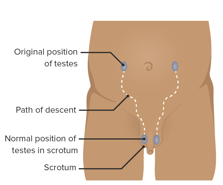
Vía de descenso testicular desde la pared abdominal posterior al escroto: Los testículos pasan a través del canal inguinal.

Imagen por Lecturio.

Anatomía del Canal Inguinal

Pared abdominal anterior

  • La pared abdominal anterior está compuesta por varios músculos:
    • Recto abdominal
    • Oblicuo externo
    • Oblicuo interno
    • Transverso del abdomen
  • La fascia Fascia Layers of connective tissue of variable thickness. The superficial fascia is found immediately below the skin; the deep fascia invests muscles, nerves, and other organs. Cellulitis transversal interna continúa hacia el canal inguinal, formando la fascia Fascia Layers of connective tissue of variable thickness. The superficial fascia is found immediately below the skin; the deep fascia invests muscles, nerves, and other organs. Cellulitis espermática interna.

Recorrido del canal inguinal

  • Aproximadamente 4 cm de largo desde la espina ilíaca anterosuperior hasta el tubérculo púbico (superolateralmente a inferomedialmente)
  • Anillo inguinal profundo: una evaginación de la fascia Fascia Layers of connective tissue of variable thickness. The superficial fascia is found immediately below the skin; the deep fascia invests muscles, nerves, and other organs. Cellulitis transversal (que rodea el cordón espermático como la fascia Fascia Layers of connective tissue of variable thickness. The superficial fascia is found immediately below the skin; the deep fascia invests muscles, nerves, and other organs. Cellulitis espermática interna)
  • Anillo inguinal superficial: una fisura en EN Erythema nodosum is an immune-mediated panniculitis (inflammation of the subcutaneous fat) caused by a type IV (delayed-type) hypersensitivity reaction. It commonly manifests in young women as tender, erythematous nodules on the shins. Erythema Nodosum la aponeurosis del músculo oblicuo externo
Las capas de la pared abdominal anterior.

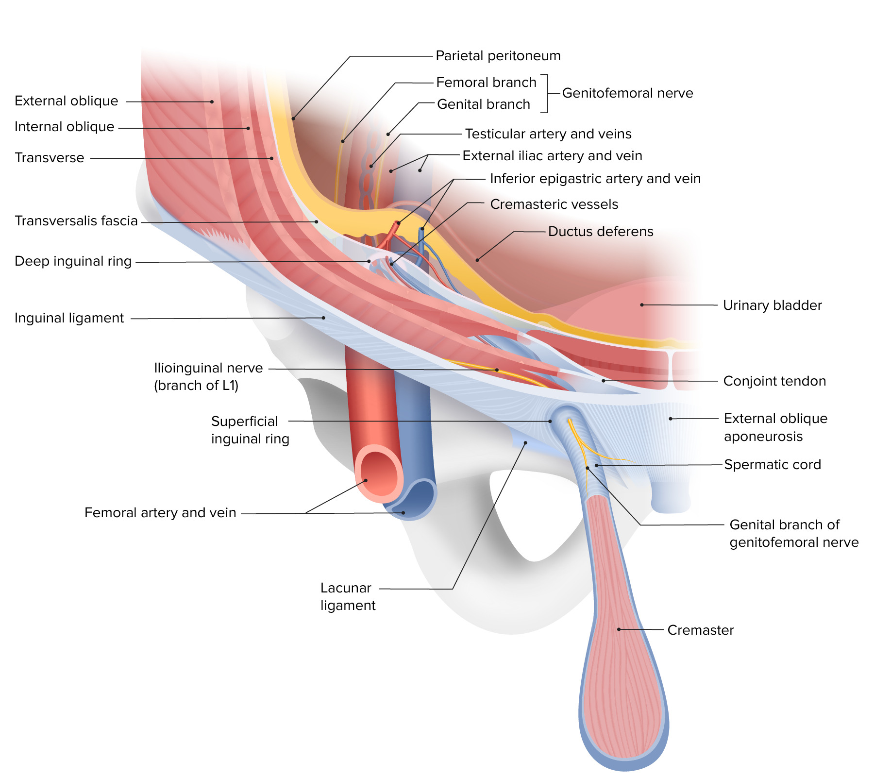
Las capas de la pared abdominal anterior, describiendo el recorrido del canal inguinal y la composición de los anillos inguinales profundo y superficial

Imagen por Lecturio. Licencia: CC BY-NC-SA 4.0

Límites del canal inguinal

Los LOS Neisseria límites del canal inguinal varían a lo largo de su recorrido.

  • A nivel del anillo profundo:
    • Pared anterior:
      • Músculo oblicuo interno
      • Músculo oblicuo externo
    • Pared posterior: fascia Fascia Layers of connective tissue of variable thickness. The superficial fascia is found immediately below the skin; the deep fascia invests muscles, nerves, and other organs. Cellulitis transversal
    • Techo: fascia Fascia Layers of connective tissue of variable thickness. The superficial fascia is found immediately below the skin; the deep fascia invests muscles, nerves, and other organs. Cellulitis transversal
    • Piso: ligamento inguinal
  • A la mitad del canal inguinal:
    • Pared anterior: aponeurosis oblicua externa
    • Pared posterior: fascia Fascia Layers of connective tissue of variable thickness. The superficial fascia is found immediately below the skin; the deep fascia invests muscles, nerves, and other organs. Cellulitis transversal
    • Techo: fibras arqueadas del músculo oblicuo interno y transverso del abdomen
    • Piso: ligamento inguinal
  • A nivel del anillo superficial:
    • Pared anterior: aponeurosis oblicua externa
    • Pared posterior: tendón conjunto
    • Techo: pilar medial del músculo oblicuo externo
    • Suelo: ligamento lacunar
Límites y contenido del canal inguinal masculino

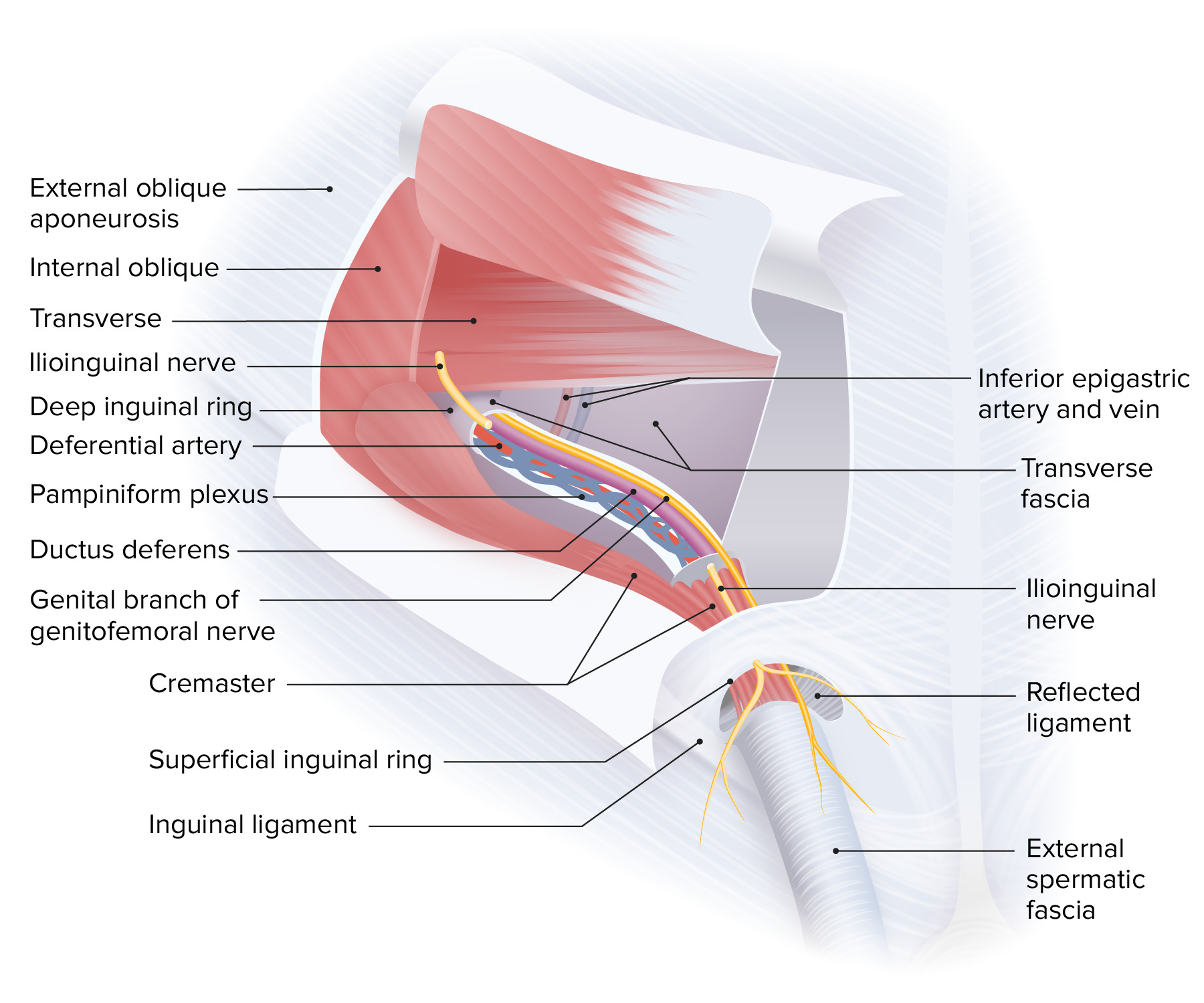
Límites y contenido del canal inguinal masculino:
Obsérvese que el nervio ilioinguinal corre a lo largo del canal inguinal externo al cordón espermático.

Imagen por Lecturio. Licencia: CC BY-NC-SA 4.0

Límites del triángulo de Hesselbach

  • Se refiere a un triángulo de la pared abdominal anterior
  • Límites:
    • Medial: margen lateral del recto abdominal (línea semilunar)
    • Superolateral: vasos epigástricos inferiores
    • Inferior: ligamento inguinal y pectíneo
  • Las hernias directas ocurren dentro del triángulo y las hernias indirectas ocurren lateral al AL Amyloidosis triángulo.

Epidemiología y Etiología de las Hernias Inguinales

Epidemiología

  • Tipo de hernia Hernia Protrusion of tissue, structure, or part of an organ through the bone, muscular tissue, or the membrane by which it is normally contained. Hernia may involve tissues such as the abdominal wall or the respiratory diaphragm. Hernias may be internal, external, congenital, or acquired. Abdominal Hernias más común (> 75% de los LOS Neisseria casos)
  • Hernias inguinales indirectas > hernias inguinales directas
  • Edades de máxima incidencia:
    • 0‒5 años
    • 75‒80 años
  • Riesgo a lo largo de la vida de aproximadamente 25% para los LOS Neisseria hombres y < 5% para las mujeres

Factores de riesgo

  • Antecedentes de hernia Hernia Protrusion of tissue, structure, or part of an organ through the bone, muscular tissue, or the membrane by which it is normally contained. Hernia may involve tissues such as the abdominal wall or the respiratory diaphragm. Hernias may be internal, external, congenital, or acquired. Abdominal Hernias o reparación de hernia Hernia Protrusion of tissue, structure, or part of an organ through the bone, muscular tissue, or the membrane by which it is normally contained. Hernia may involve tissues such as the abdominal wall or the respiratory diaphragm. Hernias may be internal, external, congenital, or acquired. Abdominal Hernias previa
  • Sexo masculino
  • Edad avanzada
  • Enfermedad pulmonar obstructiva crónica (EPOC)
  • Constipación crónica
  • Levantamiento de pesas
  • Obesidad
  • Tabaquismo

Etiología

  • Congénita:
    • Debido a un desarrollo anormal
    • Cierre fallido del proceso vaginal
  • Adquirida:
    • Se desarrolla más tarde en EN Erythema nodosum is an immune-mediated panniculitis (inflammation of the subcutaneous fat) caused by a type IV (delayed-type) hypersensitivity reaction. It commonly manifests in young women as tender, erythematous nodules on the shins. Erythema Nodosum la vida debido a la debilidad progresiva de los LOS Neisseria tejidos previamente normales
    • Condiciones con aumento de la presión intraabdominal, como actividad física intensa, tos TOS Thoracic outlet syndrome (TOS) is a broad term used for a spectrum of syndromes related to the general region of the thoracic outlet, which involves the compression or irritation of elements of the brachial plexus, subclavian artery, or subclavian vein. Thoracic Outlet Syndrome u obesidad
    • A veces iatrogénico debido a una lesión o cirugía abdominal
  • Todas las hernias directas son adquiridas, mientras que las indirectas pueden ser congénitas o adquiridas.

Clasificación y Presentación Clínica

Clasificación

  • Hernias indirectas:
    • Lateral a los LOS Neisseria vasos epigástricos inferiores y al AL Amyloidosis triángulo de Hesselbach
    • El contenido atraviesa el anillo inguinal profundo, atraviesa toda la trayectoria del canal inguinal y sale del canal a través del anillo inguinal superficial.
    • El contenido está encerrado por las cubiertas del cordón espermático.
  • Hernias directas:
    • Medial a los LOS Neisseria vasos epigástricos inferiores y dentro del triángulo de Hesselbach
    • El contenido sobresale directamente a través de la pared posterior del canal inguinal y a través del anillo inguinal superficial, encerrado solo por la fascia Fascia Layers of connective tissue of variable thickness. The superficial fascia is found immediately below the skin; the deep fascia invests muscles, nerves, and other organs. Cellulitis espermática externa.
  • Hernia Hernia Protrusion of tissue, structure, or part of an organ through the bone, muscular tissue, or the membrane by which it is normally contained. Hernia may involve tissues such as the abdominal wall or the respiratory diaphragm. Hernias may be internal, external, congenital, or acquired. Abdominal Hernias en EN Erythema nodosum is an immune-mediated panniculitis (inflammation of the subcutaneous fat) caused by a type IV (delayed-type) hypersensitivity reaction. It commonly manifests in young women as tender, erythematous nodules on the shins. Erythema Nodosum pantalón: hernia Hernia Protrusion of tissue, structure, or part of an organ through the bone, muscular tissue, or the membrane by which it is normally contained. Hernia may involve tissues such as the abdominal wall or the respiratory diaphragm. Hernias may be internal, external, congenital, or acquired. Abdominal Hernias inguinal con componentes directos e indirectos
  • Hernia Hernia Protrusion of tissue, structure, or part of an organ through the bone, muscular tissue, or the membrane by which it is normally contained. Hernia may involve tissues such as the abdominal wall or the respiratory diaphragm. Hernias may be internal, external, congenital, or acquired. Abdominal Hernias de Amyand: el apéndice se encuentra dentro del saco herniario.
Anatomía de la región inguinal y la hernia.

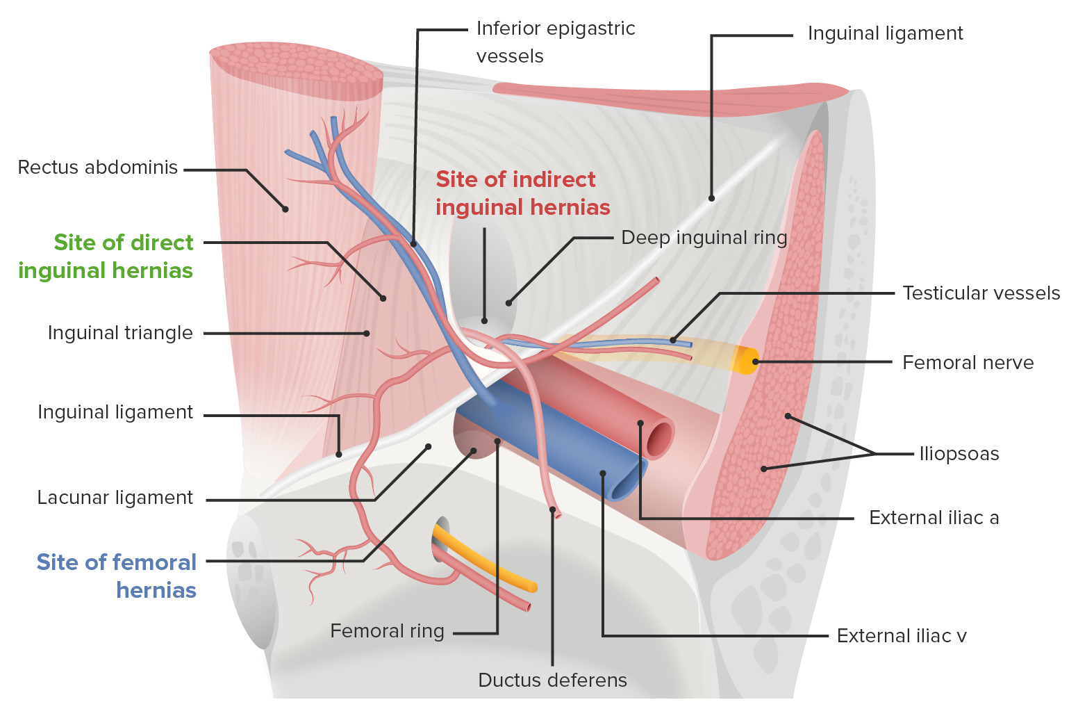
Vista del canal inguinal izquierdo: las hernias inguinales indirectas surgen lateral a los vasos epigástricos, mientras que las hernias inguinales directas surgen medial a los vasos epigástricos.

Imagen por Lecturio.

Presentación clínica

  • Dolor Dolor Inflammation o malestar inguinal leve
  • Abultamiento visible en EN Erythema nodosum is an immune-mediated panniculitis (inflammation of the subcutaneous fat) caused by a type IV (delayed-type) hypersensitivity reaction. It commonly manifests in young women as tender, erythematous nodules on the shins. Erythema Nodosum la zona de la ingle, que aumenta al AL Amyloidosis pararse o durante actividades físicas que aumentan la presión intraabdominal (toser, estornudar, levantar pesas) y se reduce al AL Amyloidosis acostarse.
  • Puede estar asociada con un hidrocele comunicante
  • Se puede observar eritema sobre la hernia Hernia Protrusion of tissue, structure, or part of an organ through the bone, muscular tissue, or the membrane by which it is normally contained. Hernia may involve tissues such as the abdominal wall or the respiratory diaphragm. Hernias may be internal, external, congenital, or acquired. Abdominal Hernias si hay estrangulación y muerte del tejido.

Complicaciones

  • Incarceración:
    • Incapacidad del contenido de la hernia Hernia Protrusion of tissue, structure, or part of an organ through the bone, muscular tissue, or the membrane by which it is normally contained. Hernia may involve tissues such as the abdominal wall or the respiratory diaphragm. Hernias may be internal, external, congenital, or acquired. Abdominal Hernias para volver a su cavidad original
    • Se presenta con dolor Dolor Inflammation severo y un bulto no reducible
    • Si los LOS Neisseria intestinos están incarcerados, se desarrollan síntomas de obstrucción intestinal.
  • Estrangulación:
    • El contenido de la hernia Hernia Protrusion of tissue, structure, or part of an organ through the bone, muscular tissue, or the membrane by which it is normally contained. Hernia may involve tissues such as the abdominal wall or the respiratory diaphragm. Hernias may be internal, external, congenital, or acquired. Abdominal Hernias debe primero estar incarcerado.
    • La irrigación a los LOS Neisseria órganos incarcerados se ve VE Ventilation: Mechanics of Breathing comprometida, lo que causa isquemia y la muerte del tejido.

Diagnóstico y Tratamiento

Diagnóstico

  • Antecedentes clínicos y examen físico
  • Palpación del canal inguinal:
    • Con el paciente de pie, palpe desde la piel del escroto hacia el anillo inguinal superficial.
    • Pida al AL Amyloidosis paciente que tosa (maniobra de Valsalva).
    • El abultamiento se puede sentir en EN Erythema nodosum is an immune-mediated panniculitis (inflammation of the subcutaneous fat) caused by a type IV (delayed-type) hypersensitivity reaction. It commonly manifests in young women as tender, erythematous nodules on the shins. Erythema Nodosum la yema del dedo.
  • Imagenología:
    • Se utiliza para la confirmación en EN Erythema nodosum is an immune-mediated panniculitis (inflammation of the subcutaneous fat) caused by a type IV (delayed-type) hypersensitivity reaction. It commonly manifests in young women as tender, erythematous nodules on the shins. Erythema Nodosum casos inciertos y en EN Erythema nodosum is an immune-mediated panniculitis (inflammation of the subcutaneous fat) caused by a type IV (delayed-type) hypersensitivity reaction. It commonly manifests in young women as tender, erythematous nodules on the shins. Erythema Nodosum ocasiones para la planificación quirúrgica
    • Ultrasonido:
      • Mejor estudio de imagenología inicial en EN Erythema nodosum is an immune-mediated panniculitis (inflammation of the subcutaneous fat) caused by a type IV (delayed-type) hypersensitivity reaction. It commonly manifests in young women as tender, erythematous nodules on the shins. Erythema Nodosum pacientes sin evidencia física de hernia Hernia Protrusion of tissue, structure, or part of an organ through the bone, muscular tissue, or the membrane by which it is normally contained. Hernia may involve tissues such as the abdominal wall or the respiratory diaphragm. Hernias may be internal, external, congenital, or acquired. Abdominal Hernias
      • El hallazgo diagnóstico es un aumento del diámetro del canal inguinal (normalmente < 13 mm en EN Erythema nodosum is an immune-mediated panniculitis (inflammation of the subcutaneous fat) caused by a type IV (delayed-type) hypersensitivity reaction. It commonly manifests in young women as tender, erythematous nodules on the shins. Erythema Nodosum el anillo inguinal profundo).
    • Tomografía computarizada (TC): particularmente útil para distinguir entre diferentes subtipos de hernias inguinales
    • Resonancia magnética (RM):
      • La mejor modalidad de imagenología para diferenciar entre hernias inguinales y femorales con una sensibilidad y especificidad superior al AL Amyloidosis 95%
      • Debido al AL Amyloidosis costo y la disponibilidad limitada de las RM, la TC todavía se utiliza con más frecuencia.

Tratamiento

  • Reparación quirúrgica de la hernia Hernia Protrusion of tissue, structure, or part of an organ through the bone, muscular tissue, or the membrane by which it is normally contained. Hernia may involve tissues such as the abdominal wall or the respiratory diaphragm. Hernias may be internal, external, congenital, or acquired. Abdominal Hernias:
    • Hernias complicadas
    • Hernias no complicadas con síntomas moderados
    • De forma selectiva para hernias sin complicaciones con sintomatología leve o alto riesgo de incarceración
    • Se pueden encontrar detalles específicos de las técnicas de reparación quirúrgica en EN Erythema nodosum is an immune-mediated panniculitis (inflammation of the subcutaneous fat) caused by a type IV (delayed-type) hypersensitivity reaction. It commonly manifests in young women as tender, erythematous nodules on the shins. Erythema Nodosum hernias abdominales.
  • Técnicas quirúrgicas:
    • Refuerzo de la pared posterior del canal inguinal con malla sintética
    • Reducción de los LOS Neisseria diámetros de los LOS Neisseria anillos inguinales

Referencias

  1. Sadler, T. W. (2012). Chapter 16: Urogenital system. In Langman’s Medical Embryology. 12th ed., pp. 232–259. Philadelphia: Wolters Kluwer Health/Lippincott Williams & Wilkins.
  2. Malangoni, M. A., & Rosen, M. J. (2012). Hernias. In Mattox, K. L., et al. (Eds.), Sabiston Textbook of Surgery: The Biological Basis of Modern Surgical Practice, 19th ed., pp. 1114‒1140. Elsevier.
  3. Brooks, D. V., & Hawn, M. (2019). Classification, clinical features, and diagnosis of inguinal and femoral hernias in adults. In Chen, W. (Ed.). UpToDate. Retrieved June 19, 2021, from https://www.uptodate.com/contents/classification-clinical-features-and-diagnosis-of-inguinal-and-femoral-hernias-in-adults

¡Crea tu cuenta gratis o inicia una sesión para seguir leyendo!

Regístrate ahora y obtén acceso gratuito a Lecturio con páginas de concepto, videos médicos y cuestionarios para tu educación médica.

User Reviews

Details