


A hipertensão pode resultar em várias complicações a nível ocular, das quais a retinopatia é a situação clínica mais MAIS Androgen Insensitivity Syndrome comum. Retinopatia hipertensiva consiste no desenvolvimento de alterações vasculares na retina Retina The ten-layered nervous tissue membrane of the eye. It is continuous with the optic nerve and receives images of external objects and transmits visual impulses to the brain. Its outer surface is in contact with the choroid and the inner surface with the vitreous body. The outermost layer is pigmented, whereas the inner nine layers are transparent. Eye: Anatomy como resultado direto da hipertensão arterial. Na hipertensão arterial aguda, a resposta primária das arteríolas da retina Retina The ten-layered nervous tissue membrane of the eye. It is continuous with the optic nerve and receives images of external objects and transmits visual impulses to the brain. Its outer surface is in contact with the choroid and the inner surface with the vitreous body. The outermost layer is pigmented, whereas the inner nine layers are transparent. Eye: Anatomy é a vasoconstrição. Na hipertensão arterial crónica, a arteriosclerose afeta a vasculatura da retina Retina The ten-layered nervous tissue membrane of the eye. It is continuous with the optic nerve and receives images of external objects and transmits visual impulses to the brain. Its outer surface is in contact with the choroid and the inner surface with the vitreous body. The outermost layer is pigmented, whereas the inner nine layers are transparent. Eye: Anatomy. Ocorrem lesões na parede endotelial e surgem vários sinais da doença, incluindo a presença de hemorragias, manchas algodonosas e exsudados. Nos casos de hipertensão arterial descontrolada, pode ser observado papiledema. O tratamento é direcionado para o controlo da hipertensão arterial. Os doentes com retinopatia hipertensiva grave têm um risco aumentado de doença arterial coronária e acidente vascular cerebral; portanto, a deteção e o tratamento da hipertensão arterial é fundamental.
Last updated: Dec 15, 2025
Retinopatia hipertensiva:
Hipertensão arterial:
| Níveis de pressão arterial | Pressão arterial sistólica | Pressão arterial diastólica | |
|---|---|---|---|
| Tensão arterial elevada | 120-129 mm Hg | E | <80 mm Hg |
| Hipertensão arterial estadio 1 | 130-139 mm Hg | OU | 80-89 mm Hg |
| Hipertensão estadio 2 | ≥ 140 mm Hg | OU | ≥ 90 mm Hg |
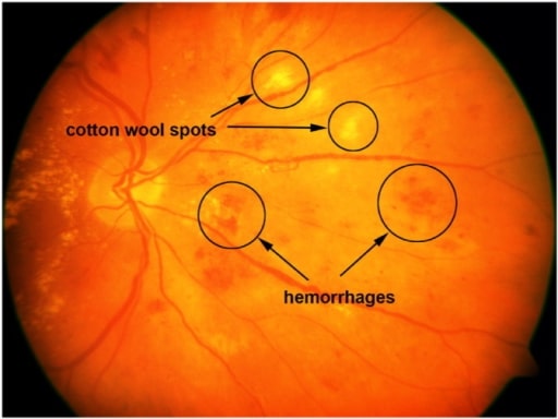
Imagem da retina com manchas algodonosas e hemorragias.
Imagem: “A sample retinal image with cotton-wool spots and hemorrhages” por the United States National Library of Medicine. Licença: CC BY 4.0.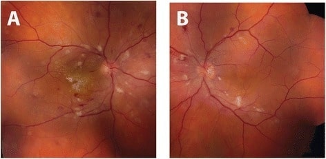
Fundoscopia do olho direito com retinopatia hipertensiva (A) e olho esquerdo (B) diminuição do calibre das arteríolas bilateralmente, manchas algodonosas, hemorragias em pontos e hemorragia intraretinal em forma de chama de vela. Exsudados intrarretinais com estrela macular também são visíveis no olho direito.
Imagem: “Initial fundus exam” por the Department of Ophthalmology and Visual Sciences, University of Michigan, 500 S State St, Ann Arbor, MI 48109 USA. Licença: CC BY 4.0.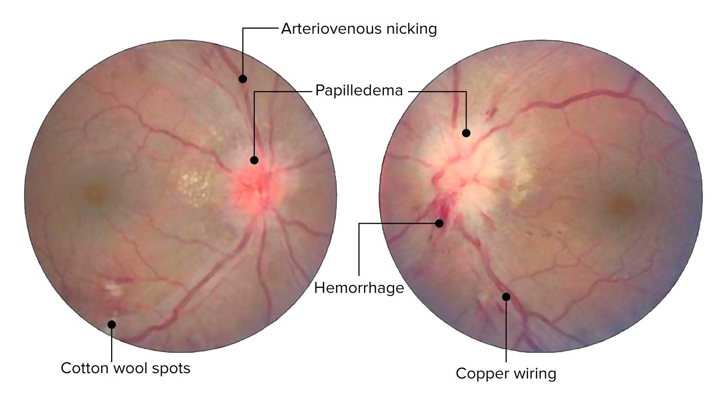
Alterações da retina no caso de retinopatia hipertensiva aguda. A imagem mostra cruzamentos arteriovenosos, alterações arteriais em fio de cobre, hemorragias e manchas algodonosas. Edema bilateral do disco ótico presente bilateralmente (mais à esquerda do que à direita).
Imagem: “Central retinal vein occlusion” por Doheny Eye Institute, University of Southern California Los Angeles, CA 90033 USA. Licença: CC BY 3.0. Editado por Lecturio.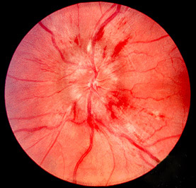
A imagem mostra papiledema (edema do disco ótico com as margens do disco embaçadas).
Imagem: “Papilledema” por Jonathan Trobe, MD. Licença: CC BY 3.0.| Grau I | Estreitamento ligeiro ou moderado das arteríolas da retina Retina The ten-layered nervous tissue membrane of the eye. It is continuous with the optic nerve and receives images of external objects and transmits visual impulses to the brain. Its outer surface is in contact with the choroid and the inner surface with the vitreous body. The outermost layer is pigmented, whereas the inner nine layers are transparent. Eye: Anatomy, com uma relação arteriovenosa de ≥ 1:2 |
|---|---|
| Grau II | Estreitamento moderado a severo das arteríolas da retina Retina The ten-layered nervous tissue membrane of the eye. It is continuous with the optic nerve and receives images of external objects and transmits visual impulses to the brain. Its outer surface is in contact with the choroid and the inner surface with the vitreous body. The outermost layer is pigmented, whereas the inner nine layers are transparent. Eye: Anatomy com uma relação arteriovenosa <1:2 ou cruzamento arteriovenoso |
| Grau III | Manchas algodonosas ou hemorragias em forma de chama de vela |
| Grau IV | Edema Edema Edema is a condition in which excess serous fluid accumulates in the body cavity or interstitial space of connective tissues. Edema is a symptom observed in several medical conditions. It can be categorized into 2 types, namely, peripheral (in the extremities) and internal (in an organ or body cavity). Edema óptico bilateral |
| Grau de retinopatia | Alterações na retina Retina The ten-layered nervous tissue membrane of the eye. It is continuous with the optic nerve and receives images of external objects and transmits visual impulses to the brain. Its outer surface is in contact with the choroid and the inner surface with the vitreous body. The outermost layer is pigmented, whereas the inner nine layers are transparent. Eye: Anatomy | Riscos sistémicos |
|---|---|---|
| Nenhum | Sem alterações detetaveis | Nenhum |
| Ligeira |
|
Risco médio de acidentes vasculares cerebrais clínicos, acidentes vasculares cerebrais subclínicos, doenças coronárias e mortalidade |
| Moderada |
|
Alto risco de acidentes vasculares cerebrais clínicos e subclínicos, declínio cognitivo, doença coronária e mortalidade |
| Maligna | Sinais de retinopatia moderada com presença de edema Edema Edema is a condition in which excess serous fluid accumulates in the body cavity or interstitial space of connective tissues. Edema is a symptom observed in several medical conditions. It can be categorized into 2 types, namely, peripheral (in the extremities) and internal (in an organ or body cavity). Edema do disco ótico | Alto risco de mortalidade |
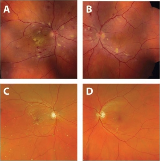
Melhoria das imagens da fundoscopia após a instituição de tratamento anti-hipertensivo.
Fundoscopias tiradas no momento da apresentação (A, B): pode-se observar constrição arteriolar, hemorragias da retina, manchas algodonosas e exsudados duros (à direita).
Quatro meses após o diagnóstico e tratamento da hipertensão arterial (C, D): A normalização da pressão arterial resultou na resolução das hemorragias e das manchas algodonosas.
Melhoria temporal do aspeto dos exsudados duros no olho direito com uma estrela macular residual.