Las venas son conjuntos celulares tubulares que transportan sangre desoxigenada y desechos de los LOS Neisseria lechos capilares de regreso al AL Amyloidosis corazón. Las venas se clasifican en EN Erythema nodosum is an immune-mediated panniculitis (inflammation of the subcutaneous fat) caused by a type IV (delayed-type) hypersensitivity reaction. It commonly manifests in young women as tender, erythematous nodules on the shins. Erythema Nodosum 3 tipos: venas pequeñas/vénulas, venas medianas y venas grandes. Cada tipo contiene 3 capas primarias: túnica íntima, túnica media y túnica adventicia. La circulación venosa es un sistema de baja presión con cantidades mucho menores de músculo liso y tejido elástico, paredes más delgadas y lúmenes más grandes que las arterias. Las venas son vasos de capacitancia con una distensibilidad significativa y la capacidad de dilatarse y retener hasta un 70%–80% del volumen sanguíneo en EN Erythema nodosum is an immune-mediated panniculitis (inflammation of the subcutaneous fat) caused by a type IV (delayed-type) hypersensitivity reaction. It commonly manifests in young women as tender, erythematous nodules on the shins. Erythema Nodosum reposo.
Last updated: Dec 15, 2025
Las venas son colecciones tubulares de células que transportan sangre desoxigenada y productos de desecho de los LOS Neisseria capilares en EN Erythema nodosum is an immune-mediated panniculitis (inflammation of the subcutaneous fat) caused by a type IV (delayed-type) hypersensitivity reaction. It commonly manifests in young women as tender, erythematous nodules on the shins. Erythema Nodosum la periferia del cuerpo de regreso al AL Amyloidosis corazón.
Las características de las venas y el sistema venoso incluyen:
Todas las venas tienen la misma estructura básica y están formadas por 3 capas primarias: túnica íntima, túnica media y túnica adventicia.
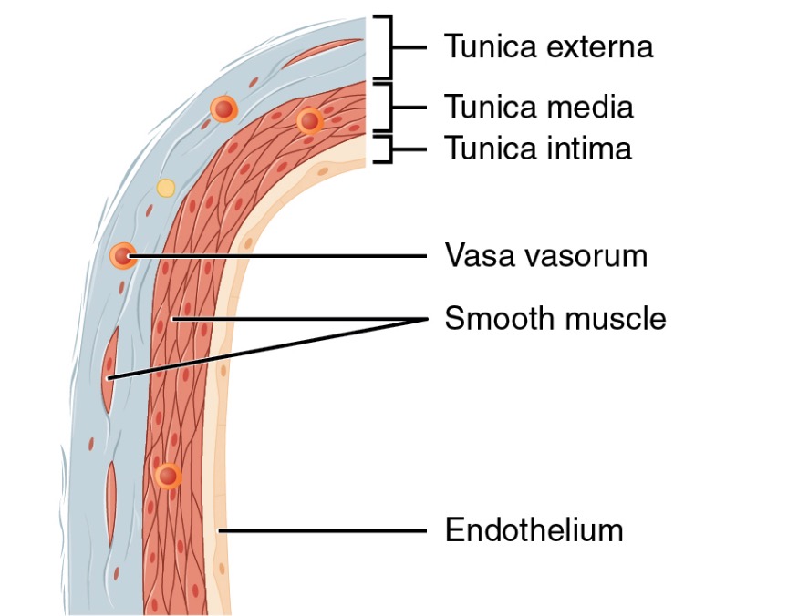
Estructura de una pared de vena
Imagen: “Structure of a vein wall” por Phil Schatz. Licencia: CC BY 4.0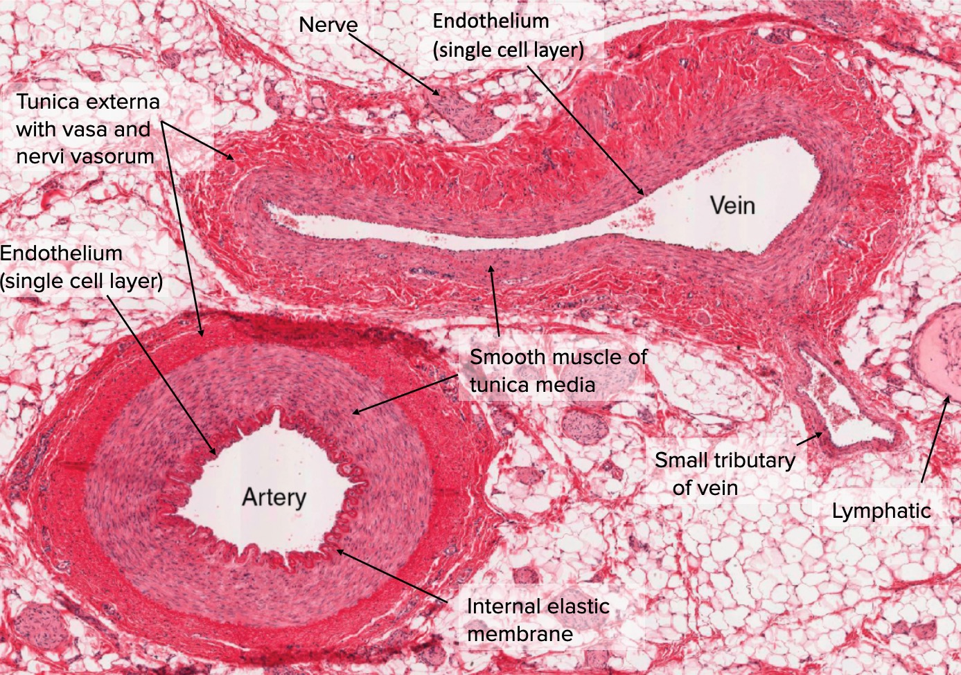
Sección transversal de arteria y vena
Imagen: “Types of Arteries and Arterioles” por Phil Schatz. Licencia: CC BY 4.0, editado por Lecturio.La diferenciación segmentaria distingue los LOS Neisseria 3 tipos principales de venas por tamaño, función y composición general. Las venas generalmente existen como una continuidad con cambios graduales en EN Erythema nodosum is an immune-mediated panniculitis (inflammation of the subcutaneous fat) caused by a type IV (delayed-type) hypersensitivity reaction. It commonly manifests in young women as tender, erythematous nodules on the shins. Erythema Nodosum la morfología de los LOS Neisseria vasos a lo largo del árbol venoso.
Los LOS Neisseria 3 tipos principales de venas son:
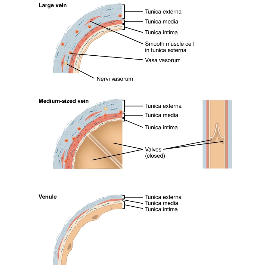
Comparación de venas grandes, medianas y pequeñas
Imagen: “Comparison of large, medium, and small veins” por Phil Schatz. Licencia: CC BY 4.0
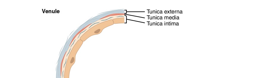
Diagrama de una vénula
Imagen: “Venule” por Phil Schatz. Licencia: CC BY 4.0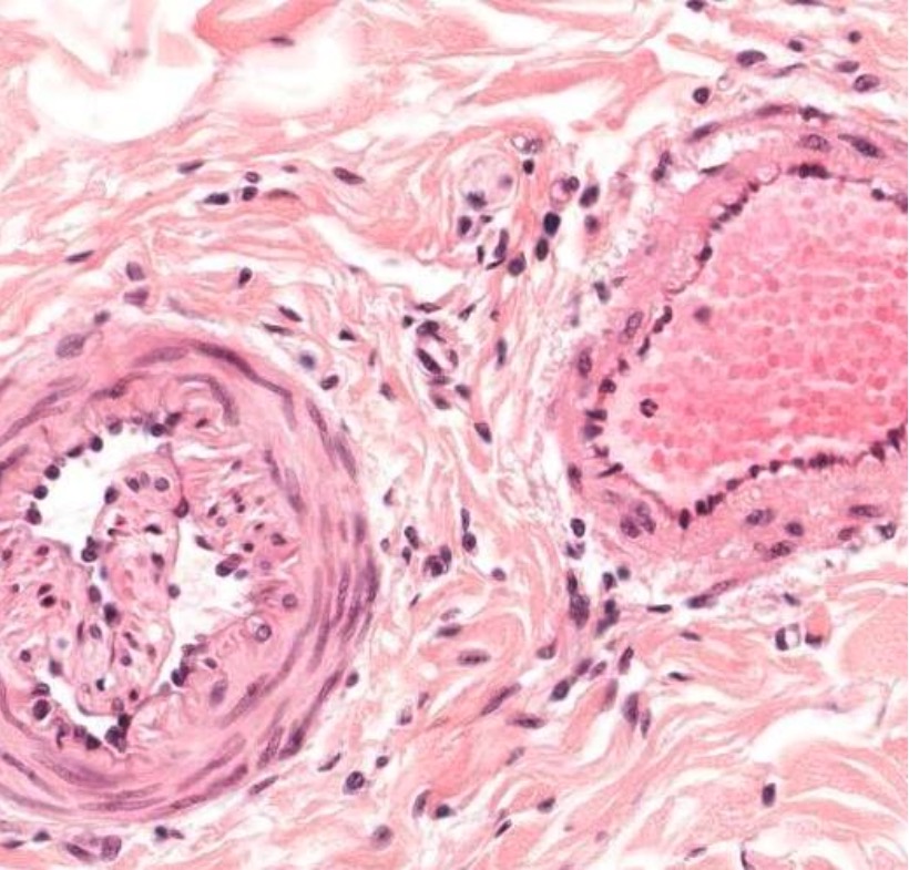
Corte transversal de una arteria pequeña (izquierda) y una vena pequeña (derecha)
Imagen por Geoffrey Meyer, PhD.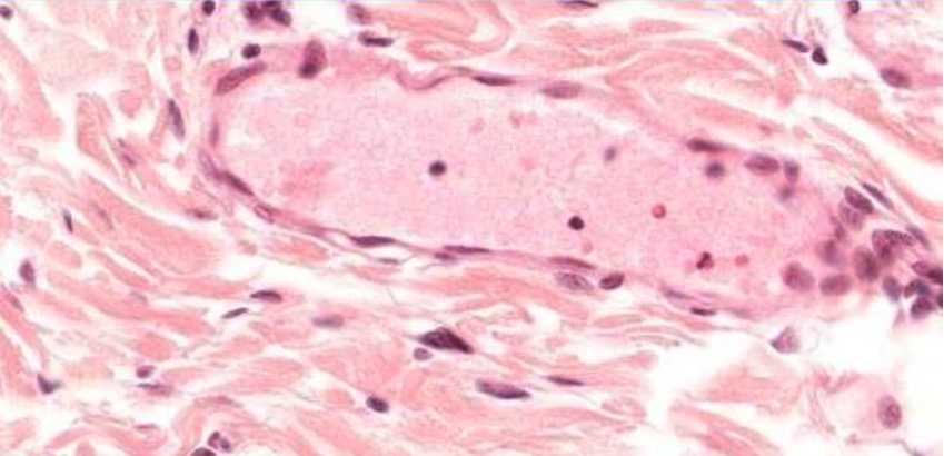
Vénula postcapilar
Imagen por Geoffrey Meyer, PhD.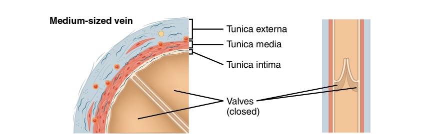
Diagrama de una vena de tamaño mediano
Imagen: “Medium-sized vein” por Phil Schatz. Licencia: CC BY 4.0
Diagrama de la bomba muscular
Imagen: “The contraction of skeletal muscles” por Phil Schatz. Licencia: CC BY 4.0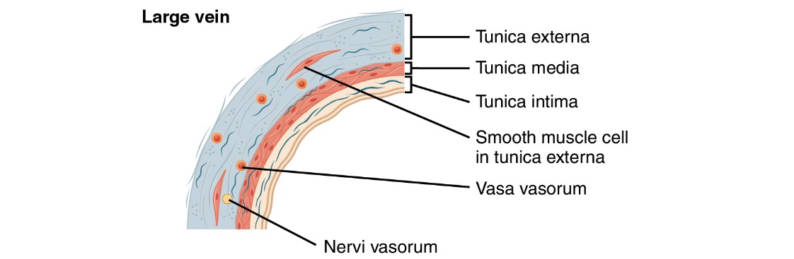
Diagrama de una vena grande
Imagen: “Large vein” por Phil Schatz. Licencia: CC BY 4.0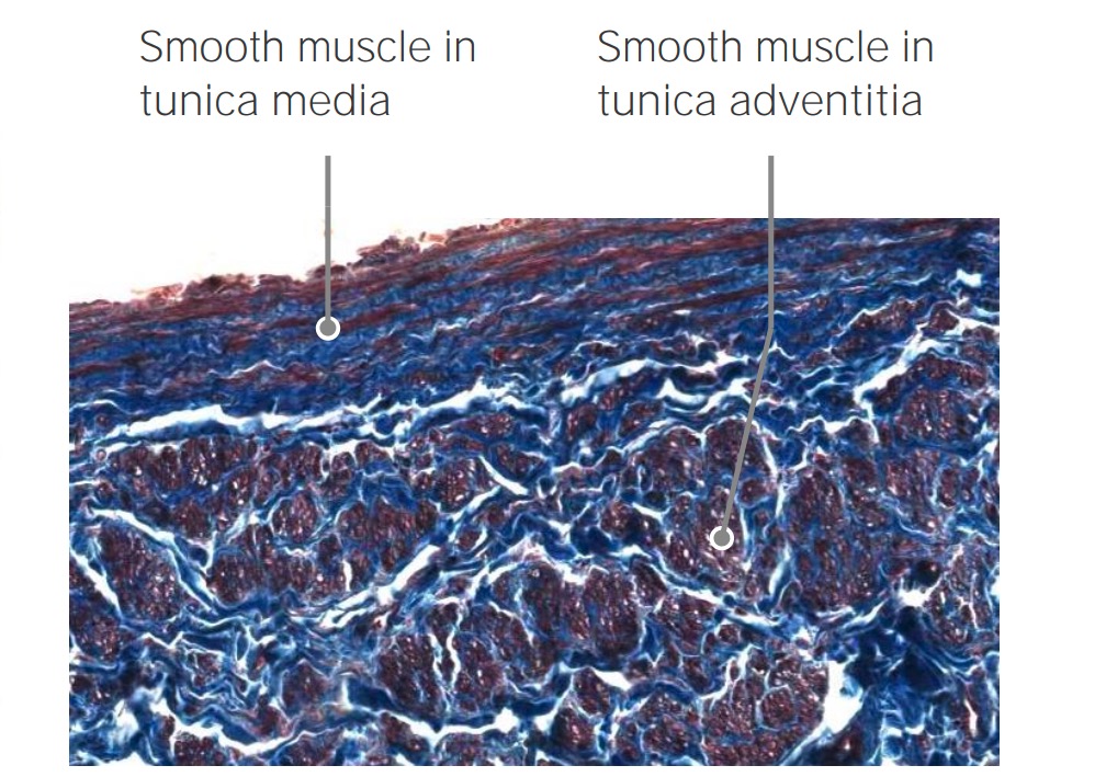
Músculos lisos longitudinales de la vena cava
Imagen por Geoffrey Meyer, PhD.