Domina Conceptos Médicos

Estudia para la escuela de medicina y tus examenes con Lecturio.

Pleura: Anatomía

La pleura Pleura The pleura is a serous membrane that lines the walls of the thoracic cavity and the surface of the lungs. This structure of mesodermal origin covers both lungs, the mediastinum, the thoracic surface of the diaphragm, and the inner part of the thoracic cage. The pleura is divided into a visceral pleura and parietal pleura. Pleura: Anatomy es una membrana serosa que recubre las paredes de la cavidad torácica y la superficie de los LOS Neisseria pulmones. Esta estructura de origen mesodérmico cubre ambos pulmones, el mediastino, la superficie torácica del diafragma y la parte interna de la caja torácica. La pleura Pleura The pleura is a serous membrane that lines the walls of the thoracic cavity and the surface of the lungs. This structure of mesodermal origin covers both lungs, the mediastinum, the thoracic surface of the diaphragm, and the inner part of the thoracic cage. The pleura is divided into a visceral pleura and parietal pleura. Pleura: Anatomy se divide en EN Erythema nodosum is an immune-mediated panniculitis (inflammation of the subcutaneous fat) caused by a type IV (delayed-type) hypersensitivity reaction. It commonly manifests in young women as tender, erythematous nodules on the shins. Erythema Nodosum la pleura Pleura The pleura is a serous membrane that lines the walls of the thoracic cavity and the surface of the lungs. This structure of mesodermal origin covers both lungs, the mediastinum, the thoracic surface of the diaphragm, and the inner part of the thoracic cage. The pleura is divided into a visceral pleura and parietal pleura. Pleura: Anatomy visceral y la pleura Pleura The pleura is a serous membrane that lines the walls of the thoracic cavity and the surface of the lungs. This structure of mesodermal origin covers both lungs, the mediastinum, the thoracic surface of the diaphragm, and the inner part of the thoracic cage. The pleura is divided into a visceral pleura and parietal pleura. Pleura: Anatomy parietal Parietal One of a pair of irregularly shaped quadrilateral bones situated between the frontal bone and occipital bone, which together form the sides of the cranium. Skull: Anatomy. Entre ambas capas hay un espacio potencial bien lubricado llamado cavidad pleural, que facilita los LOS Neisseria movimientos respiratorios de los LOS Neisseria pulmones y ayuda a evitar la fricción.

Last updated: Dec 15, 2025

Editorial responsibility: Stanley Oiseth, Lindsay Jones, Evelin Maza

Anatomía

La pleura Pleura The pleura is a serous membrane that lines the walls of the thoracic cavity and the surface of the lungs. This structure of mesodermal origin covers both lungs, the mediastinum, the thoracic surface of the diaphragm, and the inner part of the thoracic cage. The pleura is divided into a visceral pleura and parietal pleura. Pleura: Anatomy es una membrana serosa de doble capa que recubre las paredes de la cavidad torácica y la superficie de los LOS Neisseria pulmones. Por lo tanto, se extiende por toda la cavidad torácica.

Límites:

  • Superior: raíz del cuello 2–3 cm por encima de la 1ra costilla
  • Inferior: superficie torácica del diafragma
  • Medial: mediastino
  • Lateral: superficie interna de la caja torácica y músculos intercostales
Límites y partes de la pleura dentro de la cavidad torácica

Límites y partes de la pleura dentro de la cavidad torácica

Imagen por Lecturio. Licencia: CC BY-NC-SA 4.0

Cada pulmón está encerrado en EN Erythema nodosum is an immune-mediated panniculitis (inflammation of the subcutaneous fat) caused by a type IV (delayed-type) hypersensitivity reaction. It commonly manifests in young women as tender, erythematous nodules on the shins. Erythema Nodosum un saco pleural seroso que consta de 2 membranas continuas de pleura Pleura The pleura is a serous membrane that lines the walls of the thoracic cavity and the surface of the lungs. This structure of mesodermal origin covers both lungs, the mediastinum, the thoracic surface of the diaphragm, and the inner part of the thoracic cage. The pleura is divided into a visceral pleura and parietal pleura. Pleura: Anatomy visceral y parietal Parietal One of a pair of irregularly shaped quadrilateral bones situated between the frontal bone and occipital bone, which together form the sides of the cranium. Skull: Anatomy.

  • Pleura Pleura The pleura is a serous membrane that lines the walls of the thoracic cavity and the surface of the lungs. This structure of mesodermal origin covers both lungs, the mediastinum, the thoracic surface of the diaphragm, and the inner part of the thoracic cage. The pleura is divided into a visceral pleura and parietal pleura. Pleura: Anatomy parietal Parietal One of a pair of irregularly shaped quadrilateral bones situated between the frontal bone and occipital bone, which together form the sides of the cranium. Skull: Anatomy:
    • Recubre la superficie interna de la cavidad torácica
    • Separada de la pared torácica por la fascia Fascia Layers of connective tissue of variable thickness. The superficial fascia is found immediately below the skin; the deep fascia invests muscles, nerves, and other organs. Cellulitis endotorácica
    • Las partes se clasifican según las estructuras adyacentes:
      • Pleura Pleura The pleura is a serous membrane that lines the walls of the thoracic cavity and the surface of the lungs. This structure of mesodermal origin covers both lungs, the mediastinum, the thoracic surface of the diaphragm, and the inner part of the thoracic cage. The pleura is divided into a visceral pleura and parietal pleura. Pleura: Anatomy costal
      • Pleura Pleura The pleura is a serous membrane that lines the walls of the thoracic cavity and the surface of the lungs. This structure of mesodermal origin covers both lungs, the mediastinum, the thoracic surface of the diaphragm, and the inner part of the thoracic cage. The pleura is divided into a visceral pleura and parietal pleura. Pleura: Anatomy diafragmática
      • Pleura Pleura The pleura is a serous membrane that lines the walls of the thoracic cavity and the surface of the lungs. This structure of mesodermal origin covers both lungs, the mediastinum, the thoracic surface of the diaphragm, and the inner part of the thoracic cage. The pleura is divided into a visceral pleura and parietal pleura. Pleura: Anatomy mediastínica: forma un tubo membranoso en EN Erythema nodosum is an immune-mediated panniculitis (inflammation of the subcutaneous fat) caused by a type IV (delayed-type) hypersensitivity reaction. It commonly manifests in young women as tender, erythematous nodules on the shins. Erythema Nodosum forma de manga llamado raíz del pulmón y cubre el corazón y los LOS Neisseria grandes vasos del corazón
      • Pleura Pleura The pleura is a serous membrane that lines the walls of the thoracic cavity and the surface of the lungs. This structure of mesodermal origin covers both lungs, the mediastinum, the thoracic surface of the diaphragm, and the inner part of the thoracic cage. The pleura is divided into a visceral pleura and parietal pleura. Pleura: Anatomy cervical: cubierta por la membrana suprapleural, una fascia Fascia Layers of connective tissue of variable thickness. The superficial fascia is found immediately below the skin; the deep fascia invests muscles, nerves, and other organs. Cellulitis en EN Erythema nodosum is an immune-mediated panniculitis (inflammation of the subcutaneous fat) caused by a type IV (delayed-type) hypersensitivity reaction. It commonly manifests in young women as tender, erythematous nodules on the shins. Erythema Nodosum forma de cúpula unida a la 1ra costilla y a C7 que sirve de lugar de inserción para algunos de los LOS Neisseria músculos profundos del cuello
  • Pleura Pleura The pleura is a serous membrane that lines the walls of the thoracic cavity and the surface of the lungs. This structure of mesodermal origin covers both lungs, the mediastinum, the thoracic surface of the diaphragm, and the inner part of the thoracic cage. The pleura is divided into a visceral pleura and parietal pleura. Pleura: Anatomy visceral:
    • Recubre la superficie externa de los LOS Neisseria pulmones
    • Cubre las fisuras pulmonares
    • No es sensible al AL Amyloidosis dolor Dolor Inflammation
    • Se continúa con la pleura Pleura The pleura is a serous membrane that lines the walls of the thoracic cavity and the surface of the lungs. This structure of mesodermal origin covers both lungs, the mediastinum, the thoracic surface of the diaphragm, and the inner part of the thoracic cage. The pleura is divided into a visceral pleura and parietal pleura. Pleura: Anatomy parietal Parietal One of a pair of irregularly shaped quadrilateral bones situated between the frontal bone and occipital bone, which together form the sides of the cranium. Skull: Anatomy en EN Erythema nodosum is an immune-mediated panniculitis (inflammation of the subcutaneous fat) caused by a type IV (delayed-type) hypersensitivity reaction. It commonly manifests in young women as tender, erythematous nodules on the shins. Erythema Nodosum el hilio de cada pulmón
  • Recesos: se denominan según los LOS Neisseria puntos de reflexión de la pleura Pleura The pleura is a serous membrane that lines the walls of the thoracic cavity and the surface of the lungs. This structure of mesodermal origin covers both lungs, the mediastinum, the thoracic surface of the diaphragm, and the inner part of the thoracic cage. The pleura is divided into a visceral pleura and parietal pleura. Pleura: Anatomy
    • Receso costodiafragmático:
      • Es el receso más grande
      • Se encuentra entre la caja torácica y la porción diafragmática de la pleura Pleura The pleura is a serous membrane that lines the walls of the thoracic cavity and the surface of the lungs. This structure of mesodermal origin covers both lungs, the mediastinum, the thoracic surface of the diaphragm, and the inner part of the thoracic cage. The pleura is divided into a visceral pleura and parietal pleura. Pleura: Anatomy a cada lado de la cavidad torácica
    • Receso costomedial:
      • Se encuentra en EN Erythema nodosum is an immune-mediated panniculitis (inflammation of the subcutaneous fat) caused by a type IV (delayed-type) hypersensitivity reaction. It commonly manifests in young women as tender, erythematous nodules on the shins. Erythema Nodosum la parte anterior entre la caja torácica y el mediastino a cada lado de la cavidad torácica
      • El receso izquierdo es mayor que el derecho debido a la escotadura cardíaca del pulmón izquierdo.
    • Receso vertebromediastinal:
      • Se encuentra posteriormente entre la caja torácica, la columna vertebral y el mediastino posterior a cada lado de la cavidad torácica
Capas de la pared torácica - haz neurovascular intercostal

Capas de la pared torácica:
Obsérvese la doble capa de pleura y la cavidad pleural, separada de la caja torácica por la fascia endotorácica.

Imagen por Lecturio. Licencia: CC BY-NC-SA 4.0

Cavidad pleural:

  • El espacio potencial entre la pleura Pleura The pleura is a serous membrane that lines the walls of the thoracic cavity and the surface of the lungs. This structure of mesodermal origin covers both lungs, the mediastinum, the thoracic surface of the diaphragm, and the inner part of the thoracic cage. The pleura is divided into a visceral pleura and parietal pleura. Pleura: Anatomy visceral y la pleura Pleura The pleura is a serous membrane that lines the walls of the thoracic cavity and the surface of the lungs. This structure of mesodermal origin covers both lungs, the mediastinum, the thoracic surface of the diaphragm, and the inner part of the thoracic cage. The pleura is divided into a visceral pleura and parietal pleura. Pleura: Anatomy parietal Parietal One of a pair of irregularly shaped quadrilateral bones situated between the frontal bone and occipital bone, which together form the sides of the cranium. Skull: Anatomy (prácticamente inexistente/indetectable)
  • Contiene una pequeña cantidad (0,1–0,2 mL/kg) de líquido seroso que ayuda a evitar la fricción entre ambas pleuras
  • La tensión superficial del líquido mantiene los LOS Neisseria pulmones expandidos y en EN Erythema nodosum is an immune-mediated panniculitis (inflammation of the subcutaneous fat) caused by a type IV (delayed-type) hypersensitivity reaction. It commonly manifests in young women as tender, erythematous nodules on the shins. Erythema Nodosum contacto con la pared torácica a través de la doble capa de pleura Pleura The pleura is a serous membrane that lines the walls of the thoracic cavity and the surface of the lungs. This structure of mesodermal origin covers both lungs, the mediastinum, the thoracic surface of the diaphragm, and the inner part of the thoracic cage. The pleura is divided into a visceral pleura and parietal pleura. Pleura: Anatomy.

Related videos

Irrigación e Inervación

Tabla: Neurovasculatura de la pleura Pleura The pleura is a serous membrane that lines the walls of the thoracic cavity and the surface of the lungs. This structure of mesodermal origin covers both lungs, the mediastinum, the thoracic surface of the diaphragm, and the inner part of the thoracic cage. The pleura is divided into a visceral pleura and parietal pleura. Pleura: Anatomy
Irrigación Inervación
Pleura Pleura The pleura is a serous membrane that lines the walls of the thoracic cavity and the surface of the lungs. This structure of mesodermal origin covers both lungs, the mediastinum, the thoracic surface of the diaphragm, and the inner part of the thoracic cage. The pleura is divided into a visceral pleura and parietal pleura. Pleura: Anatomy parietal Parietal One of a pair of irregularly shaped quadrilateral bones situated between the frontal bone and occipital bone, which together form the sides of the cranium. Skull: Anatomy La parte costal es irrigada por:
  • Ramas de las arterias intercostales
  • Ramas de las arterias torácicas internas
La porción mediastínica es irrigada por:
  • Arterias bronquiales
  • Arterias diafragmáticas superiores
  • Arterias torácicas internas
  • Arterias mediastínicas
La porción cervical es irrigada por: ramas de las arterias subclavias
La porción diafragmática es irrigada por: la parte superficial de la microcirculación diafragmática
Recibe inervación somática aferente (sensorial) de:
  • Nervios intercostales (T1–T11):
    • Porción costal
    • Porción cervical
    • Porción diafragmática periférica
  • Nervio frénico (C3–C5):
    • Porción mediastínica
    • Porción diafragmática central
Pleura Pleura The pleura is a serous membrane that lines the walls of the thoracic cavity and the surface of the lungs. This structure of mesodermal origin covers both lungs, the mediastinum, the thoracic surface of the diaphragm, and the inner part of the thoracic cage. The pleura is divided into a visceral pleura and parietal pleura. Pleura: Anatomy visceral
  • Circulación bronquial
  • Circulación pulmonar
Recibe inervación aferente visceral (autonómica) del: plexo pulmonar
Irrigación de las pleuras

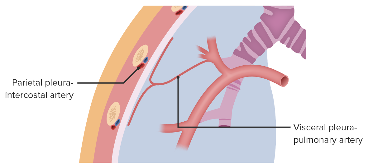
La irrigación sanguínea de las pleuras:
La pleura parietal recibe irrigación sanguínea de las arterias intercostales, diafragmáticas, mediastínicas y torácicas internas. La pleura visceral recibe irrigación sanguínea de los vasos bronquiales y pulmonares.

Imagen por Lecturio. Licencia: CC BY-NC-SA 4.0

Relevancia Clínica

Trastornos infecciosos de la pleura Pleura The pleura is a serous membrane that lines the walls of the thoracic cavity and the surface of the lungs. This structure of mesodermal origin covers both lungs, the mediastinum, the thoracic surface of the diaphragm, and the inner part of the thoracic cage. The pleura is divided into a visceral pleura and parietal pleura. Pleura: Anatomy

  • Derrame pleural: es la acumulación de líquido entre las capas de la pleura Pleura The pleura is a serous membrane that lines the walls of the thoracic cavity and the surface of the lungs. This structure of mesodermal origin covers both lungs, the mediastinum, the thoracic surface of the diaphragm, and the inner part of the thoracic cage. The pleura is divided into a visceral pleura and parietal pleura. Pleura: Anatomy parietal Parietal One of a pair of irregularly shaped quadrilateral bones situated between the frontal bone and occipital bone, which together form the sides of the cranium. Skull: Anatomy y visceral. Causada por una infección, una neoplasia, trastornos autoinmunes o una sobrecarga de volumen. Se presenta como dolor Dolor Inflammation torácico, tos TOS Thoracic outlet syndrome (TOS) is a broad term used for a spectrum of syndromes related to the general region of the thoracic outlet, which involves the compression or irritation of elements of the brachial plexus, subclavian artery, or subclavian vein. Thoracic Outlet Syndrome y disnea. Se clasifica en EN Erythema nodosum is an immune-mediated panniculitis (inflammation of the subcutaneous fat) caused by a type IV (delayed-type) hypersensitivity reaction. It commonly manifests in young women as tender, erythematous nodules on the shins. Erythema Nodosum trasudados o exudados, los LOS Neisseria derrames pleurales suelen diagnosticarse clínicamente, aunque la imagen puede confirmar el diagnóstico. El tratamiento depende de la enfermedad subyacente.
  • Pleuritis Pleuritis Pleuritis, also known as pleurisy, is an inflammation of the visceral and parietal layers of the pleural membranes of the lungs. The condition can be primary or secondary and results in sudden, sharp, and intense chest pain on inhalation and exhalation. Pleuritis: también conocida como pleuresía, una inflamación de la pleura Pleura The pleura is a serous membrane that lines the walls of the thoracic cavity and the surface of the lungs. This structure of mesodermal origin covers both lungs, the mediastinum, the thoracic surface of the diaphragm, and the inner part of the thoracic cage. The pleura is divided into a visceral pleura and parietal pleura. Pleura: Anatomy. Produce un dolor Dolor Inflammation torácico repentino e intenso al AL Amyloidosis inhalar y exhalar, y suele presentarse como parte de una neumonía. El dolor Dolor Inflammation se intensifica al AL Amyloidosis inspirar profundamente o toser. Causadas por infecciones, traumatismos, isquemia cardíaca y cáncer de pulmón. El manejo consiste en EN Erythema nodosum is an immune-mediated panniculitis (inflammation of the subcutaneous fat) caused by a type IV (delayed-type) hypersensitivity reaction. It commonly manifests in young women as tender, erythematous nodules on the shins. Erythema Nodosum el control del dolor Dolor Inflammation y el tratamiento de la enfermedad subyacente.

Trastornos traumáticos de la pleura Pleura The pleura is a serous membrane that lines the walls of the thoracic cavity and the surface of the lungs. This structure of mesodermal origin covers both lungs, the mediastinum, the thoracic surface of the diaphragm, and the inner part of the thoracic cage. The pleura is divided into a visceral pleura and parietal pleura. Pleura: Anatomy

  • Neumotórax: enfermedad potencialmente mortal en EN Erythema nodosum is an immune-mediated panniculitis (inflammation of the subcutaneous fat) caused by a type IV (delayed-type) hypersensitivity reaction. It commonly manifests in young women as tender, erythematous nodules on the shins. Erythema Nodosum la que el aire se acumula en EN Erythema nodosum is an immune-mediated panniculitis (inflammation of the subcutaneous fat) caused by a type IV (delayed-type) hypersensitivity reaction. It commonly manifests in young women as tender, erythematous nodules on the shins. Erythema Nodosum el espacio pleural, provocando el colapso parcial o total del pulmón. Puede ser traumático o espontáneo. Los LOS Neisseria pacientes se presentan con una aparición repentina de dolor Dolor Inflammation torácico agudo, disnea y disminución de los LOS Neisseria sonidos respiratorios. El diagnóstico se realiza con imágenes, aunque el neumotórax a tensión es un diagnóstico clínico. El tratamiento se basa en EN Erythema nodosum is an immune-mediated panniculitis (inflammation of the subcutaneous fat) caused by a type IV (delayed-type) hypersensitivity reaction. It commonly manifests in young women as tender, erythematous nodules on the shins. Erythema Nodosum su tamaño y en EN Erythema nodosum is an immune-mediated panniculitis (inflammation of the subcutaneous fat) caused by a type IV (delayed-type) hypersensitivity reaction. It commonly manifests in young women as tender, erythematous nodules on the shins. Erythema Nodosum la estabilidad del paciente.
  • Hemotórax: es la acumulación de sangre en EN Erythema nodosum is an immune-mediated panniculitis (inflammation of the subcutaneous fat) caused by a type IV (delayed-type) hypersensitivity reaction. It commonly manifests in young women as tender, erythematous nodules on the shins. Erythema Nodosum la cavidad pleural. Lo más habitual es que se deba a daños en EN Erythema nodosum is an immune-mediated panniculitis (inflammation of the subcutaneous fat) caused by a type IV (delayed-type) hypersensitivity reaction. It commonly manifests in young women as tender, erythematous nodules on the shins. Erythema Nodosum las arterias intercostales por un traumatismo torácico. Se presenta con dificultad para respirar y dolor Dolor Inflammation torácico. Los LOS Neisseria hallazgos al AL Amyloidosis examen físico incluyen hipotensión, taquicardia, disminución de los LOS Neisseria sonidos pulmonares y matidez a la percusión del tórax. El diagnóstico se realiza mediante una radiografía de tórax con el paciente de pie. El tratamiento es usualmente con drenaje por tubo de toracostomía. La cirugía toracoscópica o toracotomía pueden estar indicadas en EN Erythema nodosum is an immune-mediated panniculitis (inflammation of the subcutaneous fat) caused by a type IV (delayed-type) hypersensitivity reaction. It commonly manifests in young women as tender, erythematous nodules on the shins. Erythema Nodosum circunstancias específicas.

Referencias

  1. Drake, R.L., Vogl, A.W., & Mitchell, A.W.M. (2014). Gray’s Anatomy for Students (3rd ed.). Philadelphia, PA: Churchill Livingstone.
  2. Standring, S. (2016). Gray’s anatomy. The Anatomical Basis of Clinical Practice (41st ed.). Edinburgh: Churchill Livingstone/Elsevier.

¡Crea tu cuenta gratis o inicia una sesión para seguir leyendo!

Regístrate ahora y obtén acceso gratuito a Lecturio con páginas de concepto, videos médicos y cuestionarios para tu educación médica.

User Reviews

Que tengas una sesión de estudio alegre y navideña 🎁 Ahorra 50% en todos los planes >>

Details