El onfalocele es un defecto congénito de la pared abdominal anterior en EN Erythema nodosum is an immune-mediated panniculitis (inflammation of the subcutaneous fat) caused by a type IV (delayed-type) hypersensitivity reaction. It commonly manifests in young women as tender, erythematous nodules on the shins. Erythema Nodosum el que los LOS Neisseria intestinos están cubiertos por el peritoneo y las membranas amnióticas. Esta enfermedad se debe a que el intestino medio no regresa a la cavidad abdominal antes de las 10 semanas de gestación. El onfalocele se asocia con frecuencia a síndromes genéticos y anomalías cromosómicas. El diagnóstico prenatal puede realizarse mediante ultrasonido prenatal, elevación de la ⍺ fetoproteína en EN Erythema nodosum is an immune-mediated panniculitis (inflammation of the subcutaneous fat) caused by a type IV (delayed-type) hypersensitivity reaction. It commonly manifests in young women as tender, erythematous nodules on the shins. Erythema Nodosum la sangre materna y análisis cromosómico fetal. El tratamiento incluye la estabilización hemodinámica inmediata seguida de una reparación quirúrgica primaria o por etapas.
Last updated: Jan 7, 2025
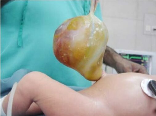
Gran onfalocele que contiene un quiste del conducto onfalomesentérico
Imagen: “Large omphalocele containing OMD cyst” por Yousuf Aziz Khan, MBBS, FCPS (Paediatric Surgeon), Department of Paediatric Surgery, National Institute of Child Health, Rafiquee Shaheed Road, Karachi – 75510, Sind, Pakistan. Licencia: CC BY 3.0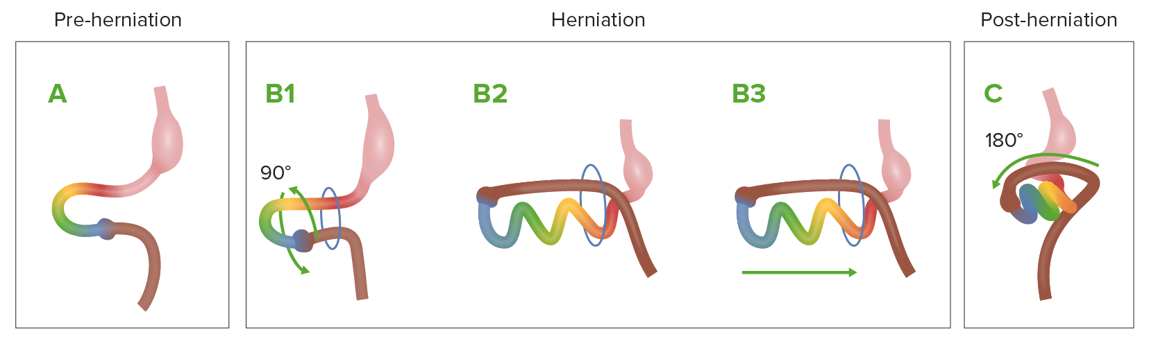
Diagrama que muestra el proceso normal de rotación y herniación intestinal durante el desarrollo embriológico
A: El intestino medio (bucle multicolor) antes de la herniación
B1–B3: Al crecer rápidamente, el intestino medio se hernia a través del anillo umbilical y comienza a rotar.
C: El intestino medio vuelve a la cavidad abdominal.
La fisiopatología no se ha HA Hemolytic anemia (HA) is the term given to a large group of anemias that are caused by the premature destruction/hemolysis of circulating red blood cells (RBCs). Hemolysis can occur within (intravascular hemolysis) or outside the blood vessels (extravascular hemolysis). Hemolytic Anemia entendido completamente; hay dos teorías principales:
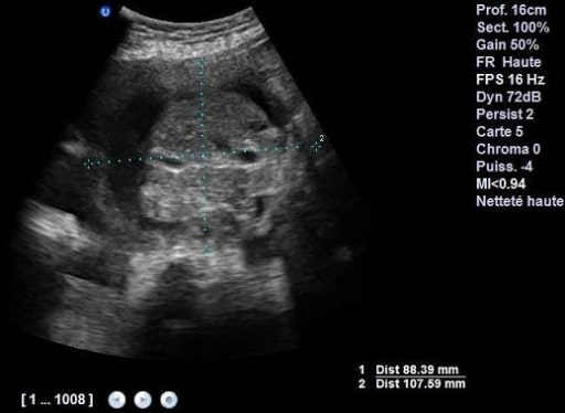
Ultrasonido obstétrico a las 37 semanas que muestra un onfalocele que contiene intestinos y parte del hígado
Imagen: “Obstetric ultrasound at 37 weeks containing intestines and part of the liver” por Gynaecology and Obstetrics Department I, University Hospital Hassan II, Fez, Morocco. Licencia: CC BY 2.0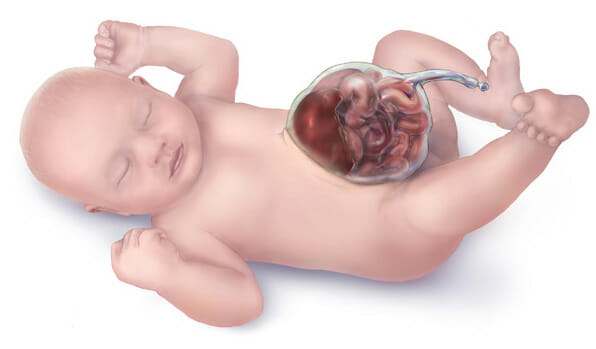
Ilustración de un onfalocele
Imagen: “Omphalocele” por Centers for Disease Control and Prevention. Licencia: Dominio PúblicoLa mayoría de los LOS Neisseria casos de onfalocele (90%) se diagnostican de forma prenatal:
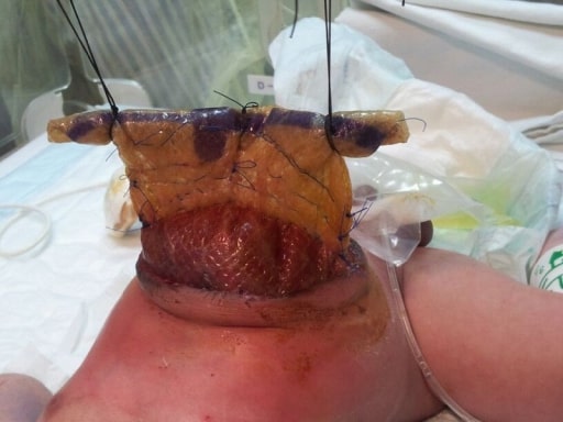
Cierre por etapas de un onfalocele gigante con malla sintética: Reducción secuencial diaria del contenido del silo de malla sintética
Imagen: “Daily sequential reduction of synthetic mesh silo content” por Department of Pediatric Surgery, GMC Hospital, Ajman, United Arab Emirates. Licencia: CC BY 3.0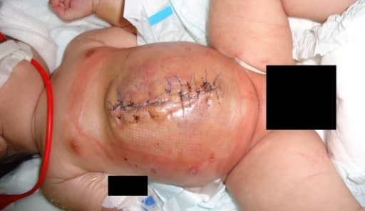
Cierre por etapas de un onfalocele gigante con malla sintética: cierre completo de un onfalocele gigante
Imagen: “Complete closure of giant omphalocele” por Department of Pediatric Surgery, GMC Hospital, Ajman, United Arab Emirates. Licencia: CC BY 3.0