El muslo es la región del miembro inferior que se encuentra entre la cadera y la articulación de la rodilla. En EN Erythema nodosum is an immune-mediated panniculitis (inflammation of the subcutaneous fat) caused by a type IV (delayed-type) hypersensitivity reaction. It commonly manifests in young women as tender, erythematous nodules on the shins. Erythema Nodosum el muslo hay un solo hueso llamado fémur, que está rodeado de grandes músculos agrupados en EN Erythema nodosum is an immune-mediated panniculitis (inflammation of the subcutaneous fat) caused by a type IV (delayed-type) hypersensitivity reaction. It commonly manifests in young women as tender, erythematous nodules on the shins. Erythema Nodosum 3 compartimentos fasciales. El muslo está irrigado principalmente por la arteria femoral y sus ramas, drenado por redes venosas profundas y superficiales, e inervado por ramas de los LOS Neisseria plexos lumbar y sacro.
Last updated: Dec 15, 2025
| Segmento | Puntos de referencia importantes |
|---|---|
| Extremo (epífisis) proximal |
|
| Diáfisis |
|
| Extremo (epífisis) distal |
|

Vista anterior del fémur derecho
Imagen por BioDigital, editado por Lecturio.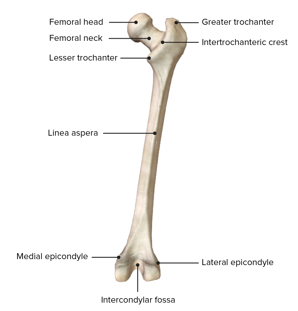
Vista posterior del fémur derecho
Imagen por BioDigital, editado por Lecturio.El tracto iliotibial o banda iliotibial es un engrosamiento de la fascia lata Fascia lata Femoral Region and Hernias: Anatomy situado en EN Erythema nodosum is an immune-mediated panniculitis (inflammation of the subcutaneous fat) caused by a type IV (delayed-type) hypersensitivity reaction. It commonly manifests in young women as tender, erythematous nodules on the shins. Erythema Nodosum la superficie lateral del muslo. El tracto iliotibial es un estabilizador de la cadera y la rodilla.
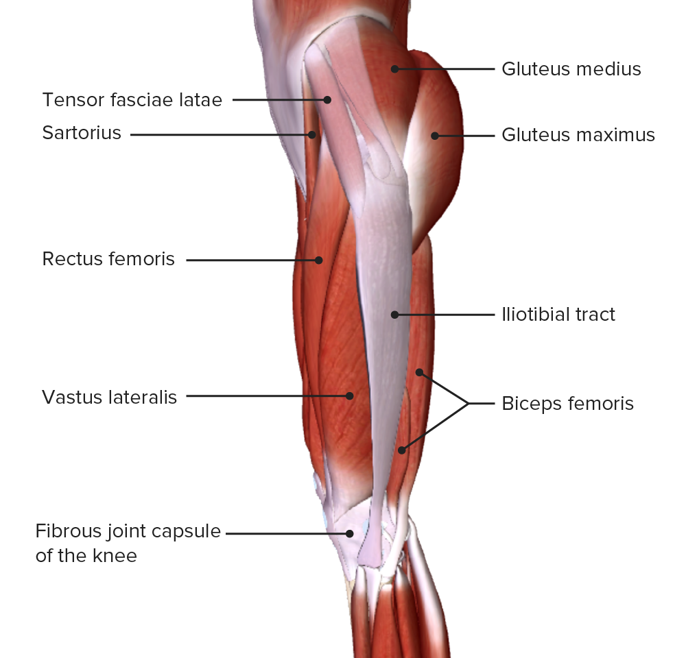
Vista lateral del muslo, mostrando el tracto iliotibial y el músculo tensor de la fascia lata.
Imagen por BioDigital, editado por Lecturio.Los LOS Neisseria 3 septos intermusculares nacen de la fascia lata Fascia lata Femoral Region and Hernias: Anatomy y se unen a la línea áspera del fémur. Los LOS Neisseria tabiques intermusculares lateral, medial y posterior dividen el muslo en EN Erythema nodosum is an immune-mediated panniculitis (inflammation of the subcutaneous fat) caused by a type IV (delayed-type) hypersensitivity reaction. It commonly manifests in young women as tender, erythematous nodules on the shins. Erythema Nodosum lo siguiente:
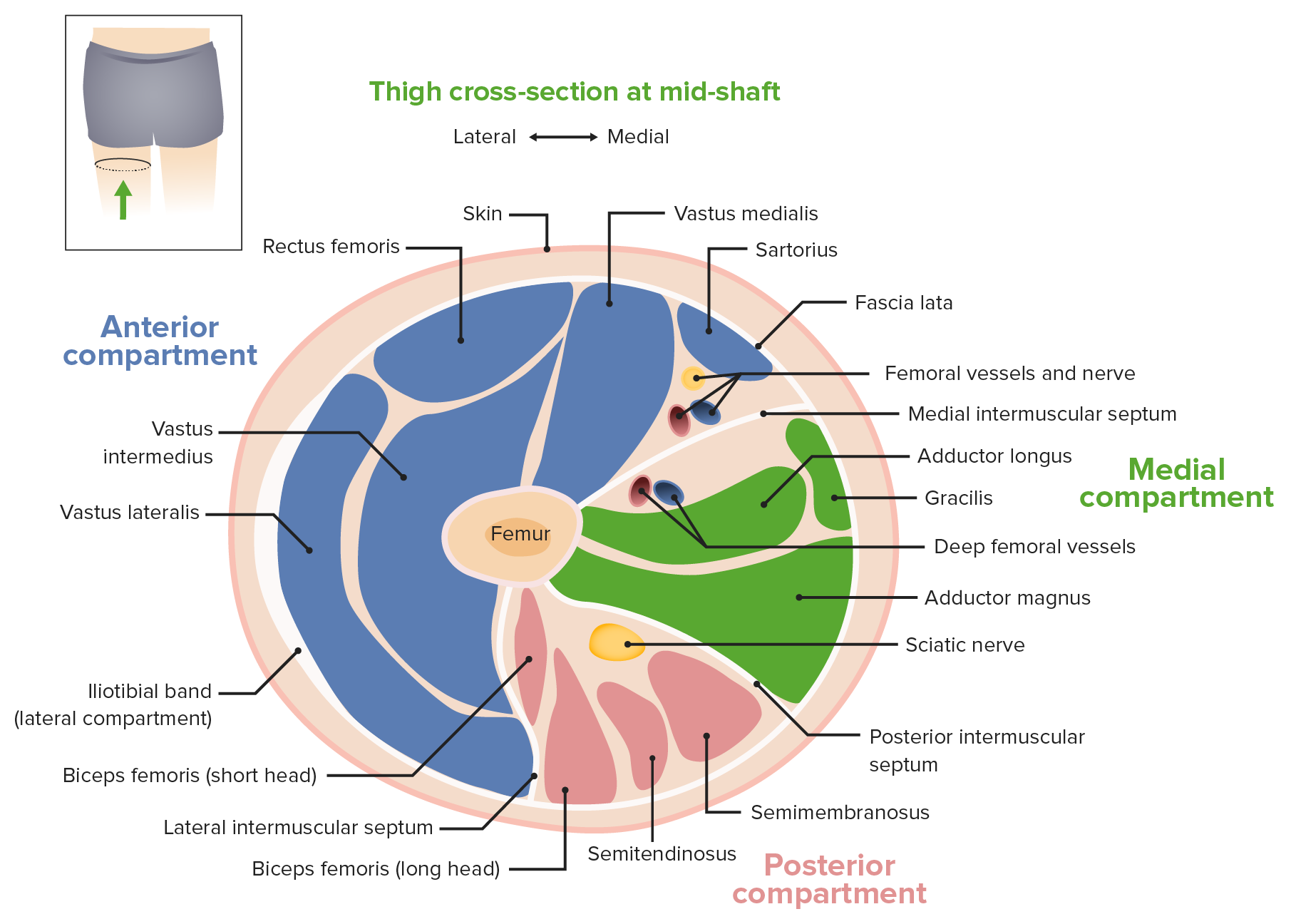
Corte transversal del muslo en la mitad de la diáfisis
Imagen por Lecturio.| Músculo | Origen | Inserción | Inervación | Función |
|---|---|---|---|---|
| Recto femoral | Espina ilíaca anteroinferior | Tuberosidad tibial a través del cuádriceps común y el ligamento rotuliano | Nervio femoral (L3, L4) |
|
| Vasto lateral | Trocánter mayor y labio lateral de la línea áspera | |||
| Vasto intermedio | Cara anterior de la diáfisis del fémur | |||
| Vasto medial | Línea intertrocantérica y labio medial de la línea áspera | |||
| Sartorio | Espina ilíaca anterosuperior | Superficie medial de la tibia Tibia The second longest bone of the skeleton. It is located on the medial side of the lower leg, articulating with the fibula laterally, the talus distally, and the femur proximally. Knee Joint: Anatomy proximal | Nervio femoral (L2) |
|
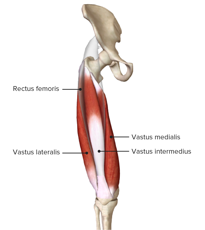
Vista anterior del muslo mostrando el origen y la inserción del cuádriceps femoral
Imagen por BioDigital, editado por Lecturio.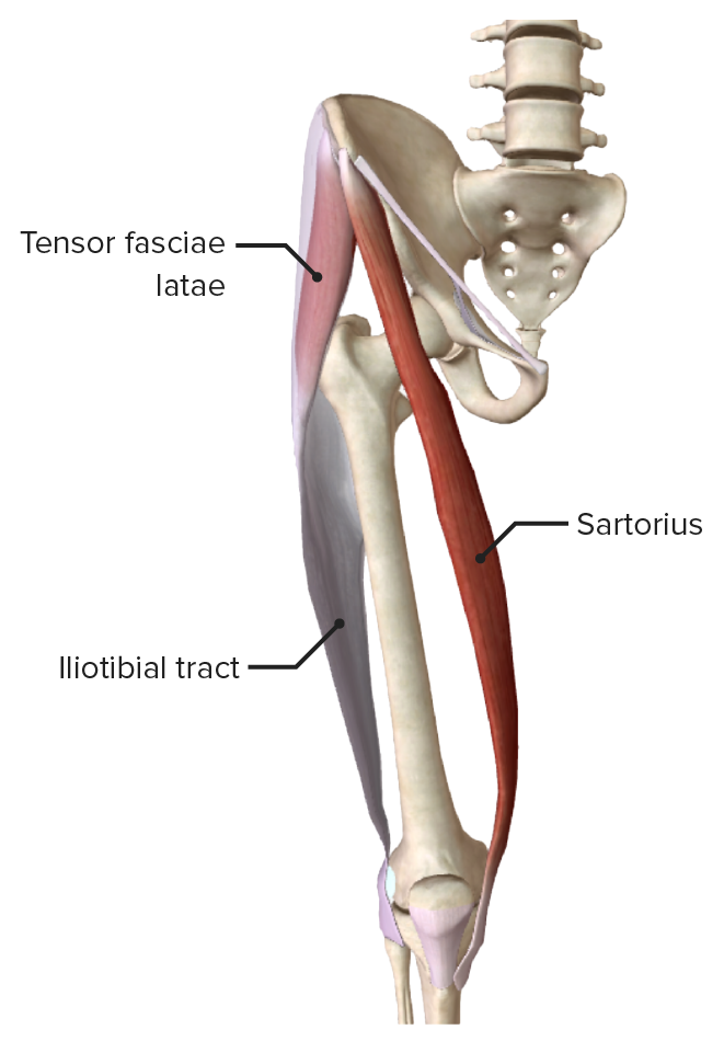
Tensor de la fascia lata, músculo sartorio y tracto iliotibial
Imagen por BioDigital, editado por Lecturio.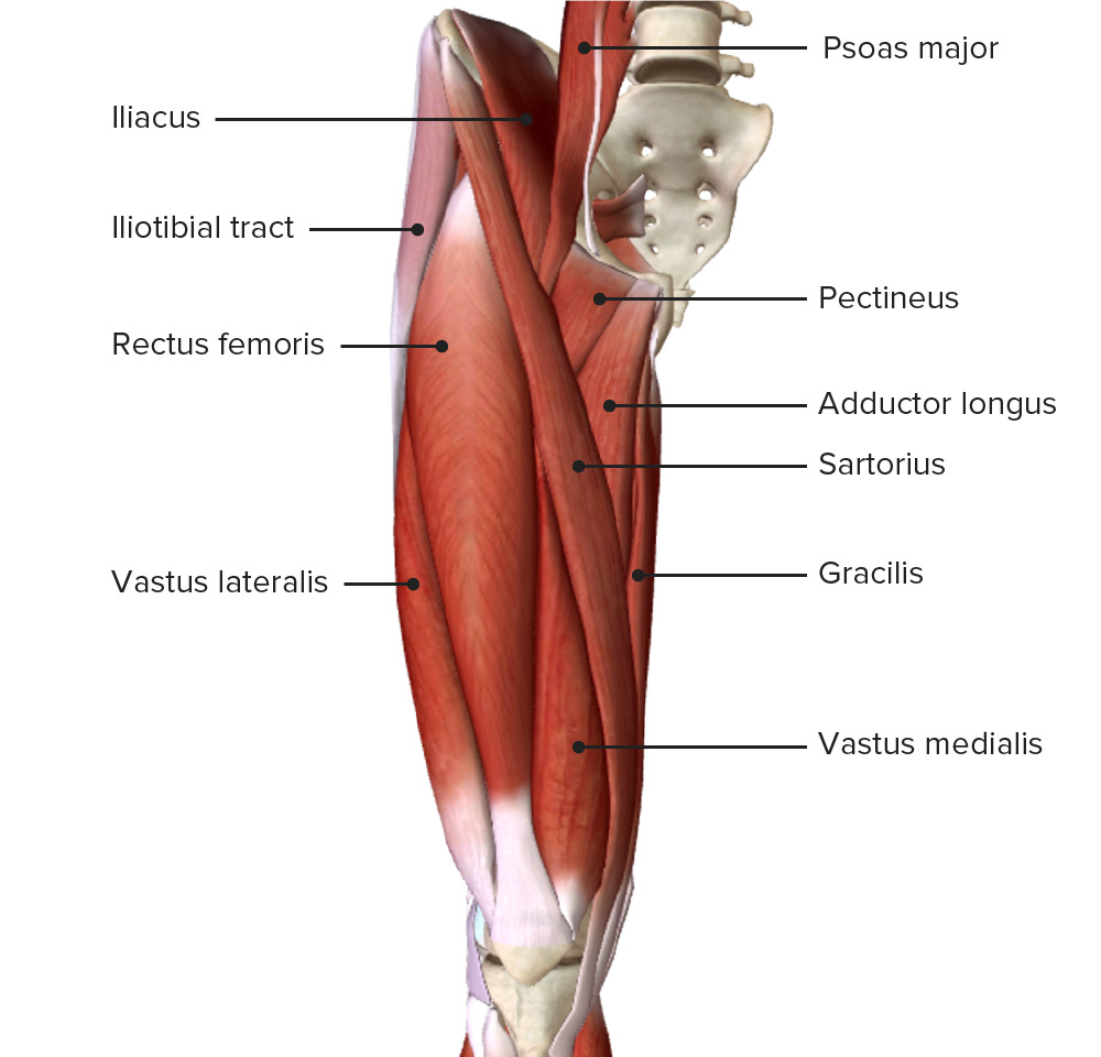
Vista anterior del muslo mostrando los músculos del compartimento anterior (cuádriceps femoral y sartorio) y su relación con los músculos vecinos y entre sí
Imagen por BioDigital, editado por Lecturio.| Músculo | Origen | Inserción | Inervación | Función |
|---|---|---|---|---|
| Bíceps femoral |
|
Superficie lateral del peroné |
|
|
| Semitendinoso | Tuberosidad isquiática | Superficie medial de la tibia Tibia The second longest bone of the skeleton. It is located on the medial side of the lower leg, articulating with the fibula laterally, the talus distally, and the femur proximally. Knee Joint: Anatomy proximal | División tibial del nervio ciático (L5, S1 S1 Heart Sounds) | Extiende la cadera, flexiona la rodilla y rota medialmente la rodilla cuando está flexionada |
| Semimembranoso | Superficie posterior del cóndilo medial de la tibia Tibia The second longest bone of the skeleton. It is located on the medial side of the lower leg, articulating with the fibula laterally, the talus distally, and the femur proximally. Knee Joint: Anatomy |
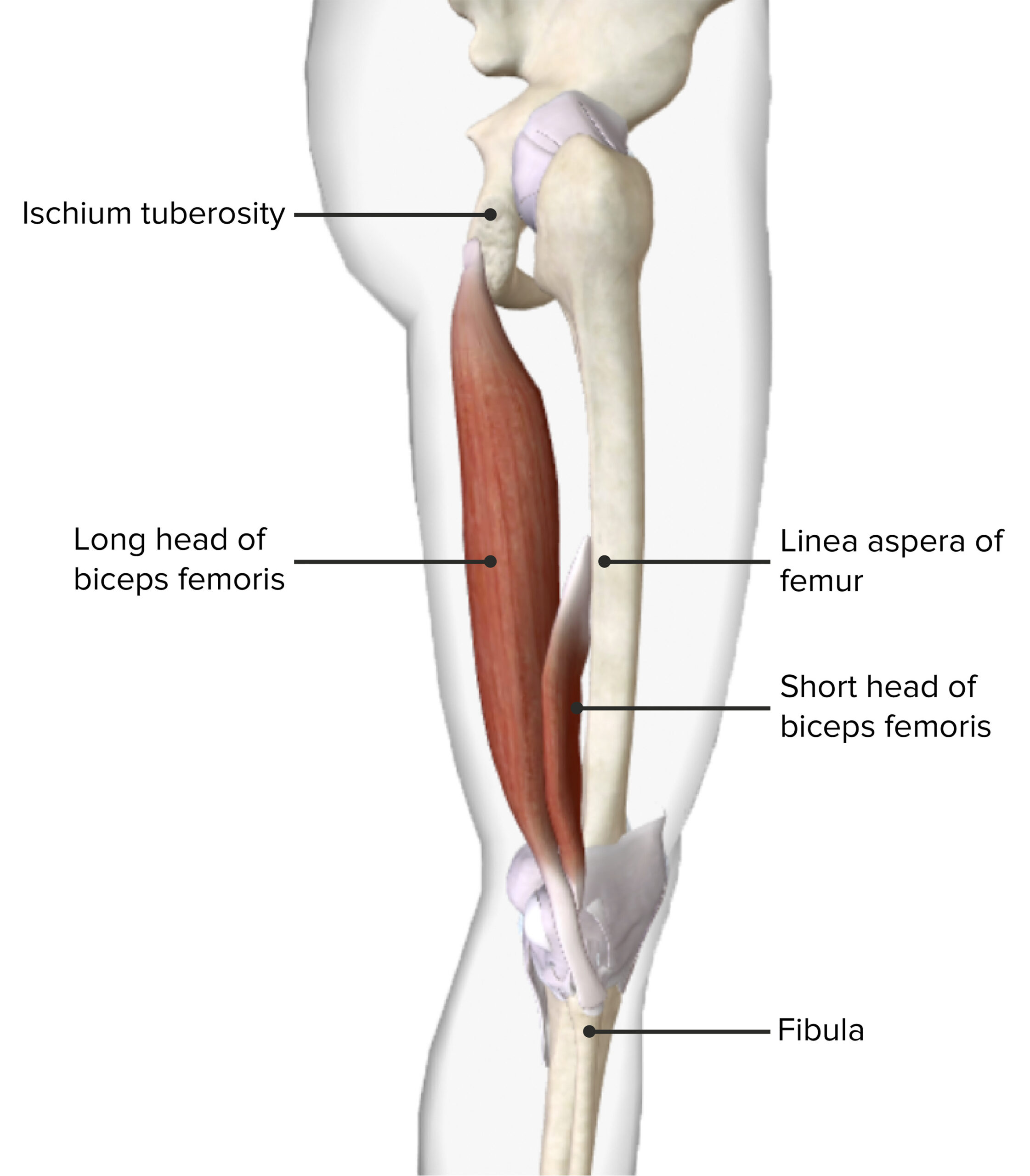
Vista lateral del muslo mostrando el origen y la inserción del músculo bíceps femoral
Imagen por BioDigital, editado por Lecturio.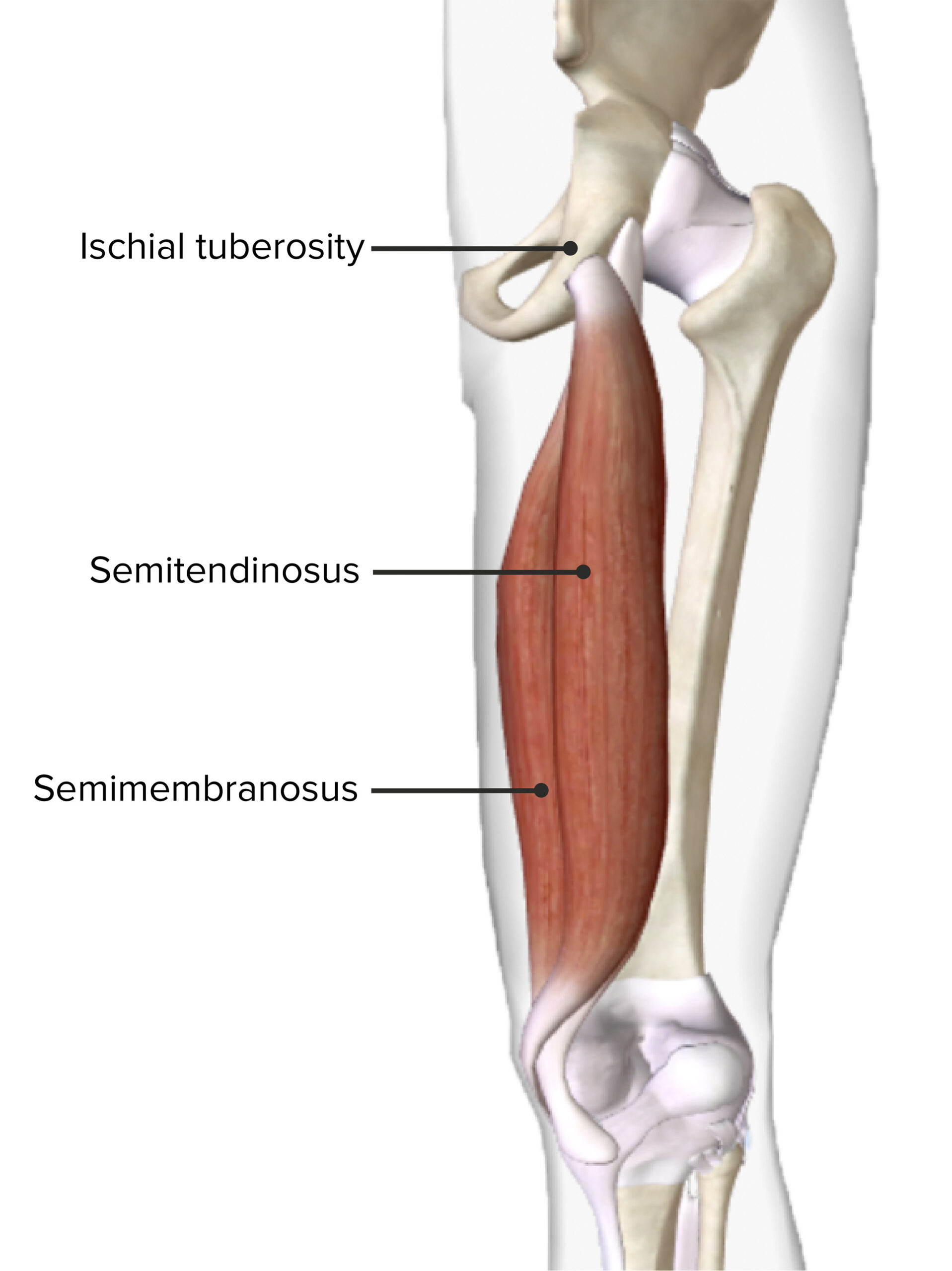
Vista posterior del muslo derecho mostrando el origen y la inserción de los músculos semitendinoso y semimembranoso
Imagen por BioDigital, editado por Lecturio.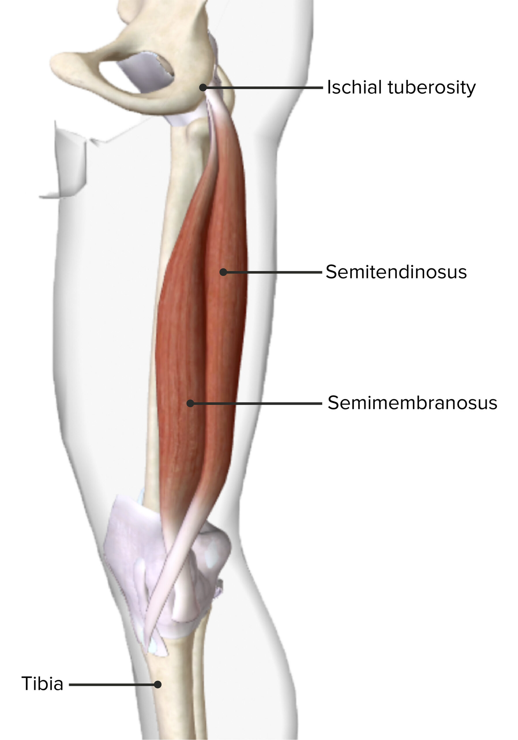
Vista medial del muslo derecho mostrando el origen y la inserción de los músculos semitendinoso y semimembranoso
Imagen por BioDigital, editado por Lecturio.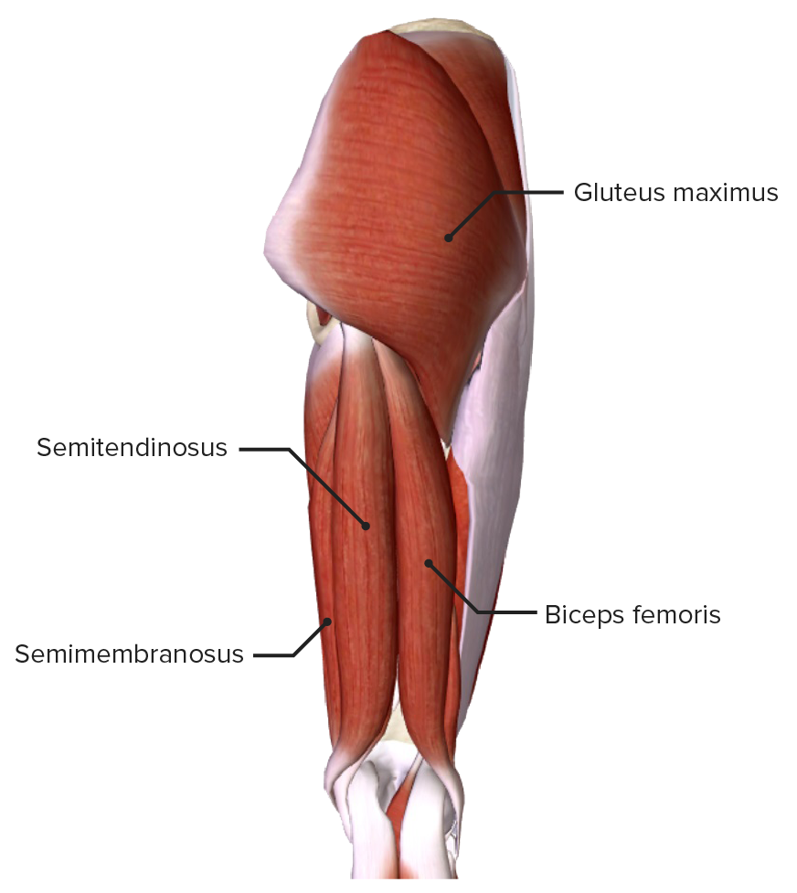
Vista posterior del muslo mostrando los músculos del compartimento posterior del muslo y su relación con otros músculos y entre sí
Imagen por BioDigital, editado por Lecturio.| Músculo | Origen | Inserción | Inervación | Función |
|---|---|---|---|---|
| Aductor mayor |
|
|
|
|
| Aductor largo | Cuerpo del pubis | Medio ⅓ de la línea áspera | Nervio obturador (L3) | Aduce la cadera |
| Aductor corto | Cuerpo y rama inferior del pubis | Línea áspera proximal | ||
| Grácil | Superficie medial de la tibia Tibia The second longest bone of the skeleton. It is located on the medial side of the lower leg, articulating with the fibula laterally, the talus distally, and the femur proximally. Knee Joint: Anatomy proximal | Nervio obturador (L2) |
|
|
| Pectíneo | Rama superior del pubis | Línea pectínea del fémur | Nervio femoral (L2) | Aduce, flexiona y estabiliza la rotación medial de la cadera |
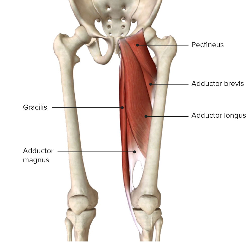
Vista anterior de los muslos mostrando el origen y la inserción de los músculos del compartimento medial
Imagen por BioDigital, editado por Lecturio.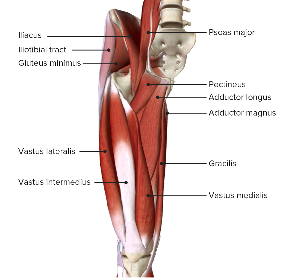
Vista anterior del muslo mostrando la capa muscular intermedia:
Observe los músculos del compartimento medial (el pectíneo, el aductor mayor, el largo y el corto, y el grácil) y su relación con otros músculos y entre sí
En EN Erythema nodosum is an immune-mediated panniculitis (inflammation of the subcutaneous fat) caused by a type IV (delayed-type) hypersensitivity reaction. It commonly manifests in young women as tender, erythematous nodules on the shins. Erythema Nodosum el extremo distal de la región medial del muslo se encuentra la pata de ganso, que comprende los LOS Neisseria tendones conjuntos de los LOS Neisseria músculos sartorio, grácil y semitendinoso que se insertan en EN Erythema nodosum is an immune-mediated panniculitis (inflammation of the subcutaneous fat) caused by a type IV (delayed-type) hypersensitivity reaction. It commonly manifests in young women as tender, erythematous nodules on the shins. Erythema Nodosum la tibia Tibia The second longest bone of the skeleton. It is located on the medial side of the lower leg, articulating with the fibula laterally, the talus distally, and the femur proximally. Knee Joint: Anatomy.
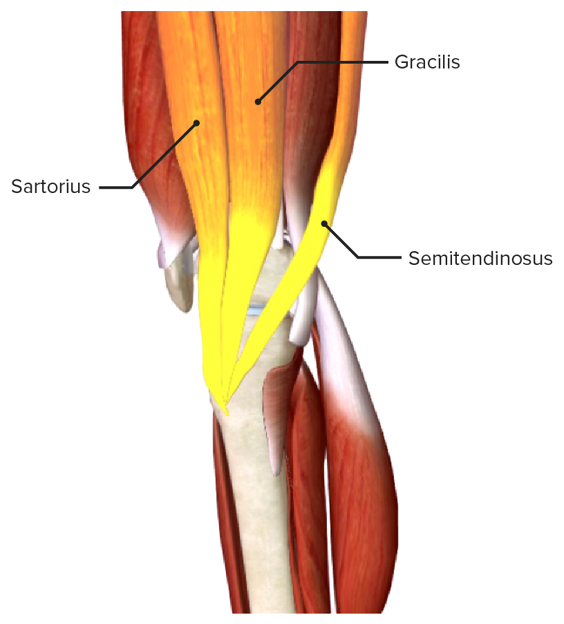
El tendón conjunto de los músculos grácil, sartorio y semitendinoso, que forman la pata de ganso
Imagen por BioDigital, editado por Lecturio.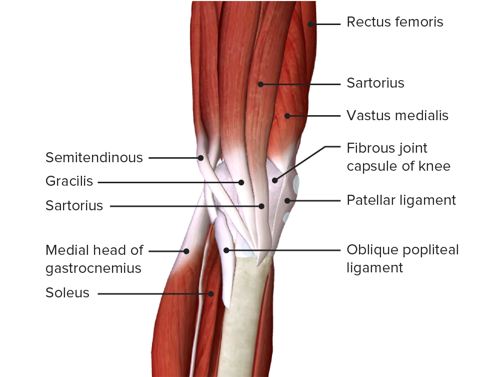
Vista medial de la parte inferior del muslo y de la articulación de la rodilla mostrando la inserción de los músculos sartorio, grácil y semitendinoso, que forman la pata de ganso.
Imagen por BioDigital, editado por Lecturio.De la pelvis Pelvis The pelvis consists of the bony pelvic girdle, the muscular and ligamentous pelvic floor, and the pelvic cavity, which contains viscera, vessels, and multiple nerves and muscles. The pelvic girdle, composed of 2 “hip” bones and the sacrum, is a ring-like bony structure of the axial skeleton that links the vertebral column with the lower extremities. Pelvis: Anatomy salen tres arterias: la femoral, la obturadora y la glútea inferior. Sin embargo, las arterias femoral y obturadora continúan hacia el muslo, mientras que la glútea inferior irriga y termina en EN Erythema nodosum is an immune-mediated panniculitis (inflammation of the subcutaneous fat) caused by a type IV (delayed-type) hypersensitivity reaction. It commonly manifests in young women as tender, erythematous nodules on the shins. Erythema Nodosum la región glútea.
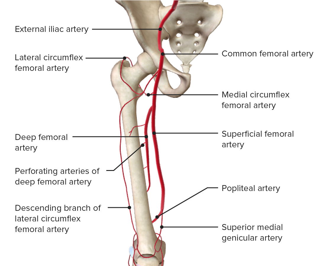
Irrigación arterial del muslo mostrando la trayectoria y las principales ramas de la arteria femoral
Imagen por BioDigital, editado por Lecturio.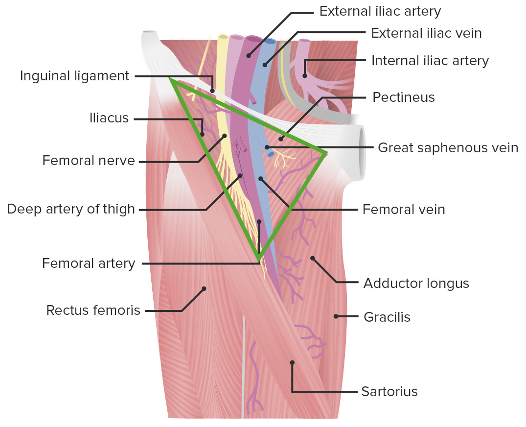
Vista anterior del muslo mostrando el paso de la arteria femoral a través del triángulo femoral
Imagen por Lecturio.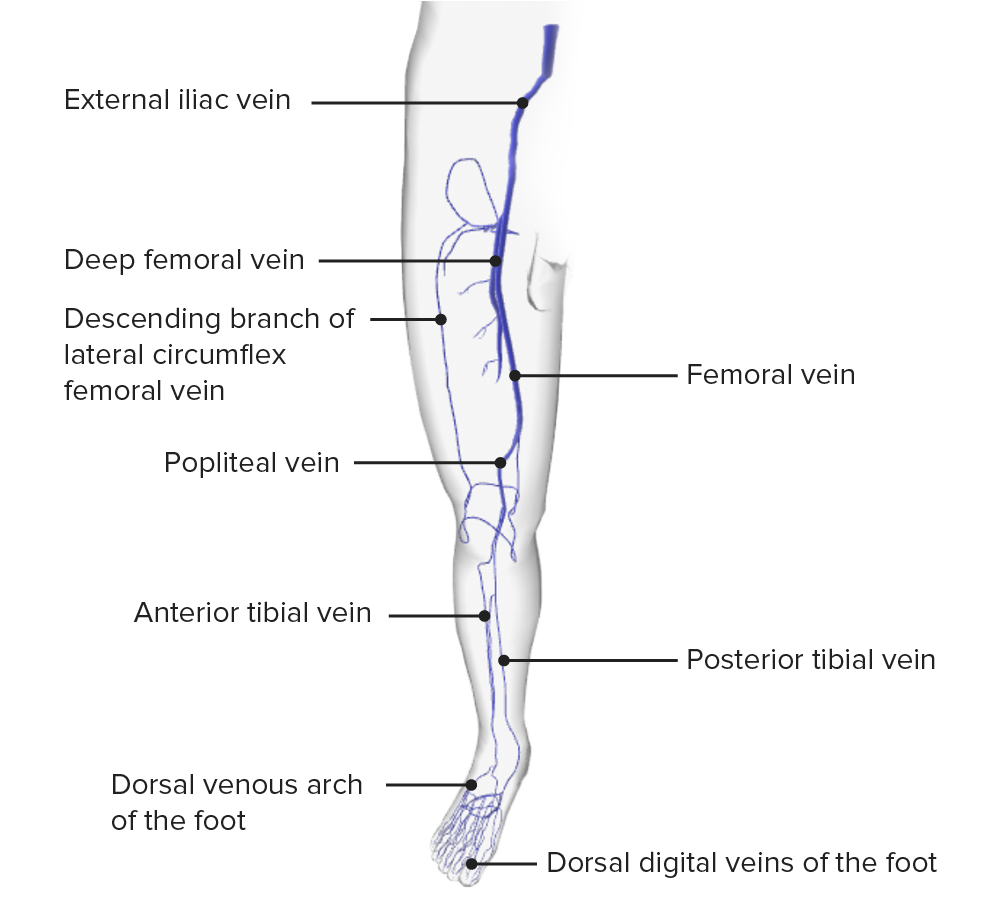
Vista anterior del drenaje venoso del miembro inferior
Imagen por BioDigital, editado por Lecturio.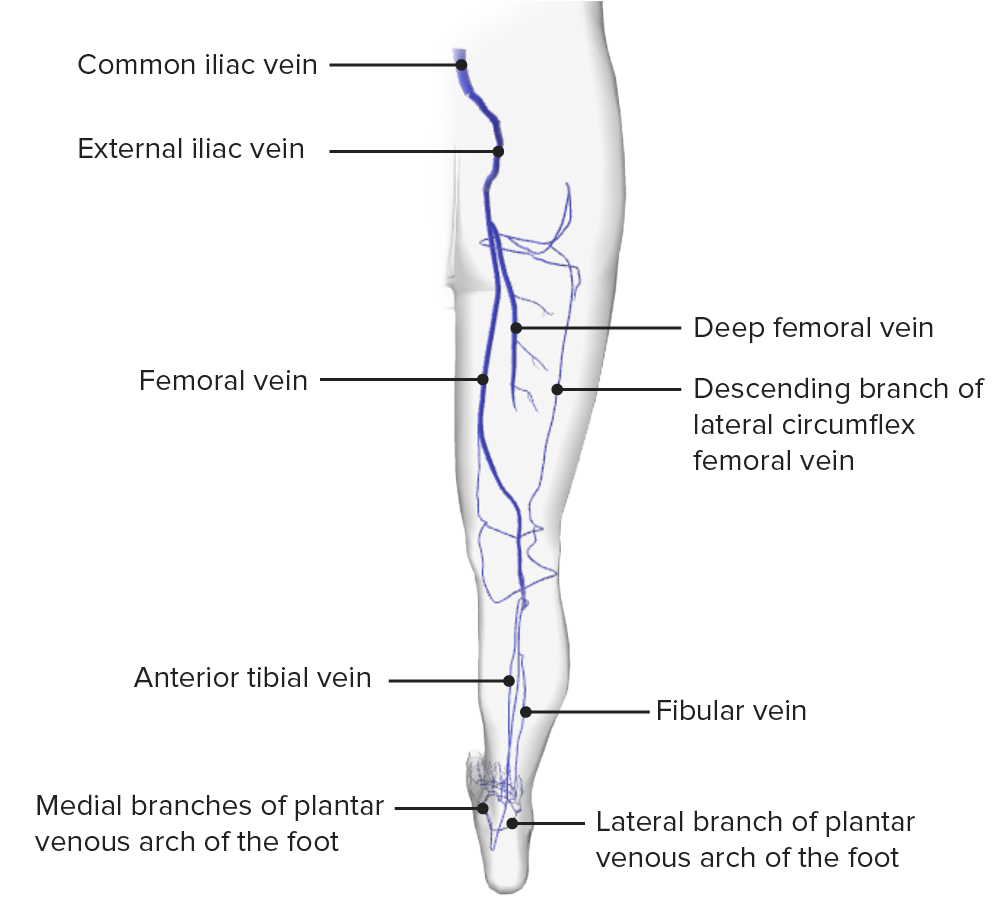
Vista posterior del drenaje venoso del miembro inferior
Imagen por BioDigital, editado por Lecturio.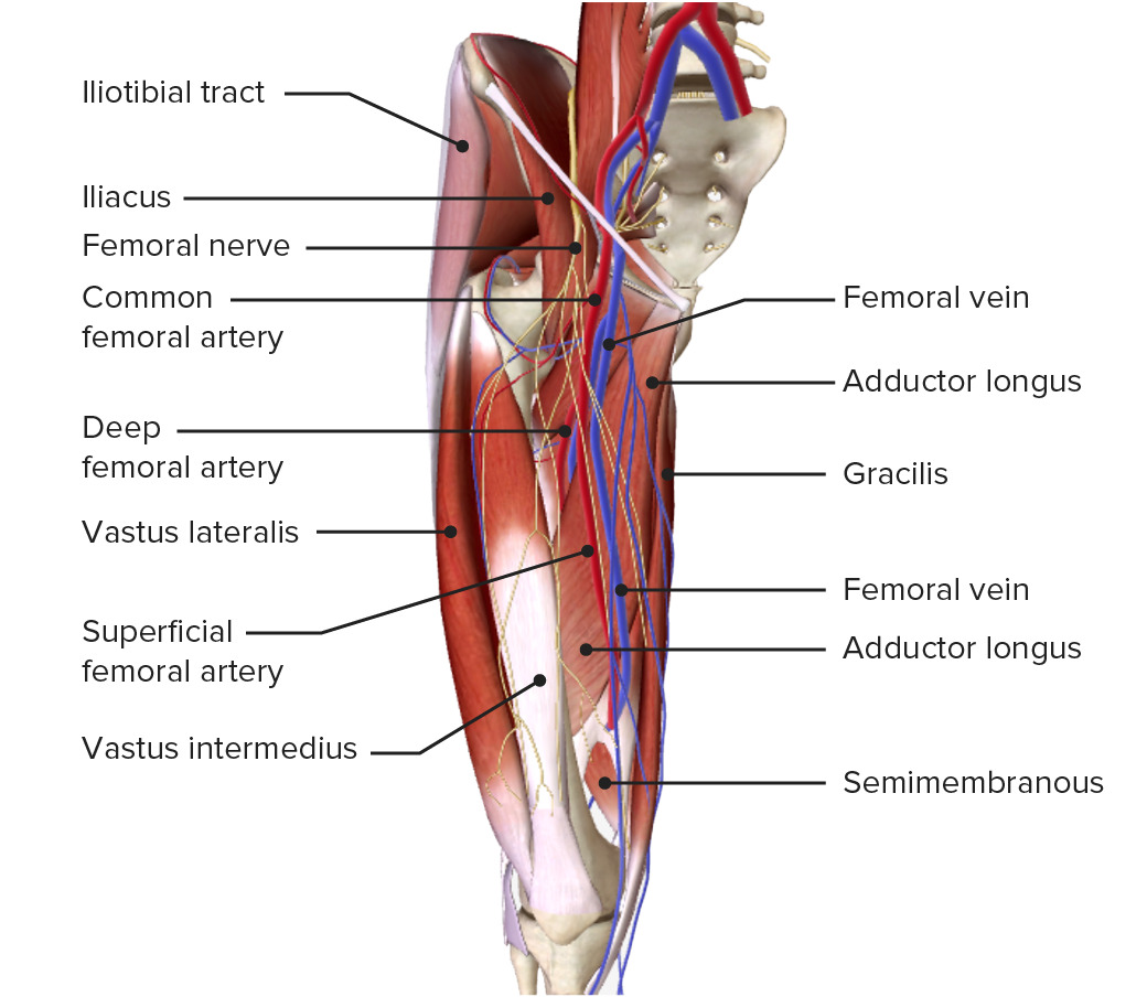
Vista anterior del muslo:
Obsérvese el curso y las ramas de la arteria profunda brachii (arteria profunda del muslo), la red venosa superficial y la relación espacial de los vasos y los músculos.
El muslo está inervado por ramas del plexo lumbar y sacro.
| Nervio | Origen | Inervación motora | Inervación sensorial |
|---|---|---|---|
| Nervio cutáneo femoral lateral | Plexo lumbar (L2–L3) | Piel anterolateral del muslo | |
| Nervio cutáneo femoral posterior | Plexo sacro ( S2 S2 Heart Sounds– S3 S3 Heart Sounds) | Piel de la región glútea, periné posterior y muslo posterior | |
| Nervio femoral |
|
|
|
| Nervio safeno | Rama del nervio femoral | Piel medial de los LOS Neisseria 2/3 inferiores del muslo, medial inferior de la pierna y pie | |
| Nervio obturador |
|
Músculos del compartimento medial | Piel medial del 1/3 superior del muslo |
| Nervio ciático (nervio más grueso del cuerpo humano) |
|
Rama tibial: Músculos del compartimento posterior | Ninguno en EN Erythema nodosum is an immune-mediated panniculitis (inflammation of the subcutaneous fat) caused by a type IV (delayed-type) hypersensitivity reaction. It commonly manifests in young women as tender, erythematous nodules on the shins. Erythema Nodosum el muslo |
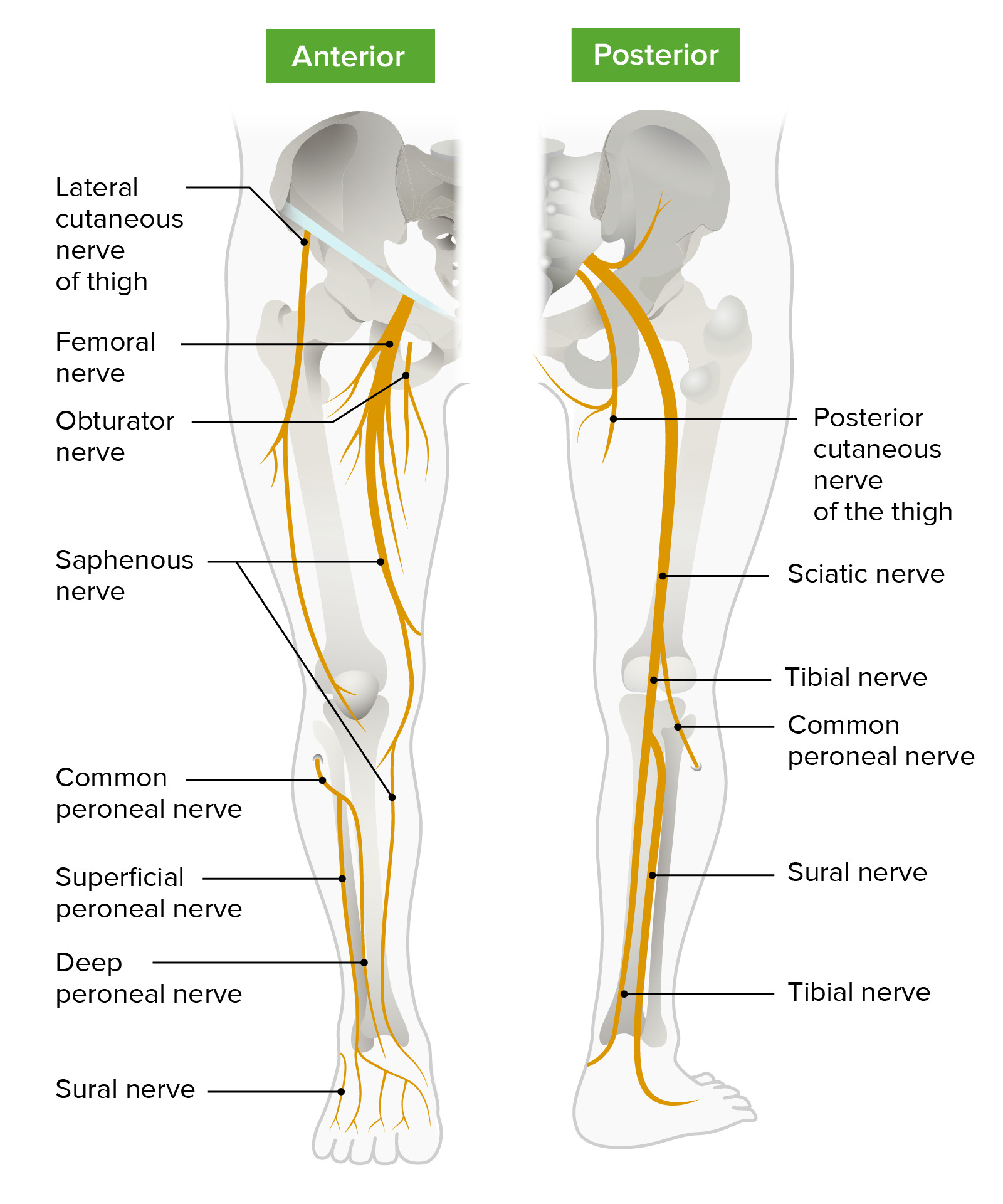
Inervación de los miembros inferiores
Imagen por Lecturio.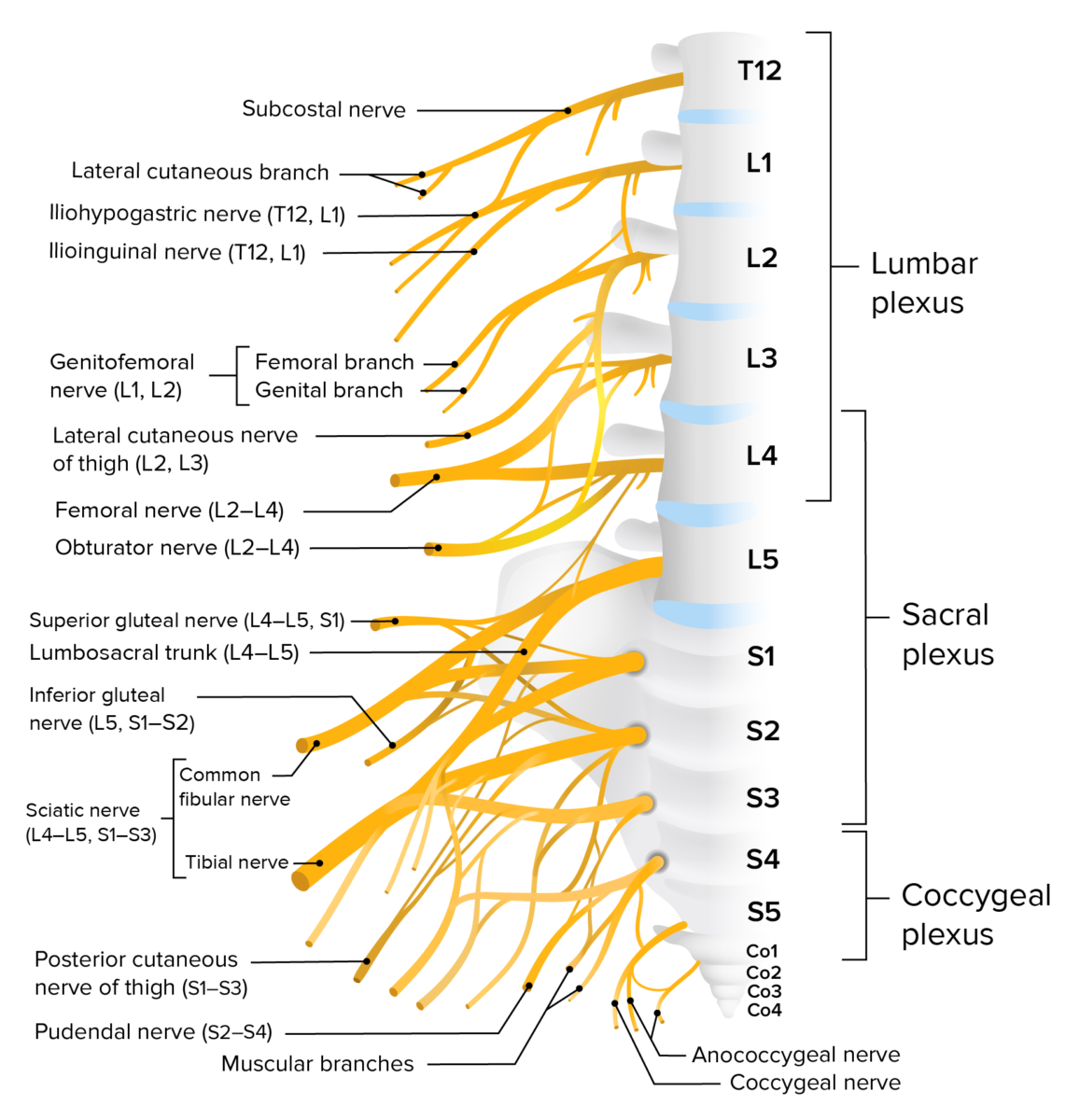
Plexo lumbosacro
Imagen por Lecturio.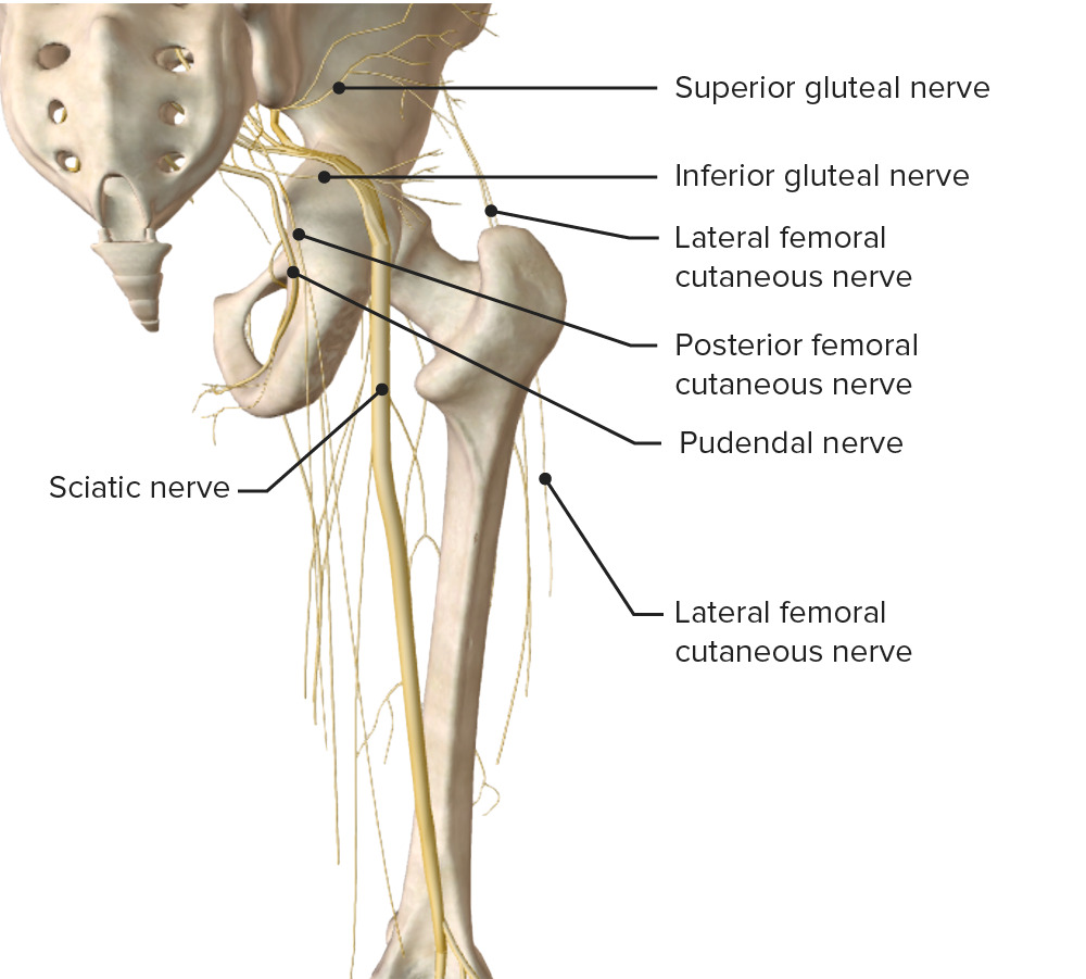
Diagrama esquemático del trayecto y las principales ramas del plexo lumbosacro
Imagen por BioDigital, editado por Lecturio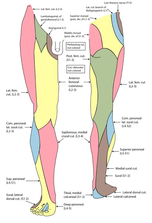
Inervación cutánea del miembro inferior
Imagen: “Gray826and831” por Henry Vandyke Carter. Licencia: Dominio PúblicoLas siguientes afecciones son clínicamente relevantes para el muslo: