Domina Conceptos Médicos

Estudia para la escuela de medicina y tus examenes con Lecturio.

Lentigo Maligno

El lentigo maligno es un melanoma Melanoma Melanoma is a malignant tumor arising from melanocytes, the melanin-producing cells of the epidermis. These tumors are most common in fair-skinned individuals with a history of excessive sun exposure and sunburns. Melanoma in situ, una lesión precancerosa que puede progresar a un melanoma Melanoma Melanoma is a malignant tumor arising from melanocytes, the melanin-producing cells of the epidermis. These tumors are most common in fair-skinned individuals with a history of excessive sun exposure and sunburns. Melanoma invasivo (específicamente el subtipo de melanoma Melanoma Melanoma is a malignant tumor arising from melanocytes, the melanin-producing cells of the epidermis. These tumors are most common in fair-skinned individuals with a history of excessive sun exposure and sunburns. Melanoma lentigo maligno). Esta condición ocurre típicamente en EN Erythema nodosum is an immune-mediated panniculitis (inflammation of the subcutaneous fat) caused by a type IV (delayed-type) hypersensitivity reaction. It commonly manifests in young women as tender, erythematous nodules on the shins. Erythema Nodosum áreas dañadas por el sol (e.g., cara y cuello) en EN Erythema nodosum is an immune-mediated panniculitis (inflammation of the subcutaneous fat) caused by a type IV (delayed-type) hypersensitivity reaction. It commonly manifests in young women as tender, erythematous nodules on the shins. Erythema Nodosum pacientes de edad avanzada. El lentigo maligno se presenta como una mácula marrón con colores variados y bordes asimétricos que crece lentamente. Se debe tomar una biopsia para confirmar el diagnóstico y la extirpación quirúrgica con un margen de seguridad es el tratamiento de 1ra línea.

Last updated: Dec 15, 2025

Editorial responsibility: Stanley Oiseth, Lindsay Jones, Evelin Maza

Descripción General

Definición

El lentigo maligno (también conocido como peca melanótica de Hutchinson) es un melanoma Melanoma Melanoma is a malignant tumor arising from melanocytes, the melanin-producing cells of the epidermis. These tumors are most common in fair-skinned individuals with a history of excessive sun exposure and sunburns. Melanoma in situ. Este tipo de lesión precancerosa puede progresar a melanoma Melanoma Melanoma is a malignant tumor arising from melanocytes, the melanin-producing cells of the epidermis. These tumors are most common in fair-skinned individuals with a history of excessive sun exposure and sunburns. Melanoma lentigo maligno.

Epidemiología

  • Pico de incidencia entre los LOS Neisseria 65 y los LOS Neisseria 80 años
  • Precursor del 3er subtipo de melanoma Melanoma Melanoma is a malignant tumor arising from melanocytes, the melanin-producing cells of the epidermis. These tumors are most common in fair-skinned individuals with a history of excessive sun exposure and sunburns. Melanoma más común ( melanoma Melanoma Melanoma is a malignant tumor arising from melanocytes, the melanin-producing cells of the epidermis. These tumors are most common in fair-skinned individuals with a history of excessive sun exposure and sunburns. Melanoma lentigo maligno)
  • Mujeres > hombres
  • La incidencia es de 13,7 por cada 100 000.

Etiología

  • Mutaciones genéticas:
    • En EN Erythema nodosum is an immune-mediated panniculitis (inflammation of the subcutaneous fat) caused by a type IV (delayed-type) hypersensitivity reaction. It commonly manifests in young women as tender, erythematous nodules on the shins. Erythema Nodosum comparación con otros subtipos de melanoma Melanoma Melanoma is a malignant tumor arising from melanocytes, the melanin-producing cells of the epidermis. These tumors are most common in fair-skinned individuals with a history of excessive sun exposure and sunburns. Melanoma, existe una mayor probabilidad de mutaciones de KIT .
    • Otras mutaciones incluyen CCND1 CCND1 Protein encoded by the bcl-1 gene which plays a critical role in regulating the cell cycle. Overexpression of cyclin d1 is the result of bcl-1 rearrangement, a t(11; 14) translocation, and is implicated in various neoplasms. Lentigo Maligna, MITF MITF Lentigo Maligna, NRAS y p53 .
  • Factores de riesgo:
    • Radiación ultravioleta acumulada (a lo largo del tiempo)
    • Condiciones genéticas:
      • Albinismo oculocutáneo
      • Xeroderma pigmentoso
      • Porfiria cutánea tardía
    • Irradiación con rayos X
    • Uso de estrógeno/progesterona
    • Uso de tintes no permanentes para el cabello.

Related videos

Presentación Clínica

  • La mayoría ocurre en EN Erythema nodosum is an immune-mediated panniculitis (inflammation of the subcutaneous fat) caused by a type IV (delayed-type) hypersensitivity reaction. It commonly manifests in young women as tender, erythematous nodules on the shins. Erythema Nodosum la cabeza/cuello (principalmente en EN Erythema nodosum is an immune-mediated panniculitis (inflammation of the subcutaneous fat) caused by a type IV (delayed-type) hypersensitivity reaction. It commonly manifests in young women as tender, erythematous nodules on the shins. Erythema Nodosum la mejilla).
  • Color variable Variable Variables represent information about something that can change. The design of the measurement scales, or of the methods for obtaining information, will determine the data gathered and the characteristics of that data. As a result, a variable can be qualitative or quantitative, and may be further classified into subgroups. Types of Variables:
    • Marrón claro a negro (puede tener variedad de colores)
    • Puede tener áreas rosadas/blancas que indican inflamación o regresión
  • Asimétrica
  • Bordes mal definidos
  • Lisa (no palpable)
  • Crecimiento lento → moteado (a medida que el melanoma Melanoma Melanoma is a malignant tumor arising from melanocytes, the melanin-producing cells of the epidermis. These tumors are most common in fair-skinned individuals with a history of excessive sun exposure and sunburns. Melanoma crece)
  • Indicadores de posible progresión a melanoma Melanoma Melanoma is a malignant tumor arising from melanocytes, the melanin-producing cells of the epidermis. These tumors are most common in fair-skinned individuals with a history of excessive sun exposure and sunburns. Melanoma invasivo:
    • Áreas elevadas/palpables
    • Bordes nítidos
    • Pigmentación más oscura
  • La piel circundante suele manifestar daño cutáneo solar:
    • Queratosis actínicas
    • Elastosis solar
    • Lentigos solares
Lentigo maligno

Lentigo maligno: una mácula marrón asimétrica con variedad de colores presente en la mejilla izquierda

Imagen: “Lentigo maligna” por kilbad. Licencia: CC BY 3.0
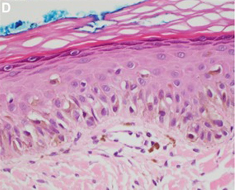
Diagnóstico

Antecedentes

  • Cantidad de exposición solar
  • Otros cánceres de piel
  • Antecedentes familiares de melanoma Melanoma Melanoma is a malignant tumor arising from melanocytes, the melanin-producing cells of the epidermis. These tumors are most common in fair-skinned individuals with a history of excessive sun exposure and sunburns. Melanoma
  • Enfermedades hereditarias

Examen físico

  • Se debe realizar un examen completo/minucioso de la piel.
  • Las lesiones malignas y premalignas sincrónicas no son infrecuentes en EN Erythema nodosum is an immune-mediated panniculitis (inflammation of the subcutaneous fat) caused by a type IV (delayed-type) hypersensitivity reaction. It commonly manifests in young women as tender, erythematous nodules on the shins. Erythema Nodosum personas con daño solar extenso en EN Erythema nodosum is an immune-mediated panniculitis (inflammation of the subcutaneous fat) caused by a type IV (delayed-type) hypersensitivity reaction. It commonly manifests in young women as tender, erythematous nodules on the shins. Erythema Nodosum la piel.

Dermatoscopia

  • Aberturas foliculares pigmentadas asimétricas (pseudored)
  • Progresión de los LOS Neisseria hallazgos:
    • Etapa temprana: pigmentación “en pimienta” alrededor de los LOS Neisseria orificios foliculares (puntos azul-grisáceos)
    • Coalescencia de puntos azul-grisáceos en EN Erythema nodosum is an immune-mediated panniculitis (inflammation of the subcutaneous fat) caused by a type IV (delayed-type) hypersensitivity reaction. It commonly manifests in young women as tender, erythematous nodules on the shins. Erythema Nodosum líneas poligonales y estructuras romboidales
    • Etapa tardía: manchas homogéneas de color marrón oscuro- negro que ocluyen los LOS Neisseria orificios foliculares

Microscopía confocal de reflectancia

  • La disponibilidad de esta técnica es limitada.
  • Requiere entrenamiento especializado
  • Más sensible, pero menos específica para el diagnóstico de lentigo maligno que la dermatoscopia

Histología

  • Estándar de oro para el diagnóstico del lentigo maligno
  • Muestra obtenida a través de:
    • Biopsia escicional (ideal)
    • Biopsia por sacabocados/biopsia incisional en EN Erythema nodosum is an immune-mediated panniculitis (inflammation of the subcutaneous fat) caused by a type IV (delayed-type) hypersensitivity reaction. It commonly manifests in young women as tender, erythematous nodules on the shins. Erythema Nodosum sitios cosméticamente sensibles
    • Biopsia por afeitado profundo
  • Hallazgos:
    • Sustitución de queratinocitos basales por nidos de melanocitos atípicos
    • Sin invasión de la membrana basal (si está presente, indica progresión a melanoma Melanoma Melanoma is a malignant tumor arising from melanocytes, the melanin-producing cells of the epidermis. These tumors are most common in fair-skinned individuals with a history of excessive sun exposure and sunburns. Melanoma invasivo)
    • Cambios de daño solar crónico:
      • Atrofia epidérmica
      • Infiltrados dérmicos inflamatorios
      • Borramiento de las crestas epidérmicas
Histopatología del lentigo maligno

Lentigo maligno:
Se puede observar un nido de melanocitos atípicos. La membrana basal está intacta. Por lo tanto, esta lesión es un lentigo maligno (in situ).

Imagen: “Lentigo maligna” por Sheliza Halani et al. Licencia: CC BY 4.0

Tratamiento y Pronóstico

Tratamiento

Escisión quirúrgica:

  • Tratamiento de 1ra linea
  • Procedimientos:
    • Escisión local amplia:
      • Deben obtenerse márgenes de 5–10 mm.
      • Para lesiones pequeñas con bordes bien delimitados
    • Escisión seriada por etapas:
      • Implica el examen histológico de las secciones permanentes
      • Las escisiones se realizan hasta obtener márgenes negativos.
      • Para lesiones grandes (> 1 cm en EN Erythema nodosum is an immune-mediated panniculitis (inflammation of the subcutaneous fat) caused by a type IV (delayed-type) hypersensitivity reaction. It commonly manifests in young women as tender, erythematous nodules on the shins. Erythema Nodosum cabeza/cuello y > 2 cm en EN Erythema nodosum is an immune-mediated panniculitis (inflammation of the subcutaneous fat) caused by a type IV (delayed-type) hypersensitivity reaction. It commonly manifests in young women as tender, erythematous nodules on the shins. Erythema Nodosum tronco/extremidades)
      • Para lesiones con bordes mal definidos
    • Cirugía micrográfica de Mohs:
      • Escisión por etapas que implica el examen de secciones congeladas
      • Puede no ser tan confiable como la técnica de escisión por etapas para el lentigo maligno

No quirúrgico:

  • Reservado para:
    • Pacientes ancianos frágiles
    • Pacientes que rechazan la cirugía
    • Lesiones grandes en EN Erythema nodosum is an immune-mediated panniculitis (inflammation of the subcutaneous fat) caused by a type IV (delayed-type) hypersensitivity reaction. It commonly manifests in young women as tender, erythematous nodules on the shins. Erythema Nodosum áreas cosméticamente sensibles donde se espera una reconstrucción problemática
  • Incluyen:
    • Radioterapia
    • Láser
    • Criocirugía
    • Imiquimod Imiquimod A topically-applied aminoquinoline immune modulator that induces interferon production. It is used in the treatment of external genital and perianal warts, superficial carcinoma, basal cell; and actinic keratosis. Hypertrophic and Keloid Scars tópico

Educación del paciente:

  • Protección contra la radiación ultravioleta (protector solar, ropa)
  • Autoexamen de la piel

Pronóstico

  • Se encuentra que hasta el 20% de las biopsias de presunto lentigo maligno tienen melanoma Melanoma Melanoma is a malignant tumor arising from melanocytes, the melanin-producing cells of the epidermis. These tumors are most common in fair-skinned individuals with a history of excessive sun exposure and sunburns. Melanoma invasivo.
  • Puede tomar < 10 hasta > 50 años para que el lentigo maligno progrese a melanoma Melanoma Melanoma is a malignant tumor arising from melanocytes, the melanin-producing cells of the epidermis. These tumors are most common in fair-skinned individuals with a history of excessive sun exposure and sunburns. Melanoma lentigo maligno.
  • Tasa de recurrencia de 6%–9% después de una escisión amplia
  • En EN Erythema nodosum is an immune-mediated panniculitis (inflammation of the subcutaneous fat) caused by a type IV (delayed-type) hypersensitivity reaction. It commonly manifests in young women as tender, erythematous nodules on the shins. Erythema Nodosum ausencia de progresión a melanoma Melanoma Melanoma is a malignant tumor arising from melanocytes, the melanin-producing cells of the epidermis. These tumors are most common in fair-skinned individuals with a history of excessive sun exposure and sunburns. Melanoma lentigo maligno, el lentigo maligno no acorta la esperanza de vida.
  • No hay muertes relacionadas con la enfermedad cuando se logra la escisión completa

Diagnóstico Diferencial

  • Lentigo solar: lesiones maculares con márgenes irregulares y pigmentación marrón uniforme en EN Erythema nodosum is an immune-mediated panniculitis (inflammation of the subcutaneous fat) caused by a type IV (delayed-type) hypersensitivity reaction. It commonly manifests in young women as tender, erythematous nodules on the shins. Erythema Nodosum la piel expuesta al AL Amyloidosis sol similar al AL Amyloidosis lentigo maligno. La biopsia de estas lesiones no muestra anidación de melanocitos atípicos. El lentigo solar es benigno y no requiere tratamiento.
  • Melanoma Melanoma Melanoma is a malignant tumor arising from melanocytes, the melanin-producing cells of the epidermis. These tumors are most common in fair-skinned individuals with a history of excessive sun exposure and sunburns. Melanoma: neoplasia maligna de la piel derivada de la transformación maligna de los LOS Neisseria melanocitos. El tipo más común de melanoma Melanoma Melanoma is a malignant tumor arising from melanocytes, the melanin-producing cells of the epidermis. These tumors are most common in fair-skinned individuals with a history of excessive sun exposure and sunburns. Melanoma es el melanoma Melanoma Melanoma is a malignant tumor arising from melanocytes, the melanin-producing cells of the epidermis. These tumors are most common in fair-skinned individuals with a history of excessive sun exposure and sunburns. Melanoma de extensión superficial, que generalmente se presenta como una mácula/parche irregular. En EN Erythema nodosum is an immune-mediated panniculitis (inflammation of the subcutaneous fat) caused by a type IV (delayed-type) hypersensitivity reaction. It commonly manifests in young women as tender, erythematous nodules on the shins. Erythema Nodosum el examen histopatológico, se detectarían melanocitos atípicos en EN Erythema nodosum is an immune-mediated panniculitis (inflammation of the subcutaneous fat) caused by a type IV (delayed-type) hypersensitivity reaction. It commonly manifests in young women as tender, erythematous nodules on the shins. Erythema Nodosum la dermis Dermis A layer of vascularized connective tissue underneath the epidermis. The surface of the dermis contains innervated papillae. Embedded in or beneath the dermis are sweat glands; hair follicles; and sebaceous glands. Skin: Structure and Functions. El lentigo maligno puede progresar a melanoma Melanoma Melanoma is a malignant tumor arising from melanocytes, the melanin-producing cells of the epidermis. These tumors are most common in fair-skinned individuals with a history of excessive sun exposure and sunburns. Melanoma lentigo maligno. El pilar del tratamiento es la escisión quirúrgica.
  • Nevo atípico: neoplasia melanocítica benigna que imita el aspecto del lentigo maligno, ya que puede ser asimétrico y > 6 mm con variedad de colores. Se necesita examen por dermatoscopia y biopsia para diferenciar. No requiere escisión si el diagnóstico es certero.
  • Queratosis seborreica: neoplasia benigna que consiste en EN Erythema nodosum is an immune-mediated panniculitis (inflammation of the subcutaneous fat) caused by a type IV (delayed-type) hypersensitivity reaction. It commonly manifests in young women as tender, erythematous nodules on the shins. Erythema Nodosum queratinocitos inmaduros que ocurre comúnmente en EN Erythema nodosum is an immune-mediated panniculitis (inflammation of the subcutaneous fat) caused by a type IV (delayed-type) hypersensitivity reaction. It commonly manifests in young women as tender, erythematous nodules on the shins. Erythema Nodosum los LOS Neisseria ancianos. Esta condición es demarcada, cerosa y tiene una apariencia adherente. Una biopsia mostrará queratinocitos anormales versus melanocitos. La dermatoscopia mostraría quistes y fisuras. Esta neoplasia no requiere tratamiento.
  • Queratosis actínica: lesión precancerosa que afecta áreas expuestas al AL Amyloidosis sol (e.g., cuero cabelludo y manos) en EN Erythema nodosum is an immune-mediated panniculitis (inflammation of the subcutaneous fat) caused by a type IV (delayed-type) hypersensitivity reaction. It commonly manifests in young women as tender, erythematous nodules on the shins. Erythema Nodosum pacientes de edad avanzada. Esta condición aparece como una lesión escamosa ligeramente elevada que debe extirparse o tratarse tópicamente para prevenir el desarrollo de carcinoma de células escamosas invasivo. La única forma de diferenciarlo del lentigo maligno es mediante una biopsia que muestre queratinocitos atípicos en EN Erythema nodosum is an immune-mediated panniculitis (inflammation of the subcutaneous fat) caused by a type IV (delayed-type) hypersensitivity reaction. It commonly manifests in young women as tender, erythematous nodules on the shins. Erythema Nodosum la capa basal.
  • Dermatofibroma: crecimiento mesenquimatoso común de la piel donde los fibroblastos de la piel son los constituyentes principales. Esta afección generalmente se presenta como un nódulo móvil, firme e indurado que mide alrededor de 0,5–1 cm de tamaño. Tras la compresión lateral, se observa una depresión similar a un hoyuelo en la piel suprayacente (signo del “ojal”). No requiere tratamiento, pero puede extirparse quirúrgicamente si molesta al AL Amyloidosis paciente.

Referencias

  1. Charifa A. (2022). Lentigo Maligna Melanoma. StatPearls [Internet]. Retrieved November 10, 2024, from https://www.statpearls.com/articlelibrary/viewarticle/24188/
  2. Sober A. J., Olbricht S., Hong A.M. (2024). Lentigo maligna: Clinical manifestations, diagnosis, and management. UpToDate. Retrieved November 10, 2024, from https://www.uptodate.com/contents/lentigo-maligna-clinical-manifestations-diagnosis-and-management

¡Crea tu cuenta gratis o inicia una sesión para seguir leyendo!

Regístrate ahora y obtén acceso gratuito a Lecturio con páginas de concepto, videos médicos y cuestionarios para tu educación médica.

User Reviews

Details