El escroto es un saco que cuelga fuera del cuerpo y que contiene partes del aparato reproductor masculino. La función principal del escroto es sostener los LOS Neisseria testículos fuera del cuerpo para que la espermatogénesis pueda completarse en EN Erythema nodosum is an immune-mediated panniculitis (inflammation of the subcutaneous fat) caused by a type IV (delayed-type) hypersensitivity reaction. It commonly manifests in young women as tender, erythematous nodules on the shins. Erythema Nodosum condiciones óptimas. El escroto puede verse afectado por diversas condiciones patológicas, y la imagenología son una herramienta valiosa para llegar al AL Amyloidosis diagnóstico adecuado. La imagenología más importante es el ultrasonido +/- el modo Doppler Doppler Ultrasonography applying the doppler effect, with frequency-shifted ultrasound reflections produced by moving targets (usually red blood cells) in the bloodstream along the ultrasound axis in direct proportion to the velocity of movement of the targets, to determine both direction and velocity of blood flow. Ultrasound (Sonography) porque las estructuras escrotales son superficiales sin gas intermedio. La RM es útil cuando el ultrasonido es indeterminado.
Last updated: Dec 15, 2025
El ultrasonido suele ser la mejor imagenología para el escroto.
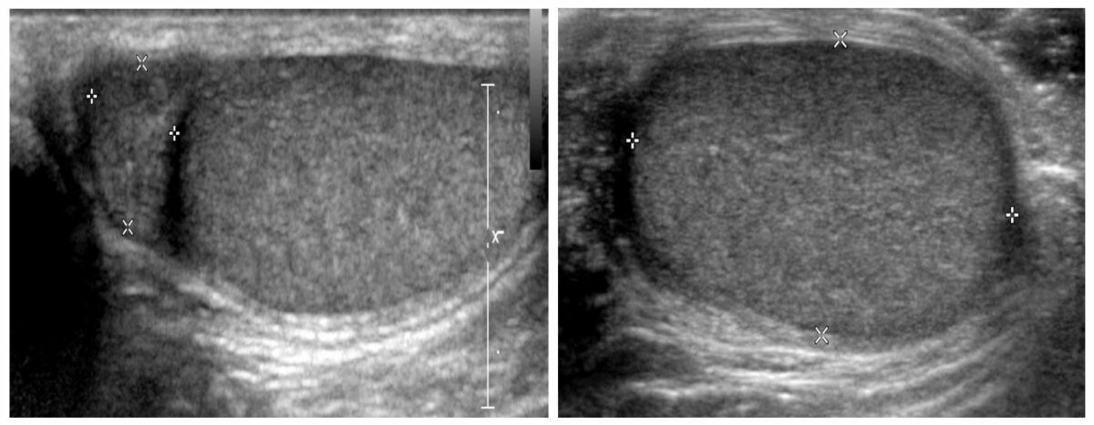
Ultrasonido escrotal que muestra el testículo (con una ecotextura homogénea sin lesiones focales ni líquido circundante) y el epidídimo
Imagen por Hetal Verma, MD.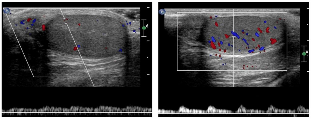
Ultrasonido Doppler que muestra un flujo arterial y venoso normal con ondas venosa (imagen izquierda) y arterial (imagen derecha) normales
Imagen por Hetal Verma, MD.Los LOS Neisseria hallazgos del ultrasonido incluyen:
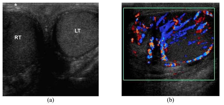
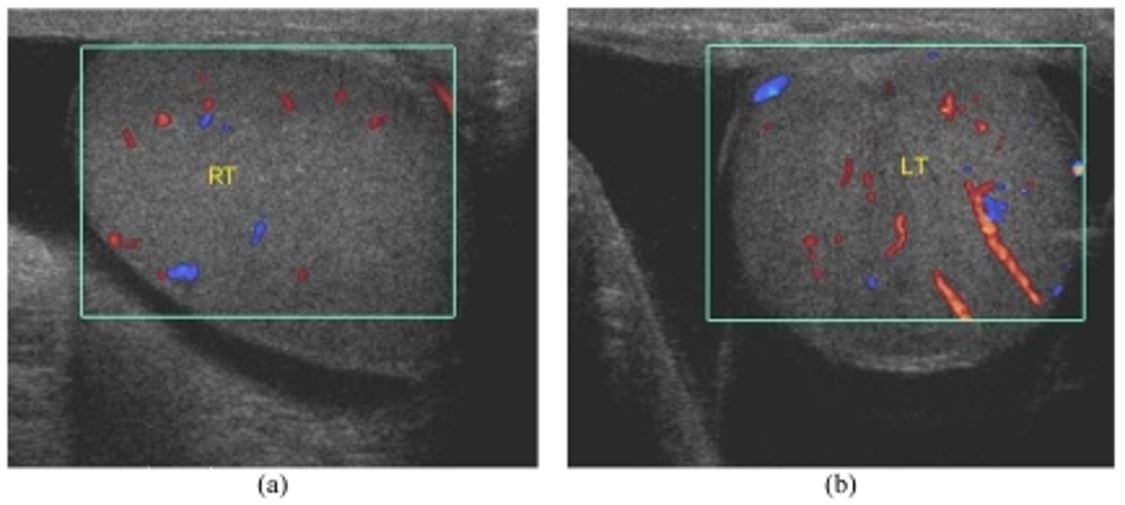
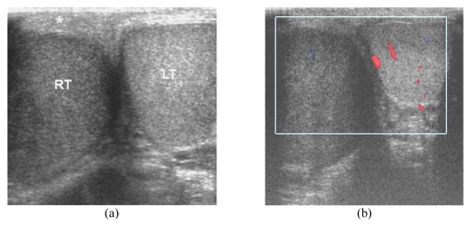
Orquiepididimitis en un hombre de 45 años que presenta una inflamación dolorosa del hemiescroto derecho desde hace 3 días:
a: Ultrasonido transversal del escroto que muestra un testículo derecho hipoecoico aumentado de tamaño (RT) y un testículo izquierdo normal (LT). La piel escrotal derecha suprayacente está engrosada (asterisco).
b: Ultrasonido Doppler color longitudinal del hemiescroto derecho muestra un aumento del flujo vascular en el epidídimo y el testículo derechos.
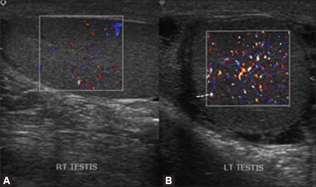
Orquitis:
El Doppler color muestra un aumento del flujo en el testículo izquierdo (flecha) con un flujo de color normal en el testículo derecho.
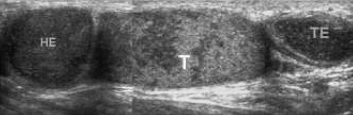
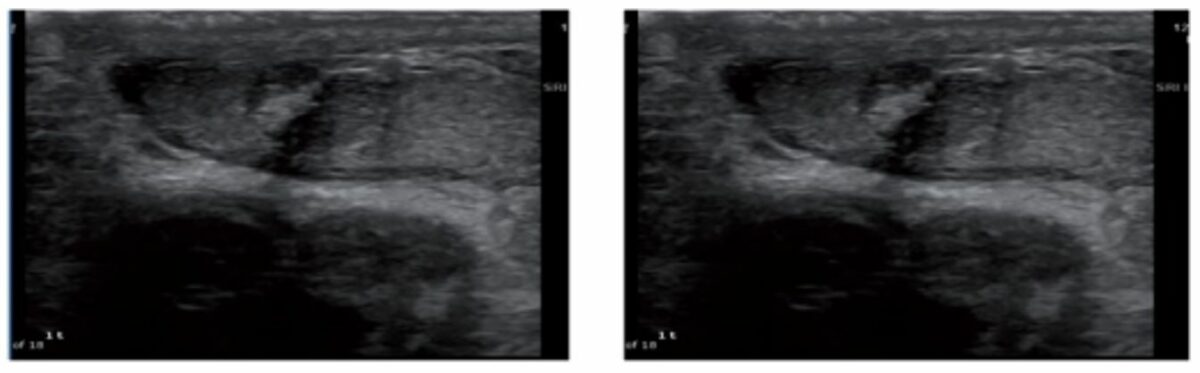
Epididimoorquitis tuberculosa en un hombre de 39 años con antecedentes de tuberculosis pulmonar, que presenta una inflamación testicular izquierda crónica e indolora desde hace 3 años:
Ultrasonido compuesto del hemiescroto izquierdo muestra una cabeza (HE) y una cola (TE) epididimarias nodulares, agrandadas y heterogéneamente hipoecoicas, y un testículo (T) heterogéneamente ecoico.
Los LOS Neisseria hallazgos del ultrasonido incluyen:
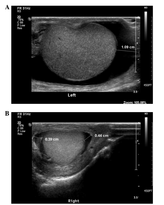
Hidrocele bilateral en un hombre de 58 años con historia de edema progresivo e indoloro del hemiescroto bilateral durante 2 años:
Las imágenes Doppler color oblicuas muestran un líquido anecoico que rodea los testículos, más a la izquierda que a la derecha. Hay un flujo vascular normal tanto en el testículo derecho (RT) como en el izquierdo (LT).
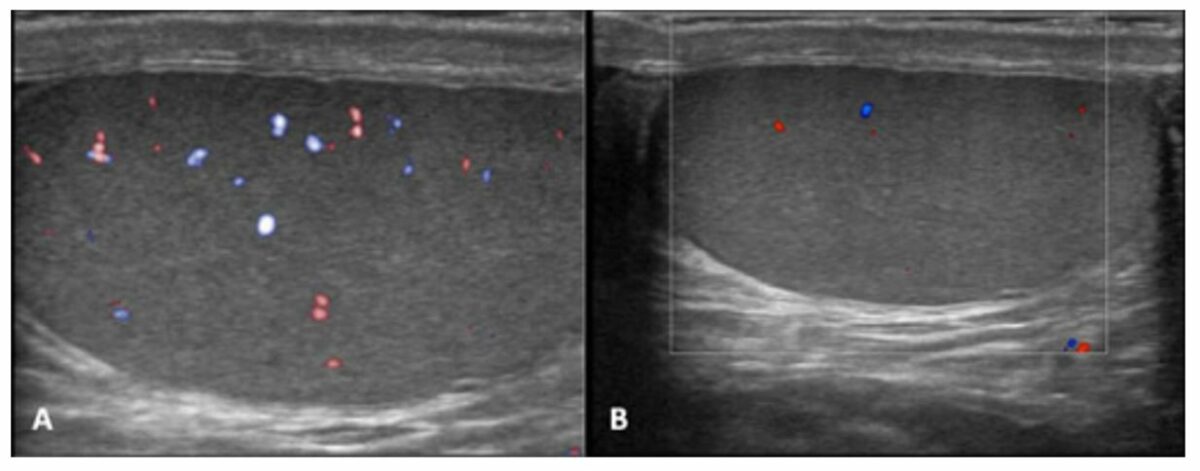
Tumor testicular metastásico que se presenta como un hidrocele escrotal:
Hallazgos del ultrasonido escrotal que muestran (A) una gran cantidad de líquido en la bolsa escrotal izquierda y (B) una pequeña cantidad de líquido con material ecogénico interno en la bolsa escrotal derecha, que indica hidroceles bilaterales
Los LOS Neisseria hallazgos del ultrasonido incluyen:
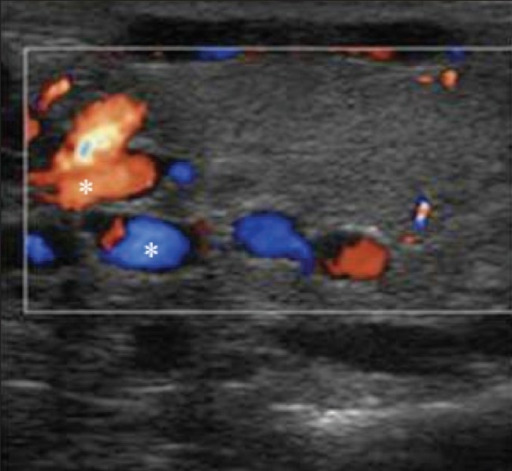
El Doppler color en varicoceles muestra un patrón venoso de flujo de color en los canales anecoicos (asterisco) confirmando el diagnóstico de varicoceles tanto intratesticulares como extratesticulares.
Imagen: “Varicoceles Color Doppler” por Patiala B. Licencia: CC BY 2.0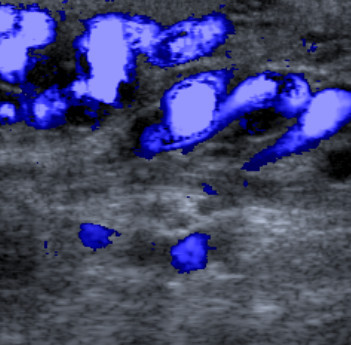
Imagen de ultrasonido Doppler color:
Vena gonadal varicosa en el canal inguinal izquierdo
Hallazgos de ultrasonido
Doppler en escala de grises y en color de la torsión testicular:
En las primeras fases de la torsión (1–3 horas), la ecogenicidad testicular parece normal. Con la progresión, el agrandamiento del testículo afectado y el aumento o la heterogeneidad de la ecogenicidad son hallazgos comunes.
Los LOS Neisseria hallazgos del ultrasonido incluyen:
Tumor de células de Leydig:
Masa heterogénea en la que se sospechaba malignidad, que finalmente resultó ser un tumor de células de Leydig con un tamaño similar al estimado en el preoperatorio
| Estructura | Ponderación T1 | Ponderación T2 | Con contraste |
|---|---|---|---|
| Testículo (estructura ovalada homogénea) | Hipointenso a isointenso | Hiperintenso | Realza |
| Epidídimo | Isointense | Hipointenso | Realza |
| Túnica albugínea | Hipointenso | Hipointenso | No aplica |
Los LOS Neisseria hallazgos de la RM incluyen:

La imagen transversal ponderada en T1 muestra una masa escrotal izquierda multilobular (flecha corta), localizada en el espacio paratesticular.
La lesión tenía una intensidad de señal similar en comparación con el parénquima testicular normal (asterisco).
Hidrocele izquierdo (flecha larga)